


 Медицина
Похожие презентации:


Neuro-oncology
1.
NEURO-ONCOLOGY2.
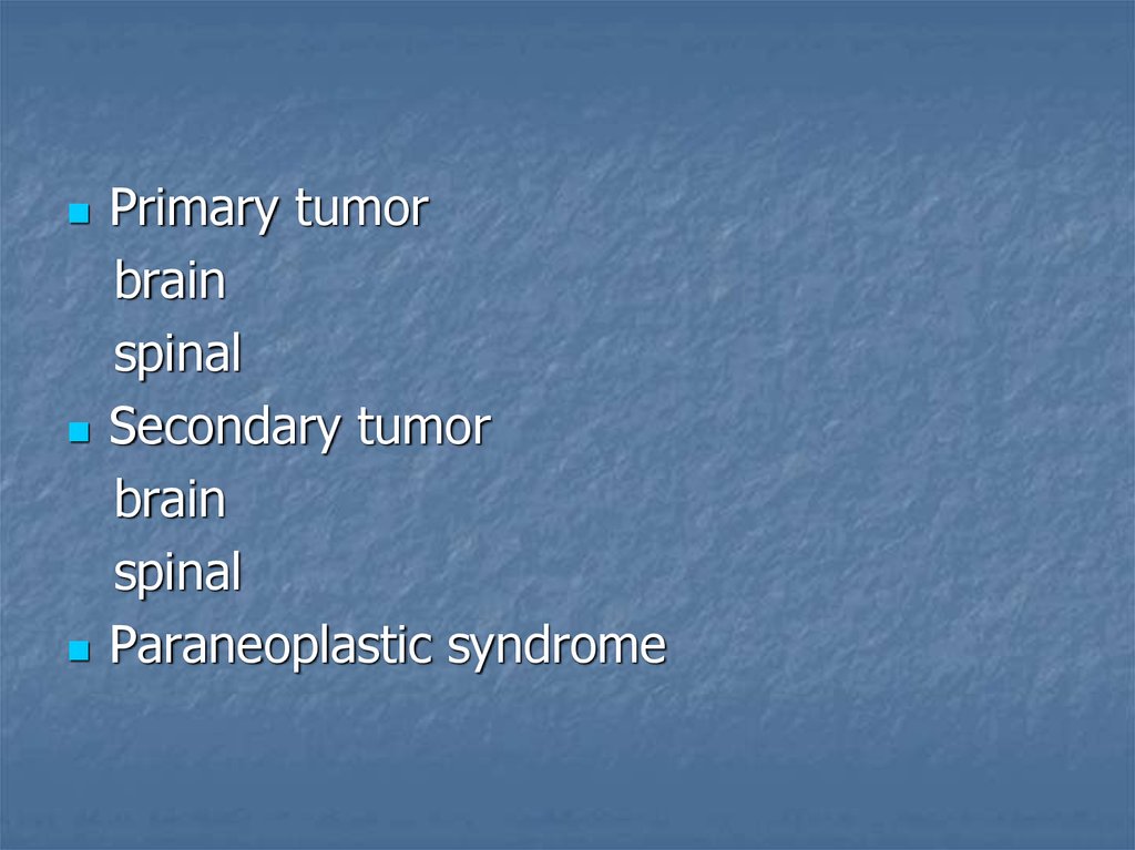
Primary tumorbrain
spinal
Secondary tumor
brain
spinal
Paraneoplastic syndrome
3.
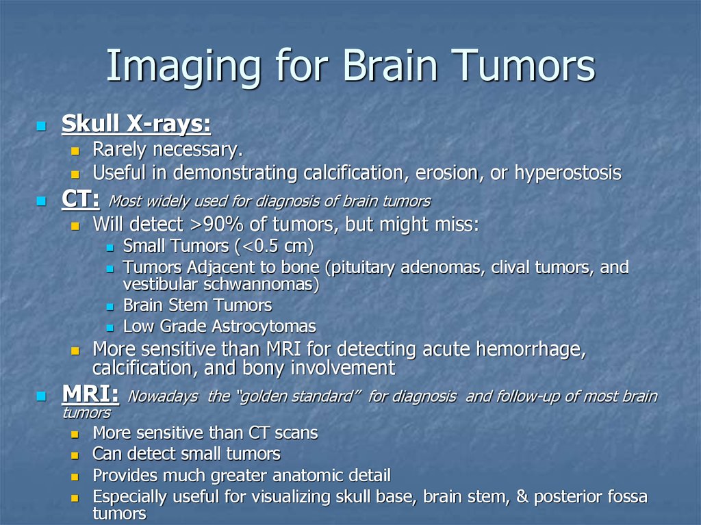
Imaging for Brain TumorsSkull X-rays:
Rarely necessary.
Useful in demonstrating calcification, erosion, or hyperostosis
CT:
Most widely used for diagnosis of brain tumors
Will detect >90% of tumors, but might miss:
More sensitive than MRI for detecting acute hemorrhage,
calcification, and bony involvement
MRI:
tumors
Small Tumors (<0.5 cm)
Tumors Adjacent to bone (pituitary adenomas, clival tumors, and
vestibular schwannomas)
Brain Stem Tumors
Low Grade Astrocytomas
Nowadays the “golden standard” for diagnosis and follow-up of most brain
More sensitive than CT scans
Can detect small tumors
Provides much greater anatomic detail
Especially useful for visualizing skull base, brain stem, & posterior fossa
tumors
4.
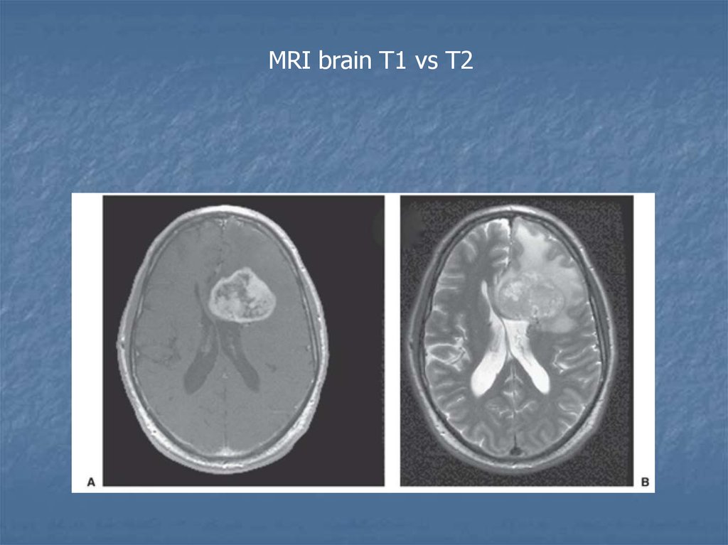
MRI brain T1 vs T25.
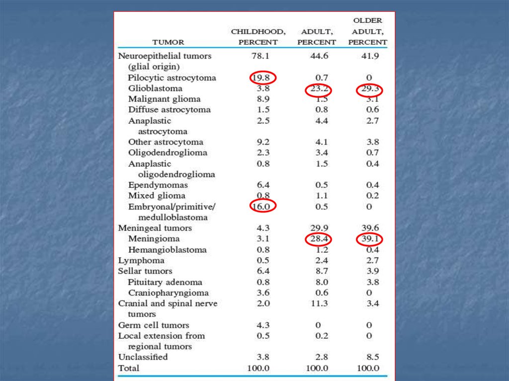
Epidemiology6.
7.
8.
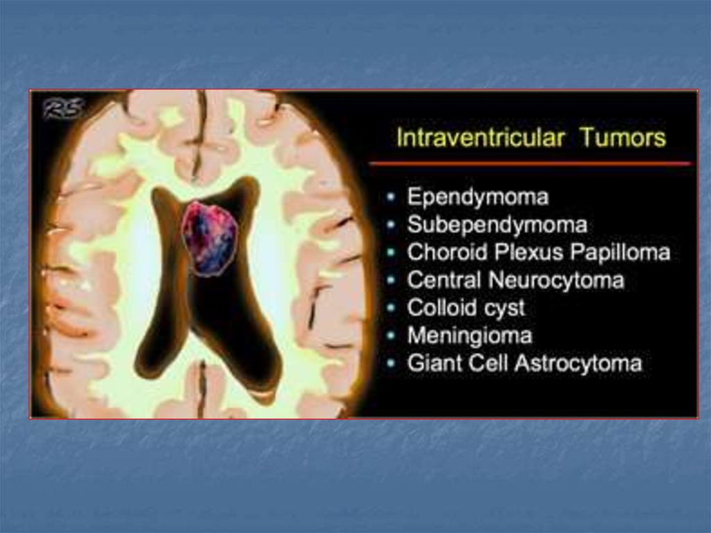
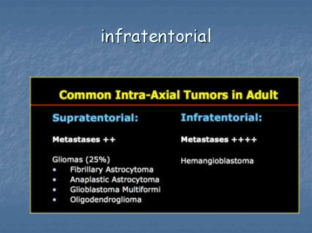
Infratentorial vs SupratentorialTumors
9.
infratentorial10.
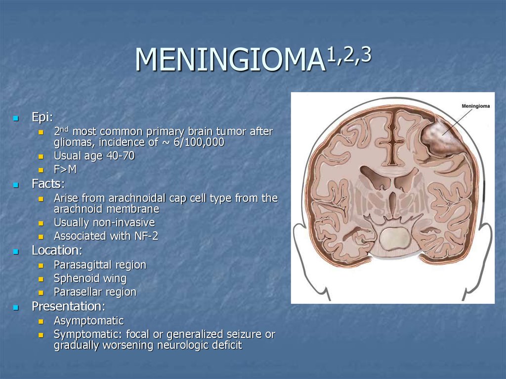
MENINGIOMA1,2,3Epi:
Facts:
Arise from arachnoidal cap cell type from the
arachnoid membrane
Usually non-invasive
Associated with NF-2
Location:
2nd most common primary brain tumor after
gliomas, incidence of ~ 6/100,000
Usual age 40-70
F>M
Parasagittal region
Sphenoid wing
Parasellar region
Presentation:
Asymptomatic
Symptomatic: focal or generalized seizure or
gradually worsening neurologic deficit
11.
On ImagingCT:
MENINGIOMA
isodense or hypodense,
homogenous extra-axial
mass with smooth or
lobulated, clearly
demarcated contours which
enhance homogenously and
densely with contrast
Frequently have areas of
calcification and produce
hyperostosis of adjacent
bone.
– MRI
• Isointense with gray matter on
T1 images
• Enhance with contrast – often
with enhancing dural trail
extending from the tumor
attachment
12.
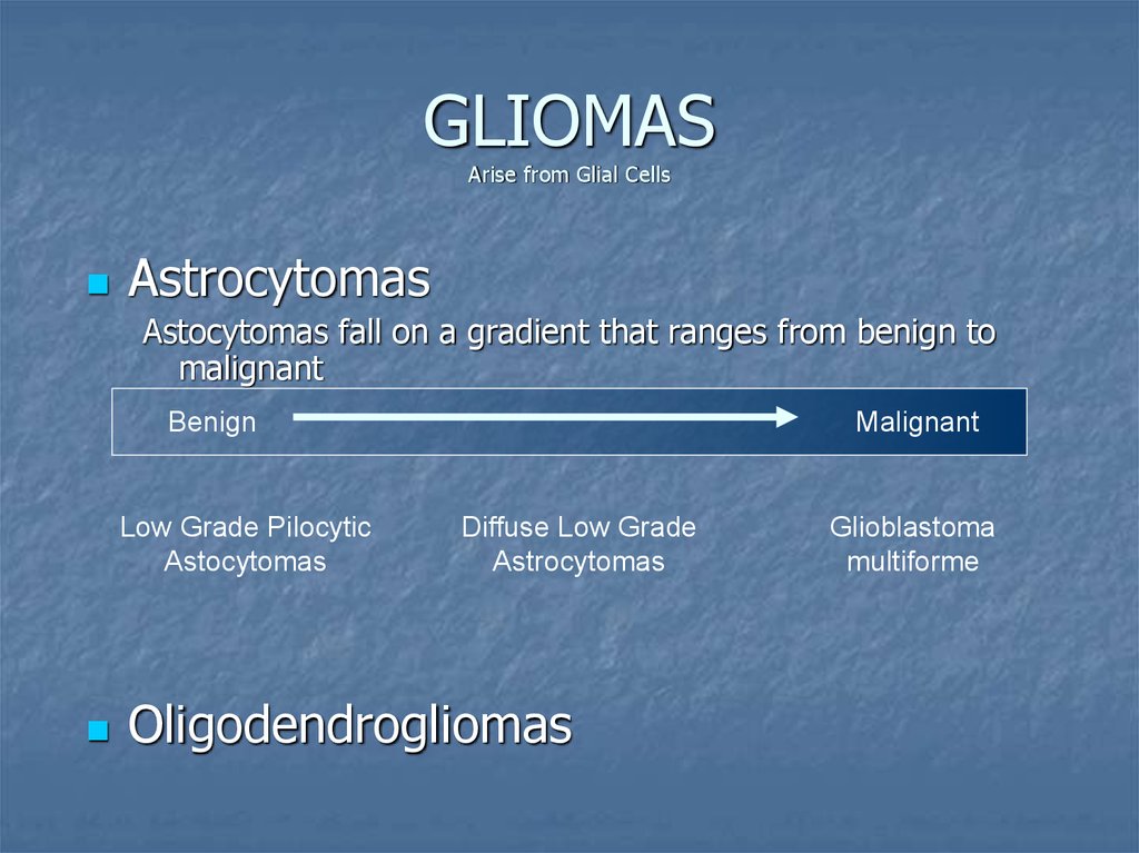
GLIOMASArise from Glial Cells
Astrocytomas
Astocytomas fall on a gradient that ranges from benign to
malignant
Benign
Low Grade Pilocytic
Astocytomas
Malignant
Diffuse Low Grade
Astrocytomas
Oligodendrogliomas
Glioblastoma
multiforme
13.
Diffuse Low Grade AstrocytomaEpi:
Facts:
Frontal Region
Subcortical white matter
Cyst
Presentation:
Widely Infiltrate surrounding tissue
Location:
15% of Astrocytomas
Young Adults
Seizures
Headache
Slowly progressive neurologic deficits
On Imaging:
T1 weighted
T2 weighted
CT: Well circumscribed, non enhancing, hypodense or isodense lesion
MRI: MRI more sensitive than CT – useful for identification and establishing extent
T1 image shows abnormal areas of decreased signal
T2 image shows abnormal areas of increased signal
Usually no enhancement
14.
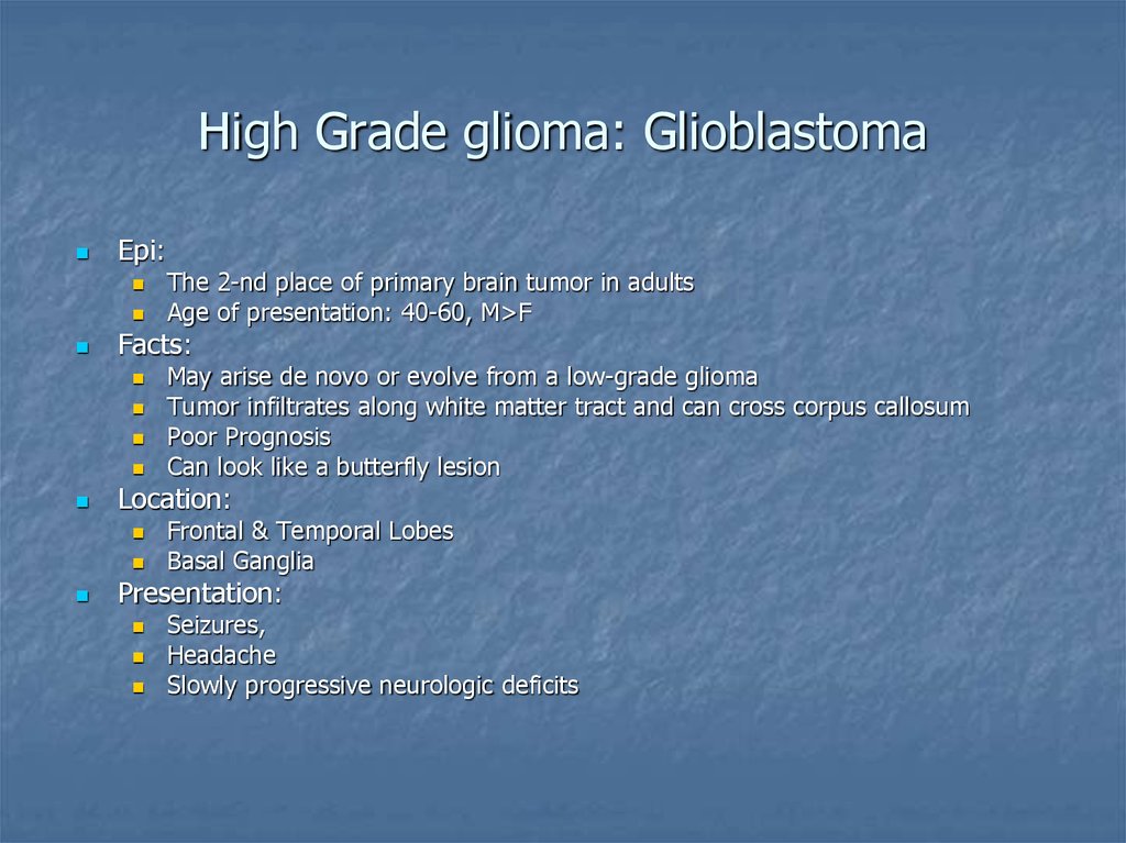
High Grade glioma: GlioblastomaEpi:
Facts:
May arise de novo or evolve from a low-grade glioma
Tumor infiltrates along white matter tract and can cross corpus callosum
Poor Prognosis
Can look like a butterfly lesion
Location:
The 2-nd place of primary brain tumor in adults
Age of presentation: 40-60, M>F
Frontal & Temporal Lobes
Basal Ganglia
Presentation:
Seizures,
Headache
Slowly progressive neurologic deficits
15.
High Grade glioma: GlioblastomaOn Imaging: Variable
CT:
Hypodense or Isodense
Central hypodense area of necrosis surrounded by thick enhancing rim
Surrounding edema
MRI:
T1 image shows low signal intensity
T2 image shows high signal intensity
16.
High Grade glioma: GlioblastomaTreatment: steroids
surgical removal
radiotherapy
chemotherapy (temozolomide)
anticonvulsive drugs
17.
Survival18.
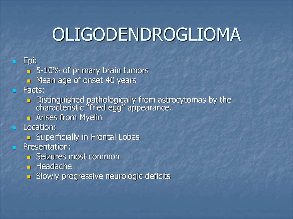
OLIGODENDROGLIOMAEpi:
5-10% of primary brain tumors
Mean age of onset 40 years
Facts:
Distinguished pathologically from astrocytomas by the
characteristic “fried egg” appearance.
Arises from Myelin
Location:
Superficially in Frontal Lobes
Presentation:
Seizures most common
Headache
Slowly progressive neurologic deficits
19.
OLIGODENDROGLIOMAOn Imaging:
CT:
Well circumscribed, hypodense lesions with heavy calcification
Cystic degeneration is common but hemorrhage & edema are uncommon
MRI:
Hypointense or isointense on T1-weighted images
Hyperintense on T2-weighted images with variable enhancement
20.
OLIGODENDROGLIOMATreatment:
Surgical excision
radiation therapy
anticonvulsive drugs
The median survival over 7 years
.
21.
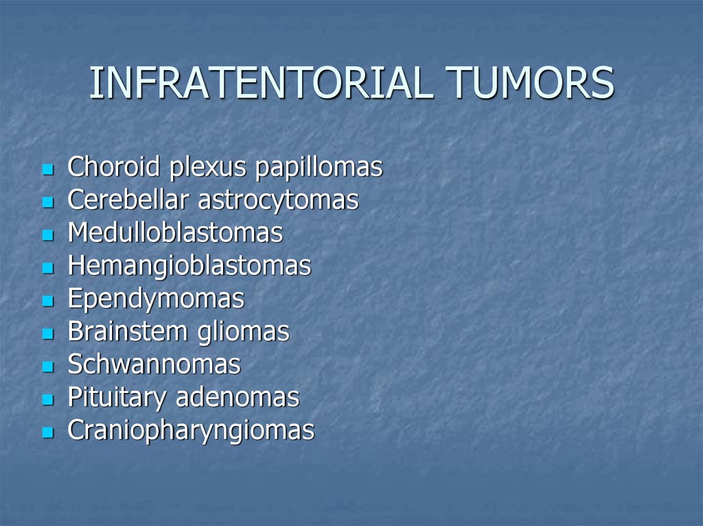
INFRATENTORIAL TUMORSChoroid plexus papillomas
Cerebellar astrocytomas
Medulloblastomas
Hemangioblastomas
Ependymomas
Brainstem gliomas
Schwannomas
Pituitary adenomas
Craniopharyngiomas
22.
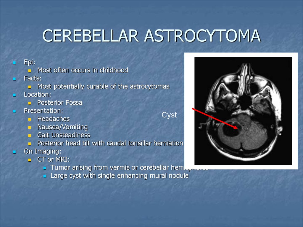
CEREBELLAR ASTROCYTOMAEpi:
Most often occurs in childhood
Facts:
Most potentially curable of the astrocytomas
Location:
Posterior Fossa
Presentation:
Cyst
Headaches
Nausea/Vomiting
Gait Unsteadiness
Posterior head tilt with caudal tonsillar herniation
On Imaging:
CT or MRI:
Tumor arising from vermis or cerebellar hemispheres
Large cyst with single enhancing mural nodule
23.
MEDULLOBLASTOMASEpi
Facts
Primitive neuroectodermal tumors (PNET)
Soft, friable tumors, often necrotic
Can metastasize via CSF tracts
Highly radiosensitive
Location
Represent 7% of primary brain tumors
2nd most common posterior fossa tumor in children
70% of patients are diagnosed prior to age 20 with peak incidence
between 5-9 years of age;
About 75% arise within the cerebellar vermis
Presentation
Most frequently present with signs of intracranial pressure
Cranial nerve deficits may also occur
24.
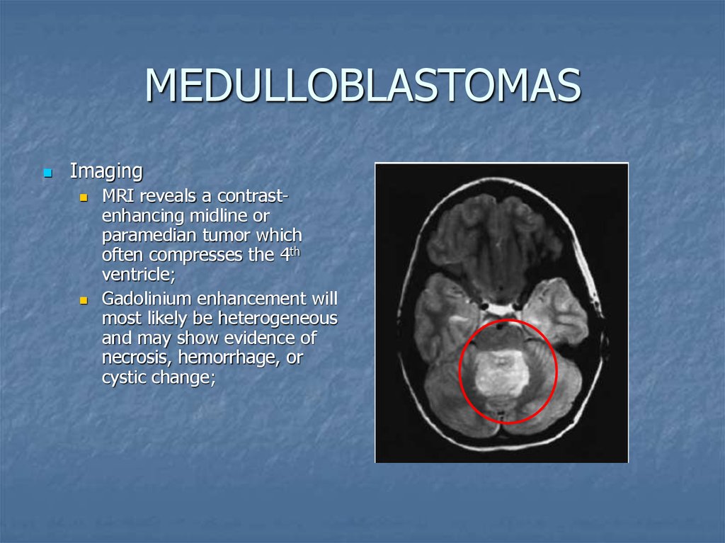
MEDULLOBLASTOMASImaging
MRI reveals a contrastenhancing midline or
paramedian tumor which
often compresses the 4th
ventricle;
Gadolinium enhancement will
most likely be heterogeneous
and may show evidence of
necrosis, hemorrhage, or
cystic change;
25.
EPENDYMOMASEpi
Facts
Derived from primitive glia
Overall survival at 10 years is 45-55%
Presentation
Accounts for 10% of CNS lesions;
Male=Female
Median age at diagnosis is 5 years old
Most patients present with symptoms of increased intracranial pressure
Location
Typically arise within or adjacent to the ependymal lining of the
ventricular system.
In children, 90% are intracranial with 60% arising in posterior fossa
(4th ventricle is the most common infratentorial site)
Most common spinal cord glioma (in adults, 75% arise within spinal
cord);;
26.
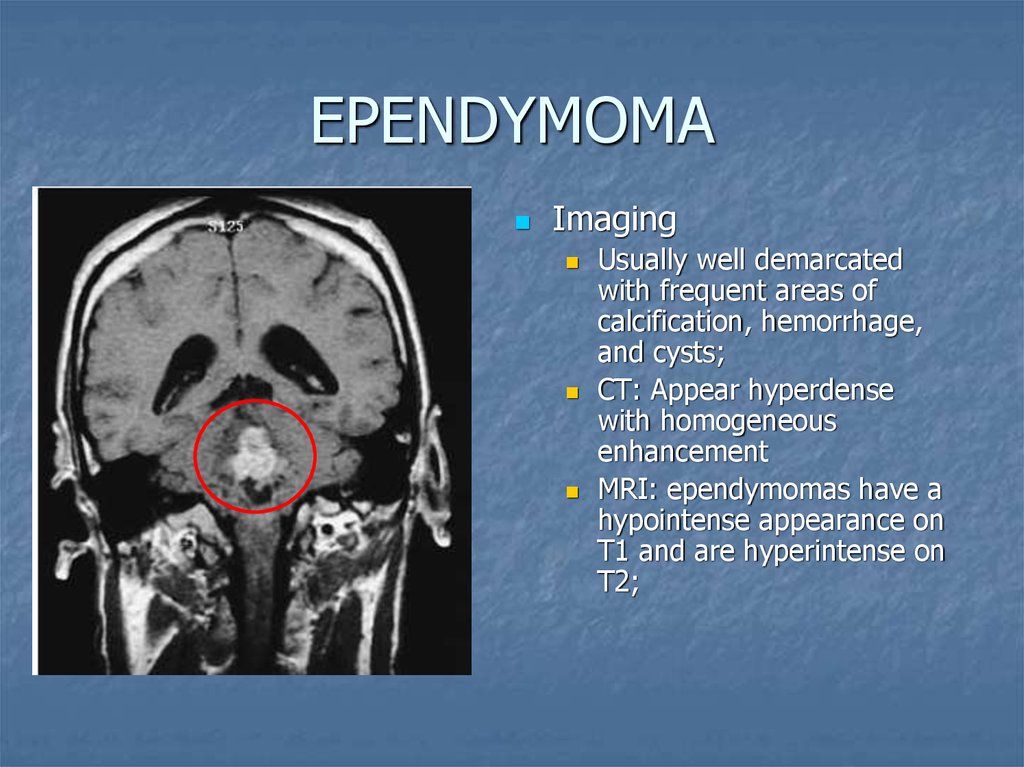
EPENDYMOMAImaging
Usually well demarcated
with frequent areas of
calcification, hemorrhage,
and cysts;
CT: Appear hyperdense
with homogeneous
enhancement
MRI: ependymomas have a
hypointense appearance on
T1 and are hyperintense on
T2;
27.
SCHWANNOMASEpi
Facts
Unilateral in 90% of cases (R=L);
Bilateral acoustic neuromas are diagnostic of NF-2;
Presentation
Female>male
Median age at diagnosis is 50
Account for 80-90% of cerebellopontine angle tumors
Comprise 8% of intracranial tumors in adults; rare in children (except with NF-2)
Patients may present with asymmetric sensorineural hearing loss, tinnitus
Fluctuating unsteadiness while walking, vertigo (although only 1% of patients
with vertigo had schwannomas);
If CN V nerve is affected, facial numbness, pain, and hyperesthesia may be
present;
If CN VII is affected, facial paresis may be present.
Tumor progression may lead to compression of brainstem or cerebellum leading
to ataxia, tonsil herniation, and hydrocephalus
Location
Arise from vestibular division of CN VIII; majority benign
28.
SCHWANNOMASImaging
MRI: with gadolinium is
more sensitive in detection
of Schwannomas (when
compared to CT); it can
detect tumors as small as
1-2 mm; seen as
enhancing lesion in the
region of CPA;
Fine-cut CT through
internal auditory canal can
detect large or medium
tumors.
29.
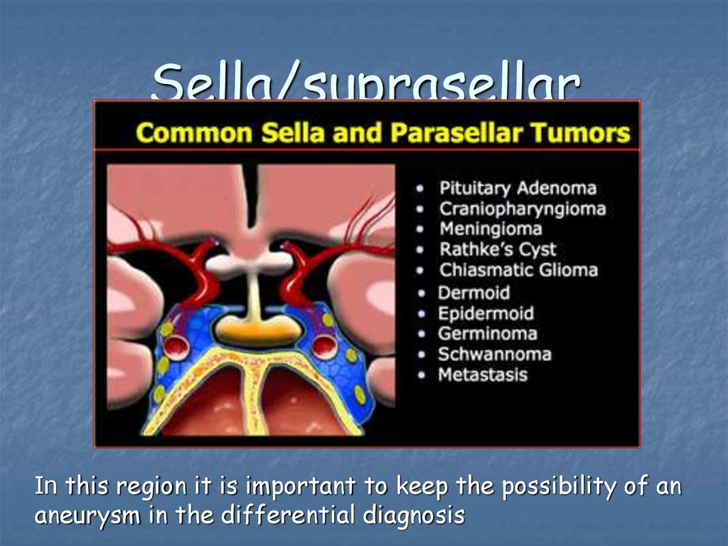
Sella/suprasellarIn this region it is important to keep the possibility of an
aneurysm in the differential diagnosis
30.
PITUITARY ADENOMASEpi
Facts
Most common tumors of pituitary gland
Represent 8% of primary brain tumors
Out of pituitary adenomas, prolactinomas are the most
common;
Presentation
May cause hypopituitarism and visual field defects;
Patients should have endocrine, radiographic, and
ophthalmologic assessments.
31.
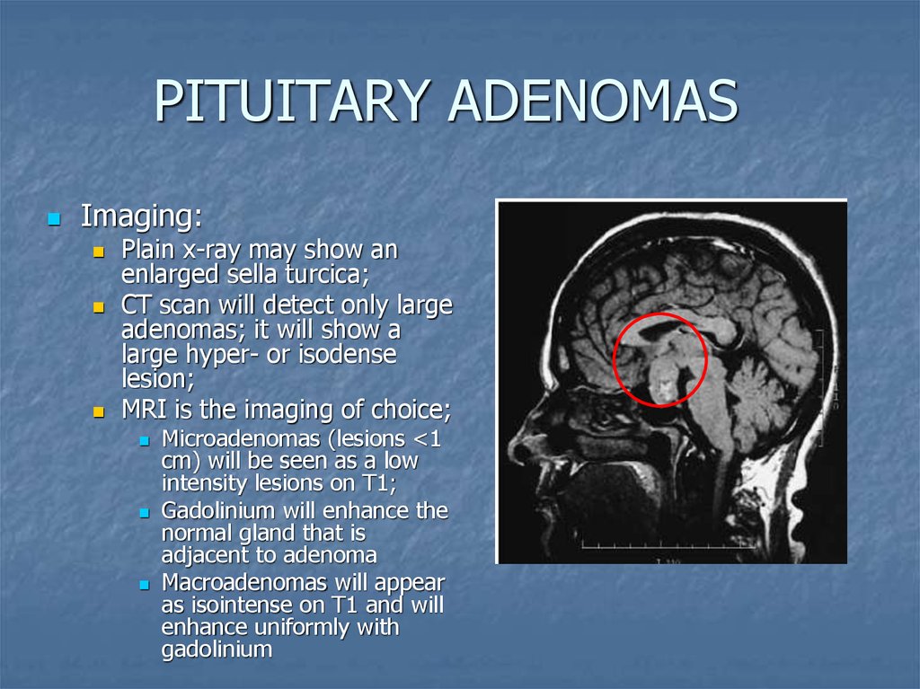
PITUITARY ADENOMASImaging:
Plain x-ray may show an
enlarged sella turcica;
CT scan will detect only large
adenomas; it will show a
large hyper- or isodense
lesion;
MRI is the imaging of choice;
Microadenomas (lesions <1
cm) will be seen as a low
intensity lesions on T1;
Gadolinium will enhance the
normal gland that is
adjacent to adenoma
Macroadenomas will appear
as isointense on T1 and will
enhance uniformly with
gadolinium
32.
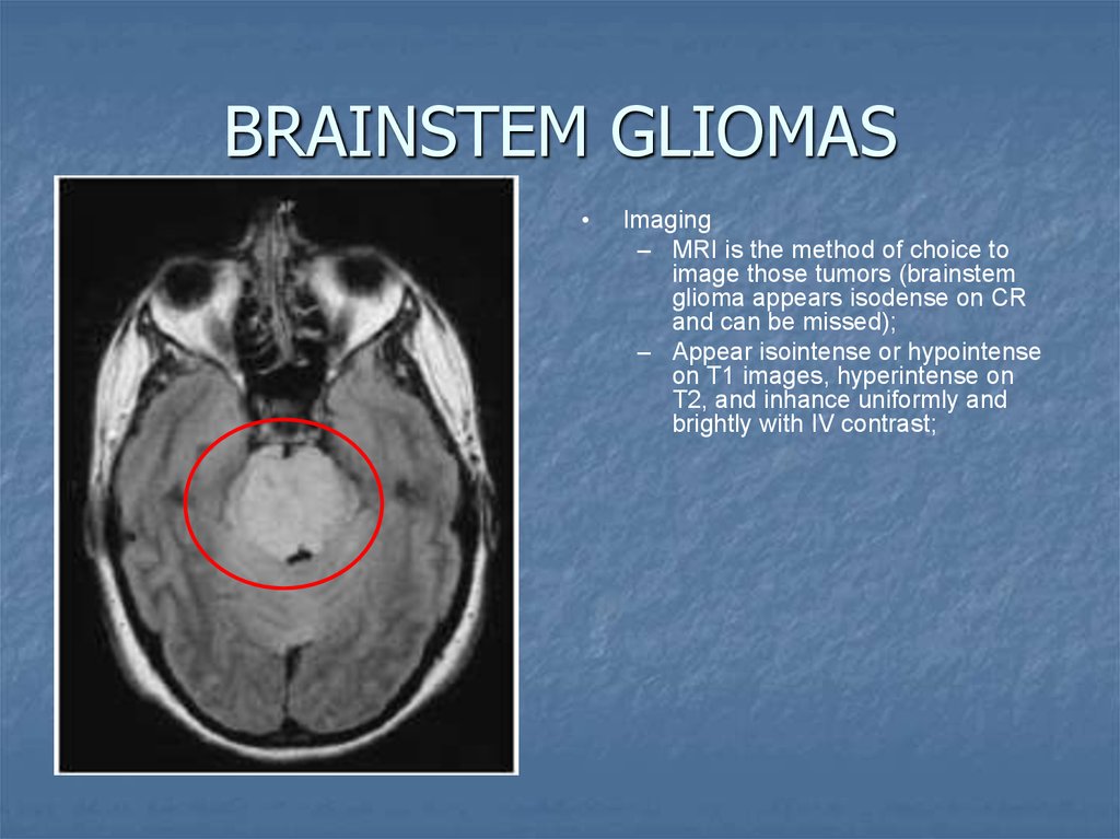
BRAINSTEM GLIOMASEpi
Male=Female
Account for 10-20% on all CNS tumors
More common in children (account for 20% of all intracranial neoplasms under the
age 15);
In children, median age at diagnosis is 5-9 years of age.
Facts
NF-1 is the only known risk factor
Mostly benign (but range from benign to very aggressive);
Long term survival for low-grade gliomas is near 100%.
Location
In peds, 80% arise in pons, with 20% arise in medula, midbrain, and
cervicomedulary junction;
Presentation
Most patients with low-grade brainstem gliomas have a long history of minor signs
and symptoms;
May present with neck pain or torticollis;
Medulary tumors may present with cranial nerve palsies, dysphagia, nasal speech
and apnea, n/v, ataxia,or weakness;
May cause “locked-in” syndrome
33.
BRAINSTEM GLIOMASImaging
– MRI is the method of choice to
image those tumors (brainstem
glioma appears isodense on CR
and can be missed);
– Appear isointense or hypointense
on T1 images, hyperintense on
T2, and inhance uniformly and
brightly with IV contrast;
34.
4th ventricleIn
adults tumors in the 4th
ventricle are uncommon.
Metastases,
followed by
hemangioblastomas, choroid
plexus papillomas and dermoid
and epidermoid cysts.
35.
Metastatic tumorsParenchymal meta – most common
masses in the in supratentorial and
infratentorial spaces (more supra)
50% solitary, 50% multiple, 20% 2
lesions
Origin – lung, (50%), breast (15%)
melanoma(11%), kidney, GIT
Cystic meta- ovary, breast, GIT
36.
Hemorrhagic metaBreast
Choriocarcinoma
lung
Melanoma
RCC
Thyroid
retinoblastoma
37.
Secondary tumors-MTSLung cancer (NSCCa)
Breast cancer
Melanoma
Kidney
Thyroid
38.
Carcinomatous Meningitis(Meningeal Carcinomatosis)
Dissemination of tumor cells throughout the meninges
and ventricles.
5 percent of cases of adenocarcinoma of breast, lung, and
gastrointestinal tract; melanoma; childhood leukemia;
and systemic lymphoma.
Manifestations:
Polyradiculopathies (particularly of the cauda equina), multiple cranial
nerve palsies, and a confusional state.
Treatment
Radiation therapy to the symptomatic areas (cranium, posterior fossa,
or spine),
Intraventricular/intratecal methotrexate
39.
Multiple brain tumors can be seenin phacomatoses:
Neurofibromatosis II: meningiomas,
ependymomas, optic nerve gliomas,
choroid plexus papillomas
Tuberous Sclerosis: subependymal
tubers, intraventricular giant cell
astrocytomas, ependymomas
von Hippel Lindau:
hemangioblastomas