


 Биология
Похожие презентации:
")


Enterobacteriaceae
1.
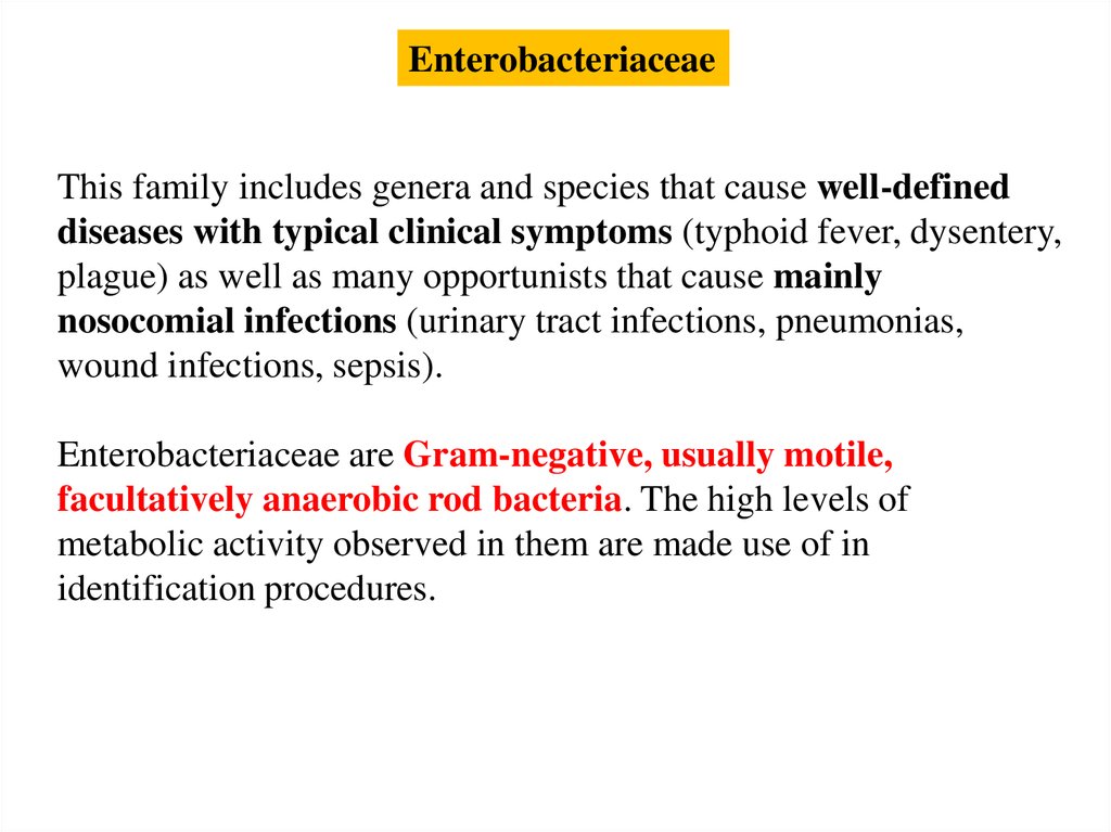
EnterobacteriaceaeThis family includes genera and species that cause well-defined
diseases with typical clinical symptoms (typhoid fever, dysentery,
plague) as well as many opportunists that cause mainly
nosocomial infections (urinary tract infections, pneumonias,
wound infections, sepsis).
Enterobacteriaceae are Gram-negative, usually motile,
facultatively anaerobic rod bacteria. The high levels of
metabolic activity observed in them are made use of in
identification procedures.
2.
The species are subdivided into epidemiologically significantserovars based on O, H, and K antigens.
The most important pathogenicity factors of Enterobacteriaceae are
colonizing factors, invasins, endotoxin, and various exotoxins.
Enterobacteriaceae are the most significant contributors to intestinal
infections, which are among the most frequent diseases of all
among the developing world populace.
3.
Definition and significanceTogether with the families Vibrionaceae and others, the
Enterobacteriaceae form the group of Gram-negative, facultatively
anaerobic rod bacteria. Their natural habitat is the intestinal tract
of humans and animals. Some species cause characteristic
diseases. While others are facultatively pathogenic, they are still
among the bacteria most frequently isolated as pathogens. They are
often responsible for nosocomial diseases.
4.
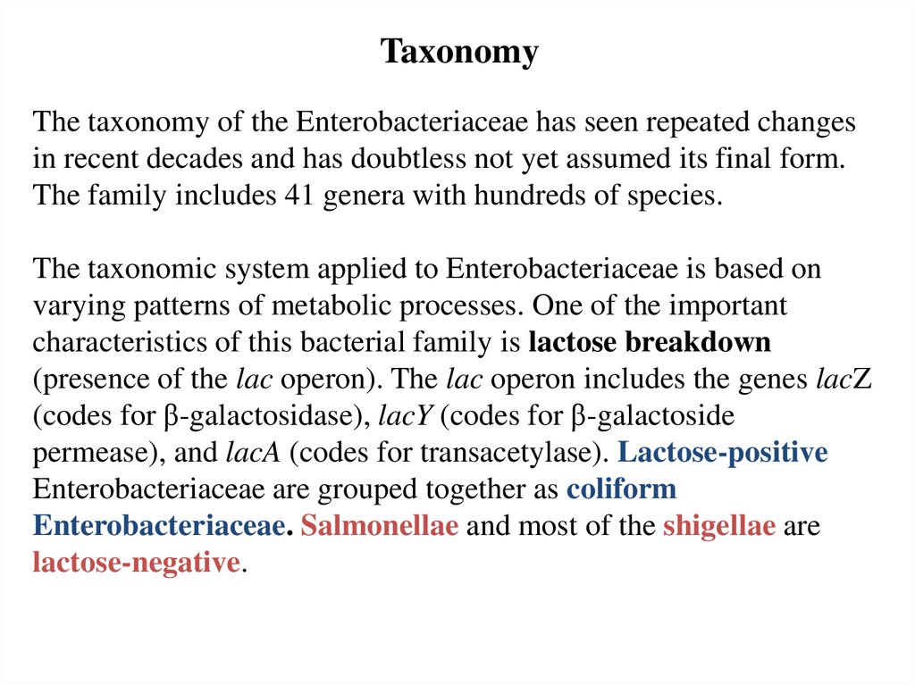
TaxonomyThe taxonomy of the Enterobacteriaceae has seen repeated changes
in recent decades and has doubtless not yet assumed its final form.
The family includes 41 genera with hundreds of species.
The taxonomic system applied to Enterobacteriaceae is based on
varying patterns of metabolic processes. One of the important
characteristics of this bacterial family is lactose breakdown
(presence of the lac operon). The lac operon includes the genes lacZ
(codes for β-galactosidase), lacY (codes for β-galactoside
permease), and lacA (codes for transacetylase). Lactose-positive
Enterobacteriaceae are grouped together as coliform
Enterobacteriaceae. Salmonellae and most of the shigellae are
lactose-negative.
5.
Enterobacteriaceae6.
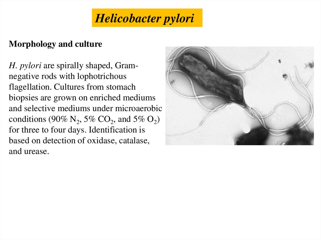
7.
Salmonella (Gastroenteritis, Typhoid Fever,Paratyphoid Fever)
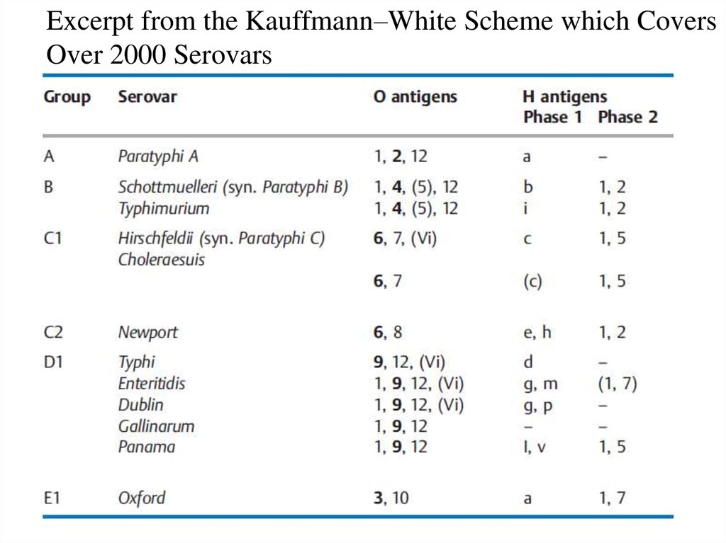
All salmonellae are classified in the species Salmonella
enterica with seven subspecies. Nearly all human
pathogen salmonellae are grouped under S. enterica,
subsp. enterica. Salmonellae are further subclassified in
over 2000 serovars based on their O and H antigens,
which used to be (incorrectly) designated as species.
8.
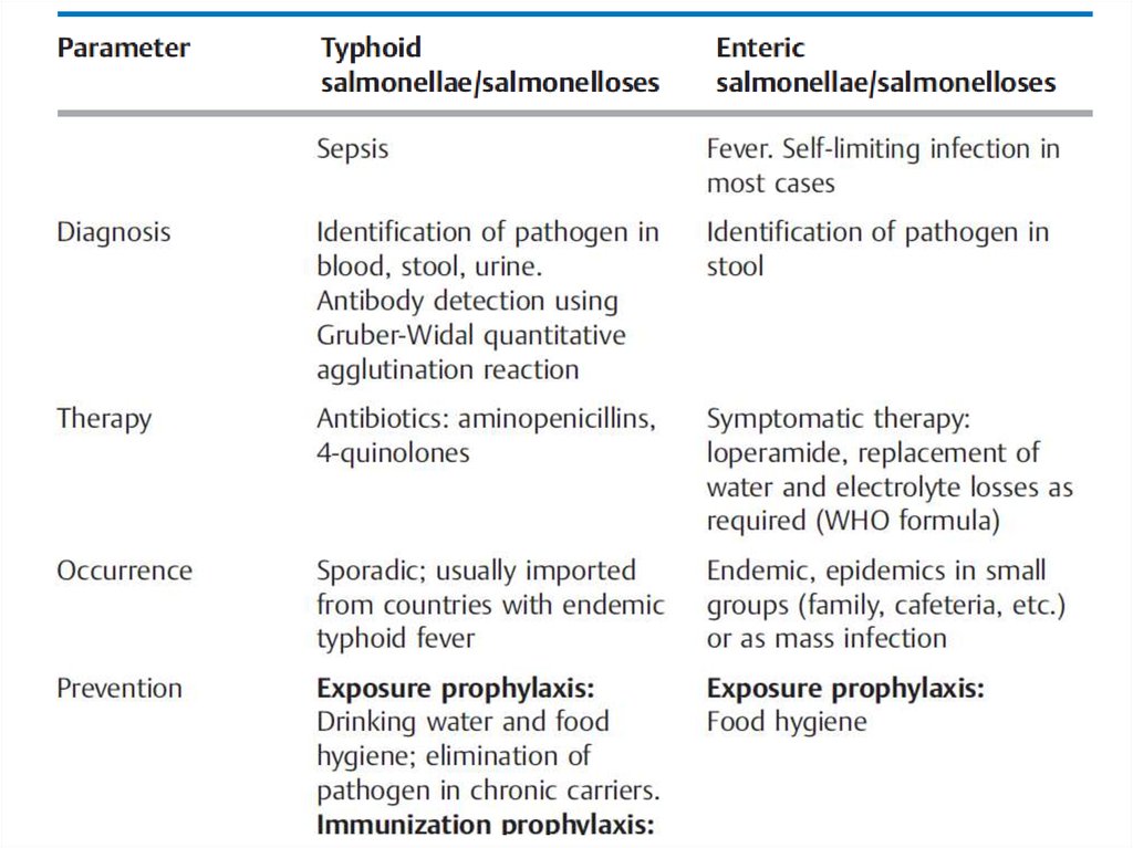
Typhoid salmonelloses are caused by the serovars typhi andparatyphi A, B, and C. The salmonellae are taken up orally and
the invasion pathway is through the intestinal tract, from where
they enter lymphatic tissue, first spreading lymphogenously,
then hematogenously.
A generalized septic clinical picture results. Human carriers are
the only source of infection. Transmission is either direct by
smear infection or indirect via food and drinkingwater. Antiinfective agents are required for therapy (ampicillin,
cotrimoxazole, 4-quinolones). An active vaccine is available to
protect against typhoid fever.
9.
Enteric salmonelloses develop when pathogens aretaken up with food. The primary infection source is
usually livestock. These relatively frequent infections
remain restricted to the gastrointestinal tract. Treatment
with anti-infective agents is necessary in exceptional
cases only.
10.
Excerpt from the Kauffmann–White Scheme which CoversOver 2000 Serovars
11.
12.
Overview of the Most Important Differences between Typhoid andEnteric Salmonellae and Salmonelloses
13.
14.
EpidemiologyThe cases of typhoid salmonelloses seen in northern and central
Europe are imported by travelers. Cases arise only sporadically
or in form of an epidemic because of a chain of unfortunate
circumstances. Humans are the only primary source of infection.
By contrast, enteric salmonelloses occur in this population both
endemically and epidemically. Case counts are steadily increasing.
Exact morbidity data are hard to come by due to the large numbers
of unreported cases. Livestock represents the most important
source of infection. The pathogens are transmitted to humans in
food.
15.
Shigella (Bacterial Dysentery)Shigella is the causative pathogen in bacterial dysentery. The genus
comprises the species S. dysenteriae, S. flexneri, S. boydii, and S.
sonnei. Shigellae are nonmotile. The three primary species can be
classified in serovars based on the fine structure of their O antigens.
Shigellae are characterized by invasive properties. They can
penetrate the colonic mucosa to cause local necrotic infections.
Humans are the sole source of infection since shigellae are
pathologically active in humans only. The pathogens are transmitted
directly, more frequently indirectly, via food and drinking water.
Antibiotics can be used therapeutically.
16.
PathogenesisShigellae are only pathogenic in humans. The pathogens
are ingested orally. Only a few hundred bacteria suffice for
an infective dose. Shigellae enter the terminal ileum and
colon, where they are taken up by the M cells in the
intestinal mucosa, which in turn are in close vicinity to the
macrophages. Following phagocytosis by the macrophages,
the shigellae lyse the phagosome and actively induce
macrophage apoptosis. The shigellae released from the
dead macrophages are then taken up by enterocytes via the
basolateral side of the mucosa (i.e., retrograde transport).
17.
PathogenesisThe invasion is facilitated by outer membrane polypeptides, the
invasins, which are coded by inv genes localized on 180–240 kb
plasmids. Adjacent enterocytes are invaded by means of lateral
transfer from infected cells. In the enterocytes, the shigellae
reproduce, finally destroying the cells. Shigella dysenteriae produces
shigatoxin, the prototype for the family of shigalike toxins (or
verocytotoxins), which also occur in several other
Enterobacteriaceae. The toxin inhibits protein synthesis in eukaryotic
cells by splitting the 23S rRNA at a certain locus. Shigatoxin
contributes to the colonic epithelial damage, the small intestine
diarrhea with watery stools at the onset of shigellosis and (less
frequent) the hemolytic-uremic syndrome (HUS).
18.
TherapyAnti-infective agents are the first line of treatment
(aminopenicillins, 4-quinolones, cephalosporins). Losses of water
and electrolytes may have to be replaced.
Epidemiology and prevention
Bacterial dysentery occurs worldwide, although it is usually seen
only sporadically in developed countries. In developing countries,
its occurrence is more likely to be endemic and even epidemic. The
source of infection is always humans, in most cases infected
persons whose stools contain pathogens for up to six weeks after
the disease has abated. Transmission is by direct contact (smear
infection) or indirect uptake via food, surface water, or flies.
Control of dysentery includes exposure prophylaxis measures
geared to prevent susceptible persons from coming into contact
with the pathogen.
19.
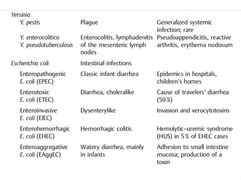
Yersinia (Plague, Enteritis)Y. pestis is the causative pathogen of plague (black death, bubonic plague).
Plague is a classic rodent zoonosis. It occurred in epidemic proportions in the
Middle Ages, but is seen today only sporadically in persons who have had
direct contact with diseased wild rodents. The pathogens penetrate into
the skin through microtraumata, from where they reach regional lymph
nodes in which they proliferate, resulting in the characteristic buboes. In
the next stage, the pathogens may enter the bloodstream or the infection
may generalize to affect other organs. Laboratory diagnosis involves isolation
and identification of the organism in pus, blood, or other material. Therapy
requires use of antibiotics.
Y. enterocolitica and Y. pseudotuberculosis cause generalized zoonoses in wild
animals and livestock. Diseased animals contaminate their surroundings.
Humans then take up the pathogens orally in water or food. The organisms
penetrate the mucosa of the lower intestinal tract, causing enteritis
accompanied by mesenteric lymphadenitis.
Extramesenteric forms are observed in 20% of infected persons (sepsis,
lymphadenopathies, various focal infections). Secondary immunopathological
complications include arthritis and erythema nodosum. Diagnosis involves
identification of the pathogen by means of selective culturing.
20.
Yersinia pestisMorphology and culture
Y. pestis is a nonflagellated, short, encapsulated, Gram-negative
rod bacteria that often shows bipolar staining. This bacterium is
readily cultured on standard nutrient mediums at 30°C.
Pathogenesis and clinical picture
The plague is primarily a disease of rodents (rats). It spreads among
them by direct contact or via the rat flea. Earlier plague epidemics
in humans resulted from these same transmission pathways. The
rare human infections seen today result from contact with rodents
that are infected with or have died of plague. The pathogen breaches
the skin through dermal injuries. From such a location, the bacteria
reach regional lymph nodes in which they proliferate. Two to five
days after infection, hemorrhagically altered, blue, and swollen
lymph nodes (buboes) are observed.
21.
Diagnosis. The pathogen must be identified in bubo punctate, sputum,or blood by means of microscopy and culturing.
Therapy. In addition to symptomatic treatment, antibiotics are the
primary method (streptomycin, tetracyclines, in the case of meningitis,
chloramphenicol). Incision of the buboes is contraindicated.
Epidemiology and prevention. Plague still occurs endemically in
wild rodents over large areas of Asia, Africa, South America, and
North America. Human plague infections have been reduced to
sporadic instances. The sources of infection are mainly diseased
rodents. Transmission of the disease is mainly via direct contact with
such animals. Prevention involves exposure prophylactic measures.
Persons with manifest disease, in particular the pulmonary form, must
be isolated. Contact persons must be quarantined for six days (=
incubation period). Cases of plague infection must be reported to
health authorities.
22.
Yersinia enterocolitica and Yersinia pseudotuberculosisOccurrence and significance
Y. enterocolitica and Y. pseudotuberculosis cause
generalized infections in domestic and wild animals, especially
rodents. The pathogens can be transmitted from animals to
humans. Y. enterocolitica is responsible for about 1% of acute
enteritis cases in Europe. Y. pseudotuberculosis is insignificant in
terms of human pathology.
23.
Escherichia coliThe natural habitat of E. coli is the intestinal tract of humans and animals.
It is therefore considered an indicator organism for fecal contamination of
water and foods. E. coli is the most frequent causative pathogen in human
bacterial infections. Extraintestinal infections include urinary tract infections,
which occur when the tract is obstructed or spontaneously caused
by the pathovar UPEC. The most important other coli infections are cholecystitis,
appendicitis, peritonitis, postoperative wound infections, and sepsis.
Intestinal infections are caused by the pathovars EPEC, ETEC, EIEC, EHEC,
and EAggEC. EPEC and EAggEC frequently cause diarrhea in infants. ETEC
produce enterotoxins that cause a choleralike clinical picture. EIEC cause a
dysenterylike infection of the large intestine. EHEC produce verocytotoxins
and cause a hemorrhagic colitis as well as the rare hemolytic-uremic syndrome.
E. coli bacteria infections are diagnosed by means of pathogen identification
24.
General characteristicsThe natural habitat of E. coli is the intestines of animals and
humans. This bacterium is therefore used as an indicator for fecal
contamination of drinking water, bathing water, and foods.
Guideline regulations: 100 ml of drinking water must not contain
any E. coli. Surface water approved for bathing should not contain
more than 100 (guideline value) to 2000 (absolute cutoff value) E.
coli bacteria per 100 ml. E. coli is also an important human
pathogen. It is the bacterial species most frequently isolated from
pathological materials
25.
Morphology, culture, and antigen structureThe Gram-negative, straight rods are peritrichously flagellated.
Lactose is broken down rapidly. The complex antigen structure
of these bacteria is based on O, K, and H antigens. Fimbrial
antigens have also been described. Specific numbers have been
assigned to the antigens, e.g., serovar O18:K1:H7.
26.
Vibrio cholerae (Cholera)Morphology and culture. Cholera vibrios are Gramnegative rod bacteria, usually slightly bent (commashaped), 1.5–2 lm in length, and 0.3–0.5 lm wide, with
monotrichous flagellation.
Culturing of V. cholerae is possible on simple nutrient
mediums at 37°C in a normal atmosphere. Owing to its
pronounced alkali stability, V. cholerae can be selectively
cultured out of bacterial mixtures at pH 9.
27.
Antigens and classification.V. cholerae bacteria are subdivided into serovars based
on their O antigens (lipopolysaccharide antigens). The
serovar pathogen is usually serovar O:1. Strains that
do not react to an O:1 antiserum are grouped together
as nonO:1 vibrios. NonO:1 strains were recently
described in India (O:139) as also causing the classic
clinical picture of cholera. O:1 vibrios
are further subclassified in the biovars cholerae and
eltor based on physiological characteristics. The var
eltor has a very low level of virulence.
28.
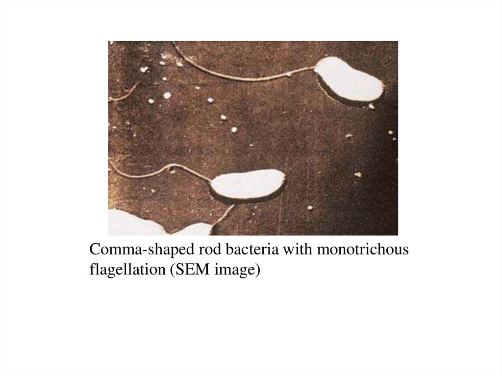
Comma-shaped rod bacteria with monotrichousflagellation (SEM image)
29.
30.
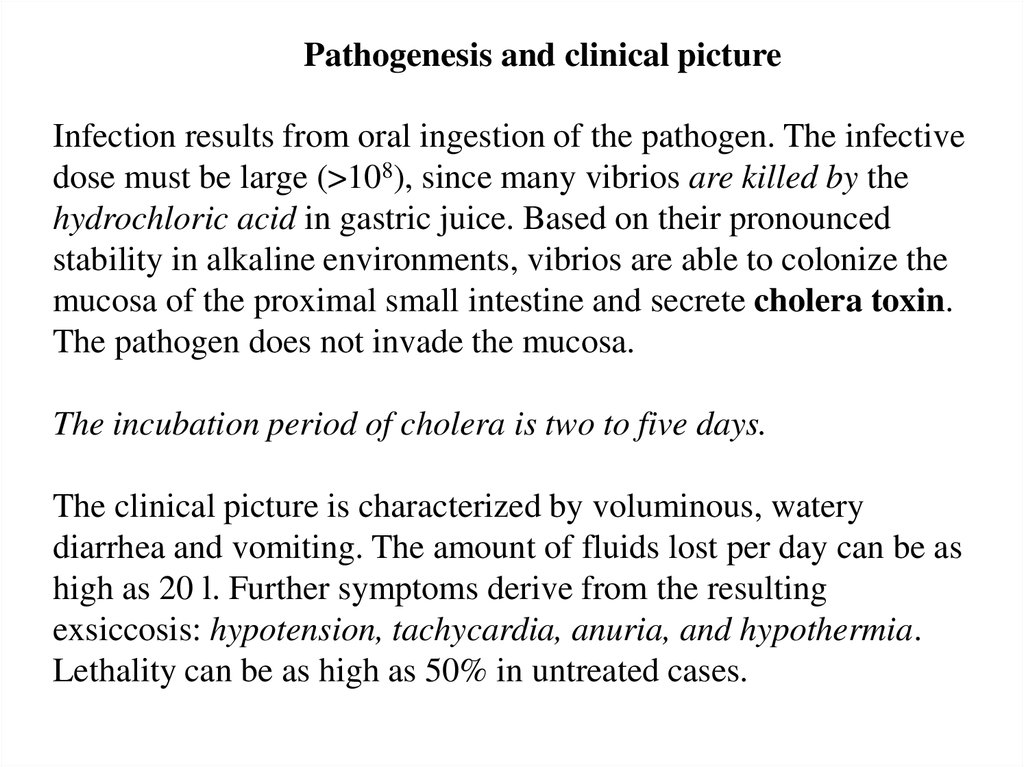
Pathogenesis and clinical pictureInfection results from oral ingestion of the pathogen. The infective
dose must be large (>108), since many vibrios are killed by the
hydrochloric acid in gastric juice. Based on their pronounced
stability in alkaline environments, vibrios are able to colonize the
mucosa of the proximal small intestine and secrete cholera toxin.
The pathogen does not invade the mucosa.
The incubation period of cholera is two to five days.
The clinical picture is characterized by voluminous, watery
diarrhea and vomiting. The amount of fluids lost per day can be as
high as 20 l. Further symptoms derive from the resulting
exsiccosis: hypotension, tachycardia, anuria, and hypothermia.
Lethality can be as high as 50% in untreated cases.
31.
DiagnosisDiagnosis requires identification of the pathogen in stool or
vomit. Sometimes a rapid microscopical diagnosis succeeds in
finding numerous Gram-negative, bent rods in swarm patterns.
Culturing is done on liquid or solid selective mediums, e.g.,
alkaline peptone water or taurocholate gelatin agar. Suspected
colonies are identified by biochemical means or by detection of
the O:1 antigen in an agglutination reaction.
32.
TherapyThe most important measure is restoration of the disturbed
waternand electrolyte balance in the body. Secondly,
tetracyclines and cotrimoxazole can be used, above all to
reduce fecal elimination levels and shorten the period of
pathogen secretion.
33.
Epidemiology and preventionNineteenth-century Europe experienced several cholera
pandemics, all of which were caused by the classic cholerae
biovar. An increasing number of cases caused by the biovar eltor,
which is characterized by a lower level of virulence, have been
observed since 1961. With the exception of minor epidemics in
Italy and Spain, Europe, and the USA have been spared major
outbreaks of cholera in more recent times. South America has for a
number of years been the venue of epidemics of the disease.
34.
Epidemiology and preventionHumans are the only source of infection. Infected persons in
particular eliminate large numbers of pathogens. Convalescents may
also shed V. cholerae for weeks or even months after the infection has
abated. Chronic carriers as with typhoid fever are very rare.
Transmission of the disease is usually via foods, and in particular
drinking water. This explains why cholera can readily spread to
epidemic proportions in countries with poor hygiene standards.
35.
Epidemiology and preventionProtection from exposure to the pathogen is the main thrust of the
relevant preventive measures. In general, control of cholera means
ensuring adequate food and water hygiene and proper elimination
of sewage. In case of an outbreak, infected persons must be
isolated. Infectious excreta and contaminated objects must be
disinfected. Even suspected cases of cholera must be reported to
health authorities without delay. The incubation period of the
cholera vibrio is reported in international health regulations to
be five days. A vaccine containing killed cells as well an attenuated
live vaccine are available. The level of immunization protection is,
however, incomplete and lasts for only six months.
36.
Haemophilus influenzaeHemophilic bacteria are so designated because they require growth
factors contained in blood. The most important human pathogen in
this genus is H. influenzae. Other Haemophilus species either infect
only animals or are found in the normal human mucosal flora. These
latter include H. parainfluenzae, H. hemolyticus, H. segnis, H.
aphrophilus, and H. paraphrophilus. These species can cause
infections on occasion.
Morphology and culture. Haemophilus are small (length: 1.0–1.5
lm, width: 0.3 lm), often encapsulated, nonmotile, Gram-negative
rods. The encapsulated strains are subclassified in serovars a-f based
on the fine structure of their capsule polysaccharides. Serovar b
(Hib) causes most Haemophilus infections in humans.
37.
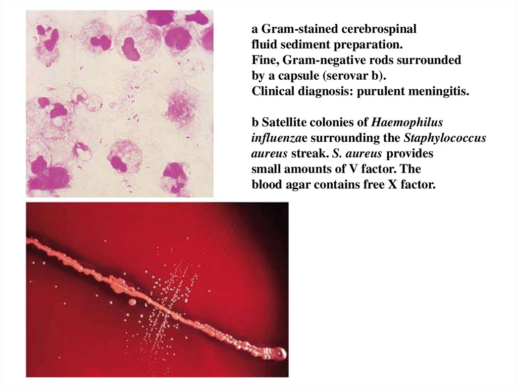
a Gram-stained cerebrospinalfluid sediment preparation.
Fine, Gram-negative rods surrounded
by a capsule (serovar b).
Clinical diagnosis: purulent meningitis.
b Satellite colonies of Haemophilus
influenzae surrounding the Staphylococcus
aureus streak. S. aureus provides
small amounts of V factor. The
blood agar contains free X factor.
38.
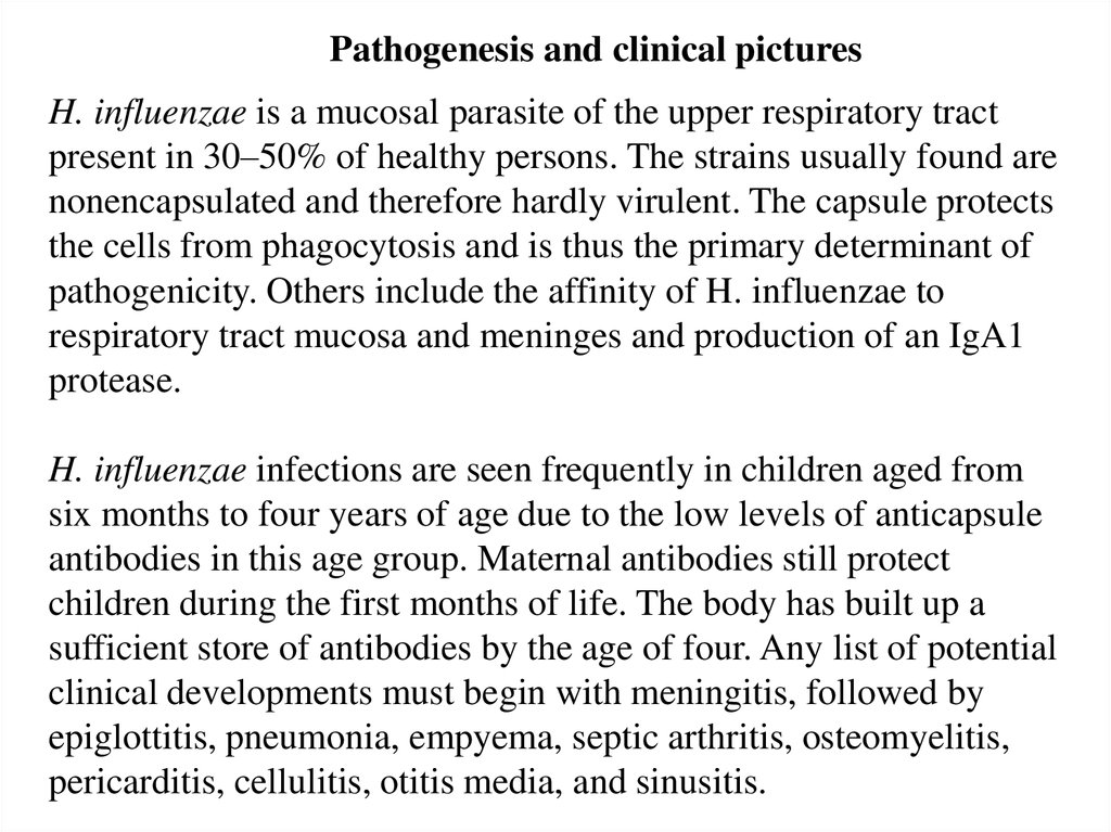
Pathogenesis and clinical picturesH. influenzae is a mucosal parasite of the upper respiratory tract
present in 30–50% of healthy persons. The strains usually found are
nonencapsulated and therefore hardly virulent. The capsule protects
the cells from phagocytosis and is thus the primary determinant of
pathogenicity. Others include the affinity of H. influenzae to
respiratory tract mucosa and meninges and production of an IgA1
protease.
H. influenzae infections are seen frequently in children aged from
six months to four years of age due to the low levels of anticapsule
antibodies in this age group. Maternal antibodies still protect
children during the first months of life. The body has built up a
sufficient store of antibodies by the age of four. Any list of potential
clinical developments must begin with meningitis, followed by
epiglottitis, pneumonia, empyema, septic arthritis, osteomyelitis,
pericarditis, cellulitis, otitis media, and sinusitis.
39.
Pathogenesis and clinical picturesHaemophilus infections in adults are usually secondary
complications of severe primary illnesses or the result of
compromised immune defenses. The most frequent
complication is an acute exacerbation of chronic bronchitis.
Pneumonias caused by H. influenzae are also observed, often as
superinfections following viral influenza. In immunocompromised
adults, even the nonencapsulated strains can cause infections of the
upper and lower respiratory tract.
40.
DiagnosisThe method of choice is identification of the pathogen in
cerebrospinal fluid, blood, pus, or purulent sputum using
microscopy and culture assays. Satelliting on blood agar is an
indication of a V factor requirement. An X factor requirement is
confirmed most readily by the porphyrin test, with a negative
result in the presence of H. influenzae.
Therapy
In view of the increasing number of betalactamase-producing H.
influenzae strains observed in recent years, penicillinase-stable
betalactam antibiotics should be used to treat these infections. The
likelihood that a strain produces betalactamase is 5–30% in most
countries. 4-quinolones are an alternative to betalactams that
should not, however, be used in children. The agent of choice in
meningitis is ceftriaxone
41.
Epidemiology and preventionH. influenzae is found only in humans. The incidence of severe invasive
infections (meningitis, sepsis, epiglottitis) in children has been reduced
drastically – to about one in 10 of the numbers seen previously—since a
vaccination program was started, and will continue to fall assuming the
vaccinations are continued .
Immunization is achieved with the conjugate vaccine Hib in which the
capsule polysaccharide epitope “b” conferring immunity is conjugated to
protein. Such a conjugate vaccine can be administered as early as the first
month of life. The immune system does not respond to pure polysaccharide
vaccines until about the age of two, since polysaccharides are T-independent
antigens against which hardly any antibodies are produced in the first two
years of life. There is also no booster response. A four-day regimen of
rifampicin has proved to be an effective chemoprophylactic treatment for nonvaccinated small children who have been exposed to the organism.
42.
Campylobacter, Helicobacter, and Spirillum belong to the groupof spiral, motile, Gram-negative, microaerophilic bacteria. C.
jejuni causes a form of enteritis. The sources of infection are
diseased animals. The pathogens are transmitted to humans in
food. The diseases are sometimes also communicable among
humans. The pathogens are identified for diagnostic purposes in
stool cultures using special selective mediums. Helicobacter pylori
contribute to the pathogenesis of type B gastritis and peptic ulcers.
Spirillum minus causes rat bite fever, known as sodoku in Japan
where it is frequent.
43.
CampylobacterCampylobacter (meaning "curved bacteria") is a genus of Gramnegative bacteria. Campylobacter typically appear comma or s-shaped
and motile. Most Campylobacter species can cause disease and can
infect humans and other animals. The bacterium's
main reservoir is poultry; humans can contract the disease from eating
food contaminated with Campylobacter species. Another source of
infection is contact with infected animals, which often
carry Campylobacter asymptomatically. At least a dozen species
of Campylobacter have been implicated in human disease, with C.
jejuni and C. coli being the most common. C. jejuni is now recognized
as one of the main causes of bacterial foodborne disease in many
developed countries.C. jejuni infection can also spread to the blood in
individuals with AIDS, while C. lari is a known cause of recurrent
diarrhea in children. C. fetus is a cause of spontaneous abortions
in cattle and sheep, as well as an opportunistic pathogen in humans.
This genus has been found to be part of the salivary microbiome.
44.
Helicobacter pyloriMorphology and culture
H. pylori are spirally shaped, Gramnegative rods with lophotrichous
flagellation. Cultures from stomach
biopsies are grown on enriched mediums
and selective mediums under microaerobic
conditions (90% N2, 5% CO2, and 5% O2)
for three to four days. Identification is
based on detection of oxidase, catalase,
and urease.
45.
Pathogenesis and clinical picturesH. pylori occurs only in humans and is transmitted by the fecal-oral
pathway. The pathogen colonizes and infects the stomach mucosa.
The pathogenicity factors include pronounced motility for efficient
target cell searching, adhesion to the surface epithelial cells of the
stomach, urease that releases ammonia from urea to facilitate
survival of the cells in a highly acidic environment and a
vacuolizing cytotoxin (VacA) that destroys epithelial cells.
46.
Once the pathogen has infected the stomach tissues an acutegastritis results, the course of which may or may not involve overt
symptoms. Potential sequelae include:
1. Mild chronic gastritis type B that may persist for years or even
decades and is often asymptomatic.
2. Duodenal ulceration, sometimes gastric ulceration as well.
3. Chronic atrophic gastritis from which a gastric adenocarcinoma
sometimes develops.
4. Rarely B cell lymphomas of the gastric mucosa (MALTomas).
47.
Diagnosis. Histopathological, cultural and, molecular identificationof the bacteria in stomach lining biopsies. Antigen detection in stool.
Antibodies can be identified with an ELISA or Western Blotting.
Therapy. In patients with ulcers and/or gastritis symptoms, a triple
combination therapy with omeprazole (proton pump blocker),
metronidazole, and clarithromycin lasting seven days is successful in
90% of cases.
Epidemiology. Based on seroepidemiological studies we know that
H. pylori occur worldwide. Generalized contamination of the
population begins in childhood and may reach 100% in adults in
areas with poor hygiene. The contamination level is about 50%
among older adults in industrialized countries. Transmission is by the
fecal-oral route.
48.
Legionella (Legionnaire’s Disease)Legionella is the only genus in the family Legionellaceae. The
species Legionella pneumophila is responsible for most legionelloses
in humans. Legionellae are difficult to stain. They are Gram-negative,
aerobic rod bacteria. Special mediums must be used to grow them in
cultures. Infections with Legionella occur when droplets containing
the pathogens are inhaled. Two clinically distinct forms are on record:
legionnaire’s disease leading to a multifocal pneumonia and
nonpneumonic legionellosis or Pontiac fever.
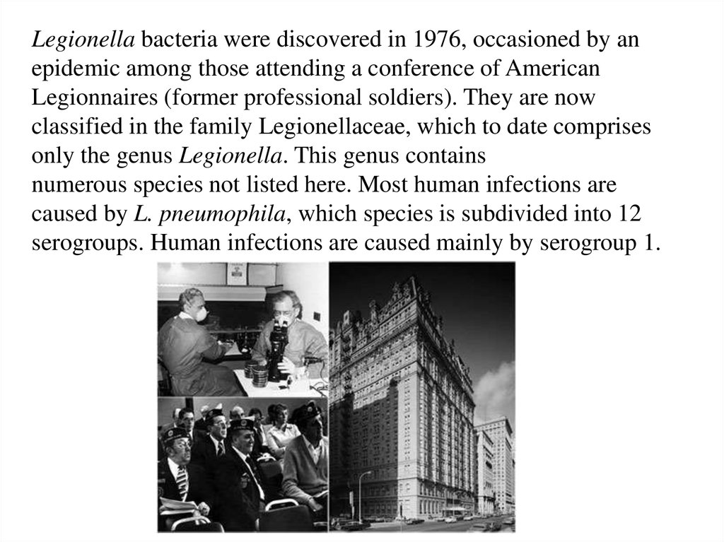
49.
Legionella bacteria were discovered in 1976, occasioned by anepidemic among those attending a conference of American
Legionnaires (former professional soldiers). They are now
classified in the family Legionellaceae, which to date comprises
only the genus Legionella. This genus contains
numerous species not listed here. Most human infections are
caused by L. pneumophila, which species is subdivided into 12
serogroups. Human infections are caused mainly by serogroup 1.
50.
The persons most likely to contract legionnaire’s disease are thosewith a primary cardiopulmonary disease and generally weakened
immune defenses. Laboratory diagnostic methods include
microscopy with direct immunofluorescence, culturing on special
mediums and antibody assays. The antibiotics of choice are the
macrolides. The natural habitat of legionellae is damp biotopes. The
sources of infection listed in the literature include hot and cold
water supply systems, cooling towers, moisturizing units in air
conditioners, and whirlpool baths. Legionelloses can occur both
sporadically and in epidemics.
51.
Morphology and cultureL. pneumophila is a rod bacterium 0.3–1 lm wide
and 2–20 lm long. Its cell wall structure is of the Gram-negative
type, but gram staining hardly “takes” with these bacteria at all.
They can be rendered visible by means of direct
immunofluorescence.
52.
Pathogenesis and clinical pictureThe pathomechanisms employed by legionellae are not yet fully clarified.
These organisms are facultative intracellular bacteria that can survive in
professional phagocytes and in alveolar macrophages. They are capable of
preventing the phagosome from fusing with lysosomes. They also produce a
toxin that blocks the oxidative burst. Two clinical forms of legionellosis have
been described:
Legionnaire’s disease. Infection results from inhalation of droplets
containing the pathogens. The incubation period is two to 10 days. The
clinical picture is characterized by a multifocal, sometimes necrotizing
pneumonia. Occurrence is more likely in patients with cardiopulmonary
primary diseases or other immunocompromising conditions. Lethality >20%.
Pontiac fever. Named after an epidemic in Michigan. Incubation period one
to two days. Nonpneumonic, febrile infection. Self-limiting. Rare.
53.
Diagnosis. Specific antibodies marked with fluorescein are used to detect thepathogens in material from the lower respiratory tract. For cultures, special
culture mediums must be used containing selective supplements to exclude
contaminants. The mediums must be incubated for three to five days. A gene
probe can also be used for direct detection of the nucleic acid (rDNA) specific to
the genus Legionella in the material. Antibodies can be assessed using the
indirect immunofluorescence technique.
Therapy. Macrolide antibiotics are now the agent of choice, having
demonstrated clinical efficacy. Alternatively, 4-quinolones can be used.
Epidemiology and prevention. Legionellosis can occur in epidemic form or in
sporadic infections. It is estimated that one third of all pneumonias requiring
hospitalization are legionelloses. Soil and damp biotopes are the natural
habitat of Legionella. Sources of infection include hot and cold water supply
systems, cooling towers, air moisturizing units in air conditioners, and whirlpool
baths. Human-to-human transmission has not been confirmed. Legionella
bacteria tolerate water temperatures as high as 50°C and are not killed
until the water is briefly heated to 70°C.
54.
Treponema pallidumBorrelia (Relapsing Fever, Lyme Disease)
Leptospira (Leptospirosis, Weil Disease)
Rickettsia
Chlamydia
Mycoplasma