


 Биология
Похожие презентации:

")


Enteric bacterial pathogens
1. ENTERIC BACTERIAL PATHOGENS
Department of Microbiology,Virology & Immunology
Ass. Prof. E. O. Kravtsova
2.
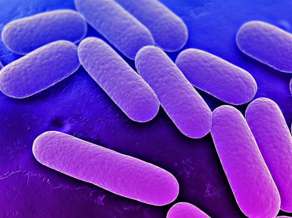
3. FAMILY ENTEROBACTERIACEAE
- are a large heterogeneous group ofGram«-» rods whose natural habitat is the intestinal
tract of humans and animals.
• Genera (20)
1. Escherichia
2. Salmonellae
3. Shigellae
4. Klebsiellae
5. Yersinia …..
4. FAMILY ENTEROBACTERIACEAE
Most of the members of Enterobacteriaceae arefacultative anaerobes, ferment a wide range of
carbohydrates, possess a complex antigenic
structure, and produce a variety of toxins and
other virulence factors.
This family is characterized biochemically by the
ability to ferment glucose with the production of
acid or acid and gas, and to reduce nitrates to
nitrites.
All members of this family are oxidase-negative.
5.
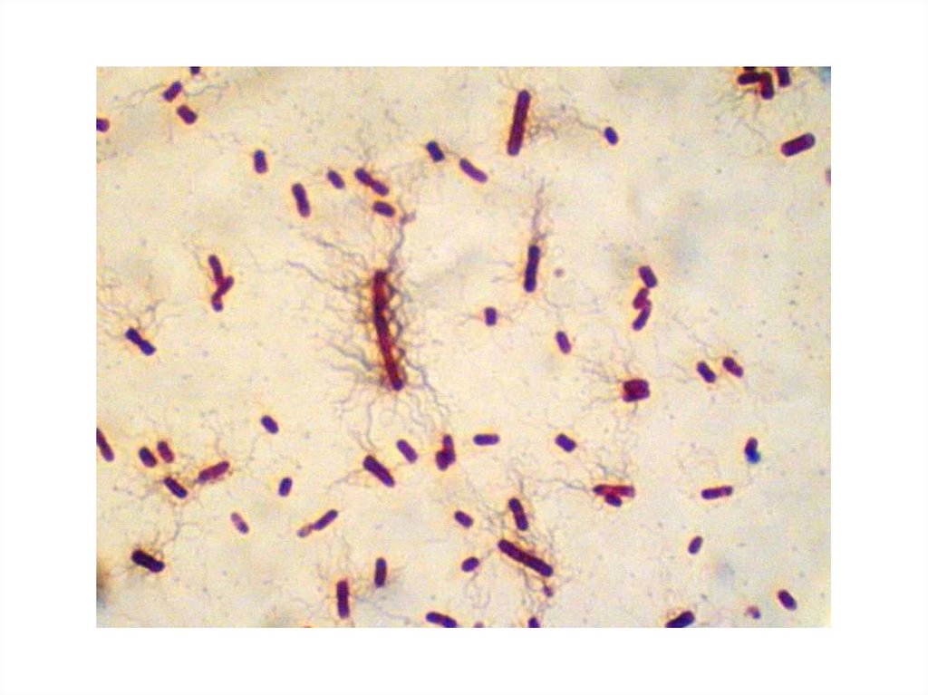
6. FAMILY ENTEROBACTERIACEAE
Gram «-» rods
Spores «-»
Capsula «+» or «-»
They are motile (E.coli) or non-motile (Shigellae)
Facultative anaerobes
T= 30-37ºC
pH= 7,2-7,5
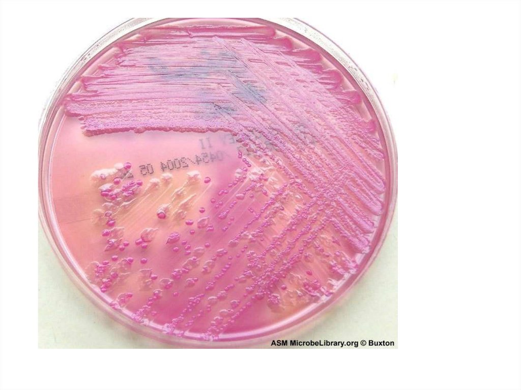
Endo medium, Ploskirev medium, Levin medium,
MacConkey agar.
Oxidase «-», Glucose «+», Lactose «-» ex.E.coli,
• Mannitol «+», Indol «-» ex.E.coli, H2S «-» or «+».
7. FAMILY ENTEROBACTERIACEAE
8. FAMILY ENTEROBACTERIACEAE
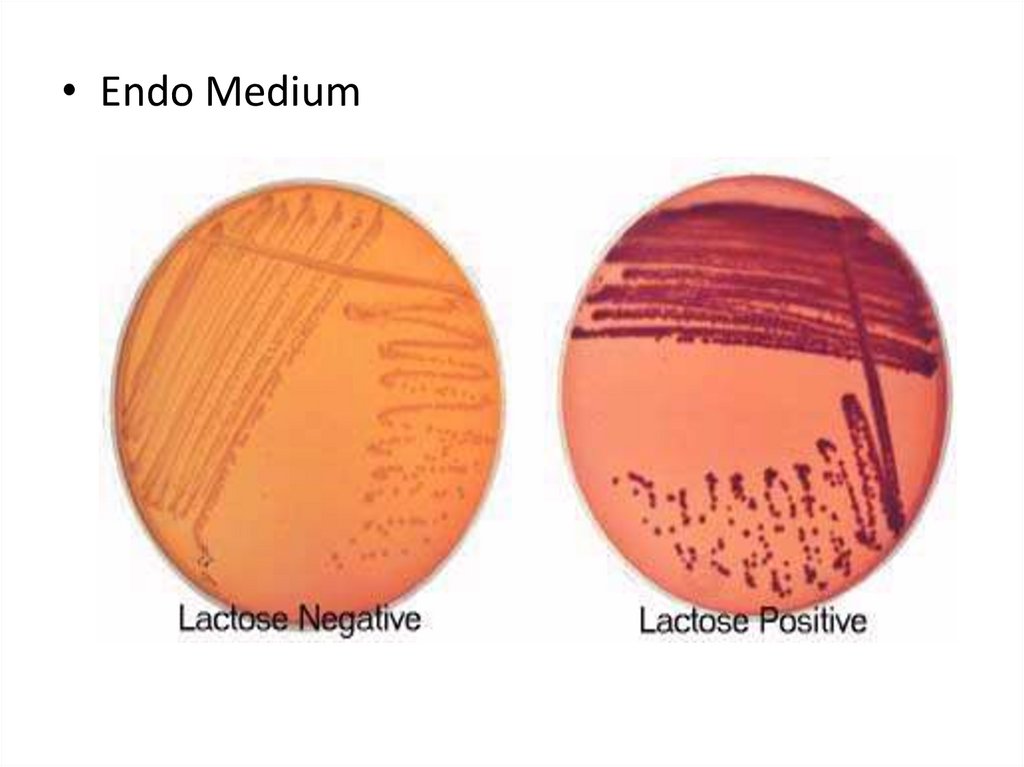
The differential culture media that containcarbohydrates and special dyes (indicators) are
used to distinguish lactose-fermenting bacteria
that form colored colonies from non-lactosefermenting microorganisms producing colorless
colonies on such differential media.
These culture media can allow rapid
identification of enteric bacteria.
9.
• Endo Medium10. Kligler Iron Agar
11. Kligler Iron Agar
• Kligler iron agar is used for the differentiation ofthe Enterobacteriaceae members on the basis of
their ability to ferment glucose and lactose and to
liberate sulfides.
• Gas formed by carbohydrates fermenters is
detected as bubbles or by splitting or
displacement of the agar.
• Hydrogen sulfide production is evidenced by a
black color either throughout the butt, or in a
ring formation near the top of the butt.
12. Kligler Iron Agar
13. Kligler Iron Agar
The lactose-positive and glucose-positive bacteriashow both the slant and the butt yellow in color (E.
coli).
The lactose-negative and glucose-positive bacteria
show the yellow butt and the red slant (Salmonella,
Shigella).
If the bacterium is glucose -negative and lactosenegative, both the butt and the slant remain red.
Hydrogen sulfide production is evidenced by a black
color either throughout the butt, or in a ring
formation near the top of the butt.
14. FAMILY ENTEROBACTERIACEAE
Enterobacteriaceae have a complex antigenicstructure.
They are classified by more than 150 different
heat-stable somatic O (lipopolysaccharide)
antigens, more than 100 heat-labile K (capsular)
antigens, and more than 50 H (flagellar) antigens.
In Salmonella Typhi the capsular antigen is called
Vi- antigen.
The antigenic classification of Enterobacteriaceae
often indicates the presence of each specific
antigen.
15. FAMILY ENTEROBACTERIACEAE
Virulence factors:
Fimbriae
Enterotoxins
Hemolysins
Endotoxins
16. FAMILY ENTEROBACTERIACEAE
Epidemiology:
They are pathogenic for human and animals.
They are transmitted by the fecal-oral route.
They may be responsible for hospital
infections.
• The main clinical symptoms are diarrhea,
vomiting, temperature.
17. FAMILY ENTEROBACTERIACEAE
• Microbiological diagnosis.• Specimens : feces, vomit, food,urine, blood.
Methods: bacteriological, serological,
biological.
18. Salmonellae Salmonella
Salmonella• Family – Enterobacteriaceae
• Genus – Salmonellae
• Species - S. enterica
• Subspecies – S. typhi, S. paratyphi A, S. paratyphi B,
S. enteritidis, S. typhimurium
• The main taxonomic groups of salmonella are:
• Family → Genus → Species → Subspecies → Serovar
S. typhi was discovered in 1880 by K. Eberth and isolated
in pure culture in 1884 by G.Gaffky.
19. Salmonella
20. Salmonella Morphology:
• Gram «-» rods• Spores «-» Capsula «-» They are motile
• Cultural properties:
• Facultative anaerobes Chemoorganotrophs
• Topt= 35-37̊º C
pH= 6,8-7,2
• Endo medium, Ploskirev medium – pale pink
colonies. Levin medium – blue colonies
• MacConkey agar- colourless colonies
• Bismuth-Sulfite agar – black colonies.
• In MPB they produce a uniform turbidity.
21.
22.
23. Bismuth Sulfite agar
24. Salmonella
Biochemical activity:Glucose «+», Maltose «+», Mannitol «+» (acid)
S.paratyphi ferments carbohydrates with acid and gas
formation.
Lactose «-» Sucrose «-»
• Indol «-», H2S «+»
• Gelatin – does not liquefy
• Oxidase «-»
25. Salmonella
• Antigenic structure:• O-somatic (serogroups), is destroyed by
formalin.
• H – flagellar (serovars) , is destroyed by phenol.
• Vi – antigen is located on the surface of the
bacterial cell , is destroyed by phenol and
temperature.
• Kauffmann and White classified Salmonellae
according their antigenic structure.
26. Salmonella. Antigenic structure
Based on the presence of O-antigens, theSalmonella have been assigned to serogroups.
The O group is designated by capital roman
letter (A, B, C, D).
Each Salmonella serogroup can be identified by
the slide agglutination test.
27. Salmonella. Antigenic structure
The H-antigens are designated by small romanletters and are kept in brackets (phase 1) and by
arabic numerals for phase 2.
The use of specific H- antisera helps to identify
the Salmonella serovars.
28. Salmonella
• Resistance:• Survive in ice for several months.
• Survive in butter, meat, cheese, bread for 1-3
months.
• Survive in water and soil for several weeks.
• Susceptible to heat, t= 60-100º C.
• Susceptible to disinfectant solutions of
phenol, chloramine.
29. Salmonella Virulence Factors
30. Salmonella Virulence Factors
The type III protein secretion system (T3SS) encodedby Salmonella pathogenicity island 1 (SPI-1) delivers
effector proteins required for intestinal invasion and the
production of enteritis.
SPI-1 encodes transcription factors that regulate the
expression of some virulence factors of Salmonella,
while other transcription factors encoded outside SPI-1
participate in the expression of SPI-1-encoded genes.
SPI-1 genes are responsible for the invasion of host cells,
regulation of the host immune response, the host
inflammatory response, apoptosis, and biofilm
formation.
31. Salmonella
Salmonella are often pathogenic for humans oranimals when acquired by the oral tract.
S. typhi, S. paratyphi A, and S. paratyphi B are
the causative agents of enteric fevers.
Other species of salmonellae are the bacteria
causing salmonellosis, a food-borne infectious
disease (enteritis).
The enteric fevers are transmitted by the fecaloral route.
32. Pathogenesis
The ingested salmonellae reach the small intestine, from which theyenter the lymphatics (Peyer’s patches) and then the bloodstream.
After an incubation period of 10-14 days, fever, malaise and
headache occur due to bacteriemia and toxicity of Salmonella byproducts.
The skin may become dotted with small hemorrhages called
“roseoles”.
Gastrointestinal symptoms appear late in the course of the disease.
Blood cultures are positive only in the first week of the disease.
At the beginning of the second week microbes are carried by the
blood to many organs, including the liver, kidneys and the intestine.
Salmonellae multiply in intestinal lymphoid tissue, kidneys and are
excreted in stools and urine.
The stools and urine cultures give positive results on the second and
third week.
33. Pathogenesis of enteric fever
1st stage – the ingestion
2nd stage – the invasion
3rd stage – bacteremia
4th stage – bacterial dissemination
5th stage – hyperergia and excretion
6th stage – final stage
Immunity acquired is relatively stable but
relapses and reinfections sometimes occur.
34. Laboratory Diagnosis
Blood cultures are positive only in the first week ofthe disease.
At the beginning of the second week microbes are
carried by the blood to many organs, including the
liver, kidneys and the intestine.
Salmonellae multiply in intestinal lymphoid tissue,
gallbladder, kidneys and are excreted in stools and
urine. The stools and urine cultures yield positive
results on the second and third week.
A positive culture of bile establishes the presence
of Salmonella genus in the biliary tract of carriers.
35. Salmonella Infections. Treatment .
Antimicrobial treatment.Replacement of fluids and electrolytes are
essential.
Susceptibility testing is an important method to
choosing a proper antibiotic, because multiple
drug resistance is a big problem in enteric
bacteria.
36.
37. SHIGELLA
The causative agent of dysentery was described in1888 by A. Chantemesse and in 1891 by A.
Grigoryev and F. Widal.
In 1898 this organism was studied in detail by K.
Shiga in Japan.
In 1900 S. Flexner in the Philippines isolated
dysentery organisms.
Later other bacteria causing dysentery were
discovered. According to the current International
Nomenclature, all dysentery bacilli are grouped
together in one genus known as Shigella.
38. SHIGELLA
Shigellosis (or bacillary dysentery) is a clinicalcondition characterized by fever, bloody
diarrhea, and fecal leukocytosis. Classical
bacterial dysentery is associated with infections
caused by any of the four Shigella species:
Sh. dysenteriae, Sh. flexneri, Sh. boydii,
Sh. sonnei.
39. Taxonomy
Family – EnterobacteriaceaeGenus – Shigella
Species - S. dysenteriae, S. flexneri, S. boydii,
S. sonnei.
40.
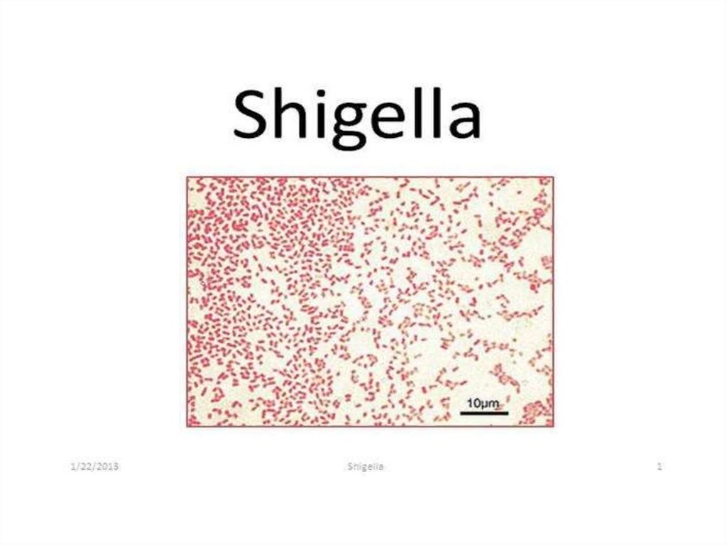
41. SHIGELLA
Shigella are slender gram-negative rods.
Spores «-»
Mirocapsula «+»
They are non-motile
They are facultative anaerobes, but grow best
aerobically.
42. Ploskirev agar
43. Endo agar
44. Levine Eosin Methylene Blue agar
45. SHIGELLA
All Shigella ferment glucose, form acid.With the exception of Shigella sonnei, they do not
ferment lactose.
Glucose «+» Lactose «-»
Mannitol «+» (S.dysenteriae «-»)
H2S «-»
Antigens: O – somatic antigen, K-surface antigen.
46. SHIGELLA
Shigella-induced infections are almost alwayslimited to the gastrointestinal tract; bloodstream
invasion is quite rare.
All Shigellae produce endotoxin.
Microabscesses in the wall of the large intestine
lead to necrosis of the mucous membrane,
superficial ulceration and bleeding.
Shigella dysenteriae also produces a heat-labile
exotoxin that affects both the gut and the central
nervous system.
47. Shigellosis
Shigellosis is the infection with fecal-oral route oftransmission.
After a short incubation period (1-2 days), there is a
sudden onset of abdominal pain, fever, and watery
diarrhea.
A day or so later, as the infection involves the ileum and
colon, the number of stools increase; they are less liquid
but often contain mucus, pus and blood.
In children and the elderly, loss of water and
electrolytes may lead to dehydration, acidosis, and even
death.
Infection is followed by a type-specific immune
response, but reinfection may occur.
48. Shigellosis. Diagnostic Laboratory Tests
SPECIMENS: Fresh stool, mucus flakes, and rectal swabsare taken for culture.
BACTERIOLOGICAL EXAMINATION:
The specimen is streaked on Ploskirev, (MacConkey or
Levine EMB agar).
Colorless (lactose-negative) colonies are inoculated into
the Kligler iron agar.
Organisms that not produce H2S and produce acid but
not gas in the butt of the Kligler agar (glucose «+»), and
that are nonmotile should be subjected to the slide
agglutination test by specific Shigella antisera.
49. Shigellosis. Treatment and Immunoprophylaxis
AB - Ciprofloxacin, ampicillin, tetracycline,chloraphenicol
The eubiotics may be effective in limiting
multiplication. There are no vaccines available
for immunoprophylaxis of bacterial dysentery.
- isolation of patients and disinfection of
excreta,
- detection of subclinical cases,
- sanitary control of water, food, and milk.