


 Медицина
Похожие презентации:

")


Deparment of infectious diseases
1.
Lugansk state medicaluniversity
Deparment of infectious
diseases
Teacher:
Nelle
2.
ABOUT MYSELFAJAY . GUNDAWAR
GROUP:18A
COURSE: 5TH
3.
4.
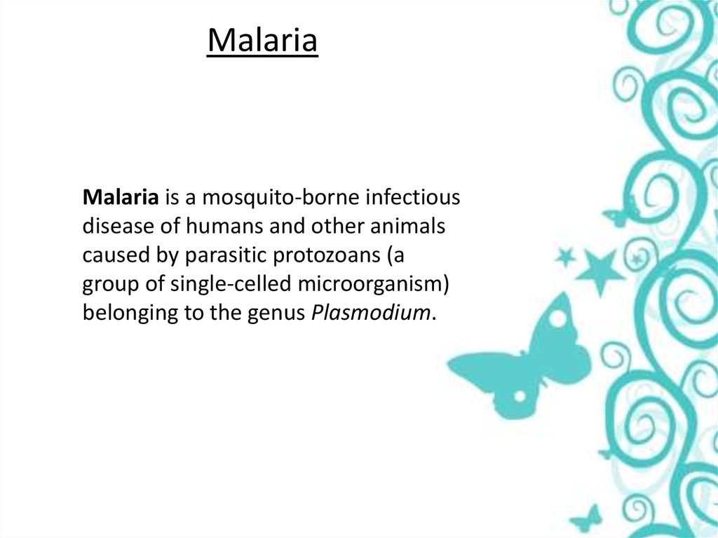
MalariaMalaria is a mosquito-borne infectious
disease of humans and other animals
caused by parasitic protozoans (a
group of single-celled microorganism)
belonging to the genus Plasmodium.
5.
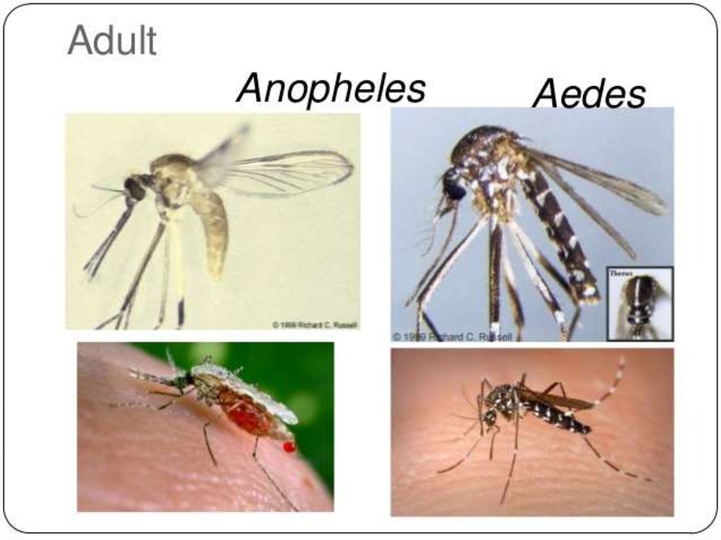
The disease is transmitted most commonly by an infected female Anophelesmosquito. The mosquito bite introduces the parasites from the mosquito's
saliva into a person's blood. The parasites travel to the liver where they
mature and reproduce.
Five species of Plasmodium can infect and be spread by humans.]Most
deaths are caused by:
- P. falciparum because
- P. vivax,
-P. ovale, and
-P. malariae
generally cause a milder form of malaria
6.
Plasmodium Falciparum - MalariaPlasmodium falciparum is the Plasmodium species
responsible for 85 % of the malaria cases. The three less
common and less dangerous Plasmodium species are: P.
ovale,P. malariae and P. vivax. Malaria infects over 200
million people annually, mostly in poor tropical and
subtropical countries of Africa. It is the deadliest parasitic
disease killing over one million people each year. 90 % of
the deaths occur south of the Sahara desert and most are
under five-year-old children. In addition to Africa, malaria
occurs in South and Southeast Asia, Central and South
America, the Caribbean and the Middle East. Even within
tropical and subtropical areas, malaria does not usually
occur at high altitudes (over 1500 meters), during colder
seasons, in countries of successful malaria programs or in
deserts.
7.
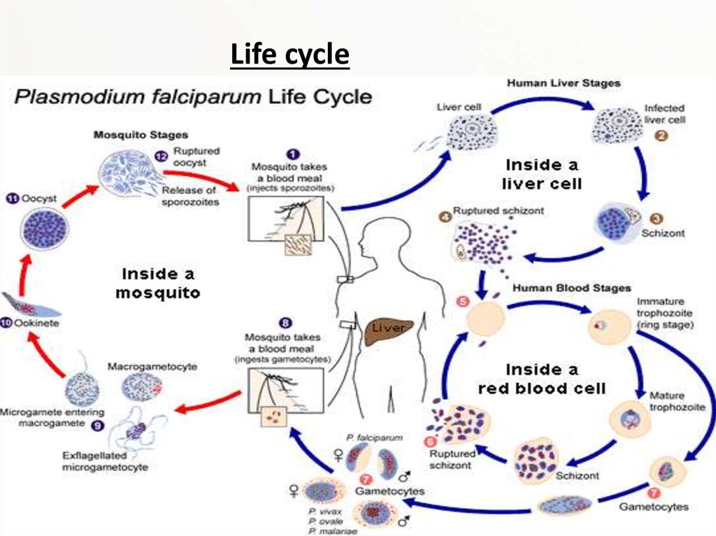
Life cycle8.
Malaria is carried by Anopheles mosquitoes. Of the over 400 Anopheles species, only 30–40 cantransmit malaria. The infection starts, when a female mosquito injects (in her saliva)
"sporozoites" (one form of P. falciparum) into human skin while taking a blood meal. A
sporozoite travels (in the bloodstream) into the liver where it invades a liver cell. It matures into
a "schizont" (mother cell) which produces 30000–40000 "merozoites" (daughter cells) within six
days. The merozoites burst out and invade red blood cells. Within two days one merozoite
transforms into a trophozoite, then into a schizont and finally 8–24 new merozoites burst out
from the schizont and the red cell as it ruptures. Then the merozoites invade new red cells. P.
falciparum can prevent an infected red cell from going to the spleen (the organ where old and
damaged red cells are destroyed) by sending adhesive proteins to the cell membrane of the red
cell. The proteins make the red cell to stick to small blood vessel walls. This poses a threat for
the human host since the clustered red cells might create a blockage in the circulation system.
A merozoite can also develop into a "gametocyte" which is the stage that can infect a mosquito.
There are two kinds of gametocytes: males (microgametes) and females (macrogametes). They
get ingested by a mosquito, when it drinks infected blood. Inside the mosquito's midgut, male
and female gametocytes merge into "zygotes" which then develop into "ookinetes." The motile
ookinetes penetrate the midgut wall and develop into "oocysts." The cysts eventually release
sporozoites, which migrate into the salivary glands where they get injected into humans. The
development inside a mosquito takes about two weeks and only after that time can the
mosquito transmit the disease. P. falciparum cannot complete its life cycle at temperatures
below 20 °C.
9.
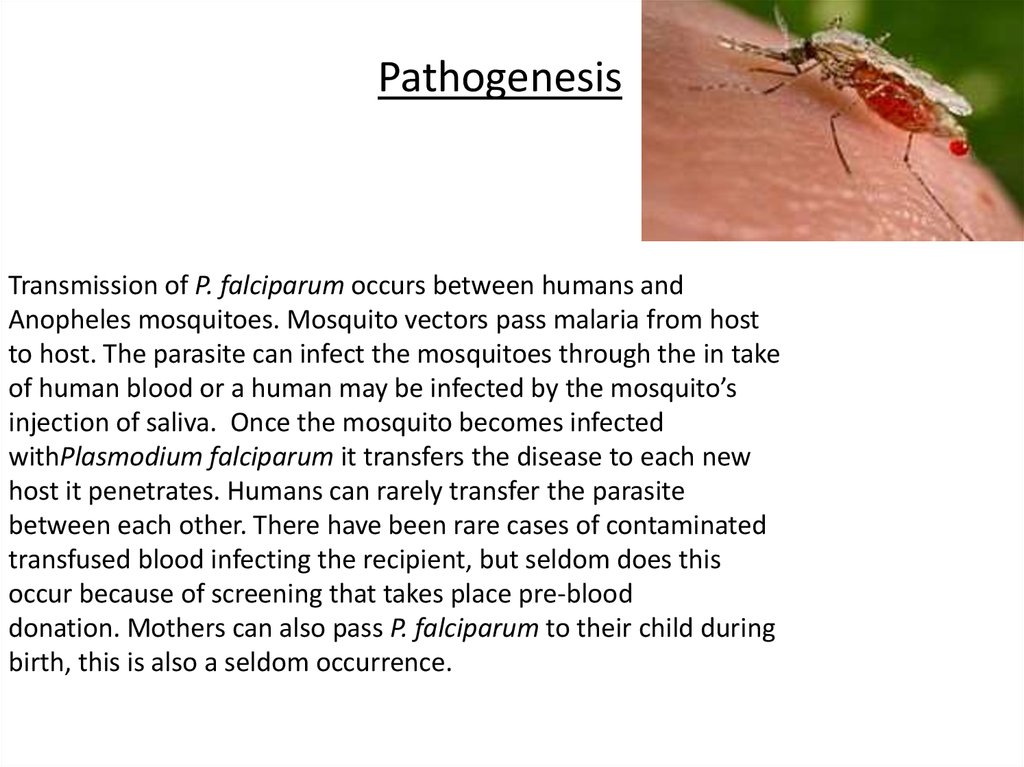
PathogenesisTransmission of P. falciparum occurs between humans and
Anopheles mosquitoes. Mosquito vectors pass malaria from host
to host. The parasite can infect the mosquitoes through the in take
of human blood or a human may be infected by the mosquito’s
injection of saliva. Once the mosquito becomes infected
withPlasmodium falciparum it transfers the disease to each new
host it penetrates. Humans can rarely transfer the parasite
between each other. There have been rare cases of contaminated
transfused blood infecting the recipient, but seldom does this
occur because of screening that takes place pre-blood
donation. Mothers can also pass P. falciparum to their child during
birth, this is also a seldom occurrence.
10.
Infectious Dose, Incubation, ColonizationSymptoms of Malaria typically begin 8-25 days following infection
however, in a few cases it can take up to a year. The late onset of
incubation is due to taking an inadequate amount of anti-malaria
medication. The infectious dose is not precisely known, but it is
understood to be a very low number. Malaria can be observed months
to years after first set of symptoms are observed. This is due to the
parasites ability to lie dormant in liver cells until the environment is
right for a relapse. This is mainly seen in P.vivax and P. ovale, rather
then P. falciparum. The parasite colonizes in the liver and is then
released into the blood stream and enters erythrocytes.
11. Epidemiology
• The key to Malaria-endemic is Anopheles the mosquito’s ability tolive in a certain area. Temperature is also important having to stay
above 20 degrees Celsius. The main areas of P. falciparum are South
America, Africa, India, and few parts of Indonesia. The ideal location
for transmission is along the equator in a warmer region.
Transmission will not occur in high altitudes, colder seasons, and
deserts. Malaria is considered to have arisen since the beginning of
mankind, but was first discovered in blood in 1880 and found to be
transmitted by mosquitoes in 1889. There are four common
species of Malaria of which P. falciparum is the most
severe. Plasmodium falciparum continues to increase in drugresistant populations and insecticide-resistant mosquitoes leading
to the prediction that the disease will only worsen over time.
12. Virulence Factors
PfEMP1,P. falciparum erythrocye membrane protein 1, is an adhesive ligand protein which is
created inside of a P. falciparum infected erythrocyte and presented on the surface. PfEMP1
is known as a knob and is encoded by the multigene segment, Var. The protein is responsible
for sequestration within the vital organs. In some case were sequestration occurs in the
brain this will lead to the cerebral form of malaria. Each Plasmodium falciparum has multiple
versions of PfEMP1 with which it can alter its appearance by changing to another PfEMP1
when the immune system begins to create antibodies for the original PfEMP1 in a process
known as antigenic variation. Changing of adherence molecules also means a change in the
receptor on the epithelial. The change in receptor is hypothesized to possibly change the
disease outcome.
RIFIN, repetitive interspersed family, is considered the most abundant multigene family.
PfEMP1 along with RIFIN is considered a crucial cornerstones for the virulence
of Plasmodium falciparum mainly due to its ability to avoid immune response through
antigenic variability. RIFIN is also presented on the outer membrane of a parasite infected
erythrocye as an adherence factor.
Rosettes are uninfected red blood cells that form clumps with Malaria-infected erythrocytes.
Clumping occurs when particularly sticky PfEMP1 attach to other red blood cells. Only a
minority of P. falciparumactually creates rosettes, but when they do they are known to be
linked to severe malaria.[
Malaria pigment (hemozoin) is released during erythrocyte rupture, causing the
uncomplicated symptoms of malaria such as chills and fever.
13.
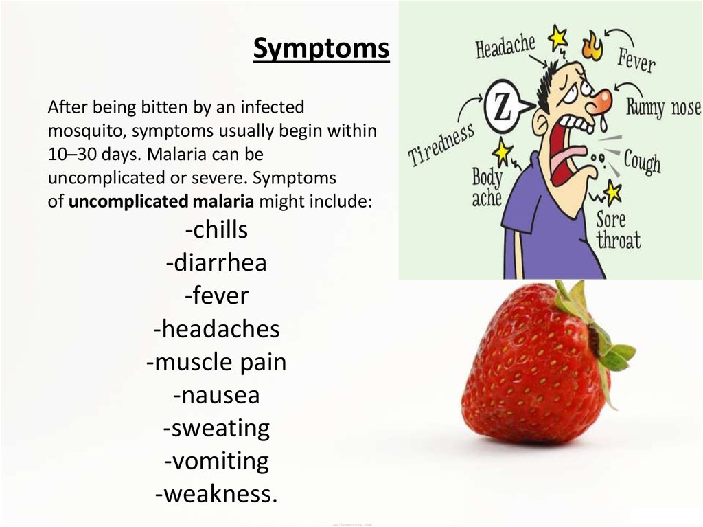
SymptomsAfter being bitten by an infected
mosquito, symptoms usually begin within
10–30 days. Malaria can be
uncomplicated or severe. Symptoms
of uncomplicated malaria might include:
-chills
-diarrhea
-fever
-headaches
-muscle pain
-nausea
-sweating
-vomiting
-weakness.
14.
• Some less noticeable manifestations:• enlargement of the spleen or liver
• increased breathing frequency
• mild anemia
• mild jaundice (yellowish eye whites and skin).
• The disease can turn into severe malaria, if there are serious organ failures or
abnormalities in the bloodstream or metabolism. Symptoms of severe
malaria might include:
• breathing difficulties
• coma
• confusion
• death
• focal neurologic signs
• seizures
• severe anemia.
15.
Some less noticeable manifestations:
abnormalities in blood coagulation
hemoglobin in the urine
high acidity of the blood
hypoglycemia (low blood glucose)
low blood pressure
kidney failure.
• During pregnancy malaria can lead to premature baby delivery or
delivery of a low-birth-weight baby. The infant can get the parasite
from the mother and develop the disease. Central nervous system
involvement (cerebral malaria) can cause (especially in small
children) blindness, deafness, speech difficulty, paralyses and
trouble with movements.
16. Diagnosis
Malaria is usually diagnosed by examining a blood sample under a microscope.
There are also test kits that detect antigens of P. falciparum in the patient's blood.
These immunologic tests are known as rapid diagnostic tests (RDTs). RDTs can
detect two different malaria antigens, one for P. falciparum and the other is found
in all four human malaria species. RDTs usually show results in about 20 minutes. It
is a good alternative to microscopy, when reliable microscopic diagnosis cannot be
done. RDT might not detect some infections, if there are not enough malaria
parasites in the patient’s blood. A negative RDT result can be followed up by
microscopy. If a patient with positive RDT result is not responding to treatment,
another blood sample should be taken. This time using microscopy to determine
whether the medicine was appropriate for thePlasmodium species.
Diagnosis can be challenging for many reasons:
Some health workers in developing countries are insufficiently trained and
supervised.
The microscopes and reagents might be of poor quality and the supply of
electricity might be unreliable.
Some health workers save blood samples until a qualified person is available to
perform the microscopy. This delay results sometimes as incorrect diagnosis.
Many malaria endemic communities do not have the proper diagnostic tools such
as microscopes and RDTs.
17.
• Rapid and accurate diagnosis using microscopicexamination of blood smears is the most precise way
to determine Plasmodium falciparum as the disease.
CDC provides various references for microscope
diagnosis along with serology, PCR, and drug resistance
testing. Each species of P. falciparum has distinctive
characteristics that can be see under a microscope. In
only early form, trophozites and gametocytes
of P.falciparumare seen in the blood as ring form inside
the erythrocyte. There are normally multiple parasites
in one erythrocytes appearing as several dots.
18. Treatment
• Most malaria deaths occur in rural areas. Quick progressionfrom illness to death can be prevented by fast and effective
medication. Patients who have uncomplicated malaria can
visit a nearby hospital to get treated and then go home to
rest. In emergency cases rectal artesunate drug can be given
as a first line treatment (if they cannot be treated orally).
Patients with severe malaria can be hospitalized for many
days. When treating a malaria patient, the following should
be taken into account:
• age and size of the person (to give the correct amount of
medication)
• drug allergies or other medications taken by the patient
• health condition, when starting the treatment
• where the person was infected (what Plasmodium species is
likely to be responsible and what drug is needed).
19.
The best line of defense against any form of malaria ispreventative treatment, antimalarial, taken properly
before, during, and after exposure to parasite.
P. falciparum and P. vivax have been confirmed to be resistant (in some
areas) to many antimalarial drugs. For example, chloroquine resistant
strain of P. falciparum has spread to most endemic areas.
Listed below are some drugs that are usually recommended by national
malaria control programs. They might not be effective in many parts of
the world due to drug resistant strains.
• artemesinin-containing combination treatments (for example,
artemether-lumefantrine, artesunate-amodiaquine)
• atovaquone-proguanil
• chloroquine
• doxycycline
• mefloquine
• quinine
• sulfadoxine-pyrimethamine.
20.
• Primaquine, is used as an adjunct againstcertain Plasmodium species. It is active against
the dormant liver forms (hypnozoites which are
rare/nonexistent with P. falciparum). Primaquine
is not recommended for people who are deficient
in glucose-6-phosphate dehydrogenase or for
pregnant women. Treating all people
simultaneously in a population can prevent major
malaria epidemics. Unfortunately it can also
increase drug resistance of the parasite and
complications in those who are glucose-6phosphate dehydrogenase deficient.
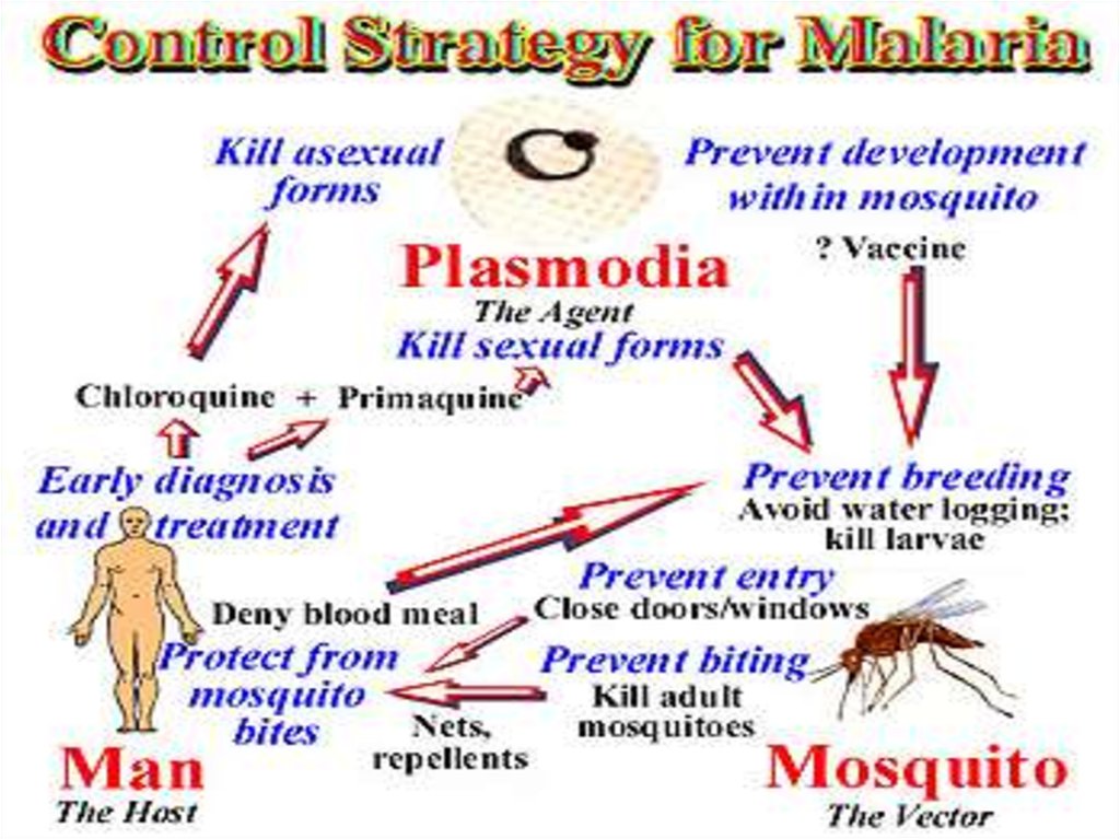
21.
PreventionInsecticide-treated bed nets may reduce deaths of children under 5 years up to 20 %
(according to trials in several African communities). Anopheles mosquitoes usually
feed during the night so you can protect yourself by sleeping under a bed net. If
everyone in a community has a bed net, the occurrence of malaria can be reduced.
Bed nets are usually made of polyester but sometimes cotton, polyethylene, or
polypropylene is used instead. All bed nets are treated with pyrethroid insecticides,
which have are low health risks to humans but are toxic to insects even at low doses.
Pyrethroids do not rapidly wear off, unless exposed to sunlight or washed. "Longlasting insecticide-treated bed nets" maintain effective levels of insecticide for three
years or more. Bed net donations can be made through organizations such as Nothing
But Nets and Malaria No More. The price of one bed net is only a few US dollars
(which is often too expensive for people in developing countries).
22.
23.
Many malaria-carrying mosquitoes are endophilic, meaning that theytypically rest inside the house after taking a blood meal. Indoor Residual
Spraying of the walls and other surfaces can kill them reducing the
chances that infected mosquitoes spread the disease from one
household to another.
Humans living in areas where malaria is common can become partially
immune. Travelers, young children, women having their first or second
pregnancy and those who are weakened by other diseases (such as
AIDS) have little to no immunity against malaria.
24.
Recommendations for pregnant women living in malaria endemic areas:•Eat iron and folate supplements to prevent anemia.
•Get a curative dose of an antimalarial drug at least twice during pregnancy
(starting from the second trimester).
•Sleep under an insecticide-treated bed net.
The number of mosquitoes may be controlled by eliminating mosquito larvae before
they reach adulthood. Rainfall forms water puddles where mosquitoes lay their eggs and
aquatic larvae develop into adults in a few days. Draining or removal of small puddles
can reduce the number of mosquitoes near populations. Chemical insecticides can also
be applied but might harm the environment. Other methods applied to water:
•insect growth regulators
•oil that suffocates the aquatic larvae
•toxins from the bacterium Bacillus thuringiensis var. israelensis (Bti)
25.
Additional personal protection methods include:-glass windows (a well-constructed house)
•repellent
•white or light-colored clothes covering most of the body.
26. Plasmodium vivax
is a protozoal parasite and a human pathogen. The most frequent andwidely distributed cause of recurring (Benign tertian) malaria, P. vivax is
one of the five species of malaria parasites that commonly infect
humans. It is less virulent thanPlasmodium falciparum, the deadliest of the
five, but vivax malaria can lead to severe disease and death due
to splenomegaly (a pathologically enlarged spleen).P. vivax is carried by the
female Anopheles mosquito, since it is only the female of the species that
bite.
27.
On the pathogenesis of Plasmodium vivax malaria: perspectives fromthe Brazilian field.
Life-threatening Plasmodium vivax malaria cases, while uncommon,
have been reported since the early 20th century. Unfortunately, the
pathogenesis of these severe vivax malaria cases is still poorly
understood. In Brazil, the proportion of vivax malaria cases has been
steadily increasing, as have the number of cases presenting serious
clinical complications. The most frequent syndromes associated with
severe vivax malaria in Brazil are severe anaemia and acute respiratory
distress. Additionally, P. vivax infection may also result in complications
associated with pregnancy. Here, we review the latest findings on severe
vivax malaria in Brazil. We also discuss how the development of targeted
field research infrastructure in Brazil is providing clinical and ex vivo
experimental data that benefits local and international efforts to
understand the pathogenesis of P. vivax.
28.
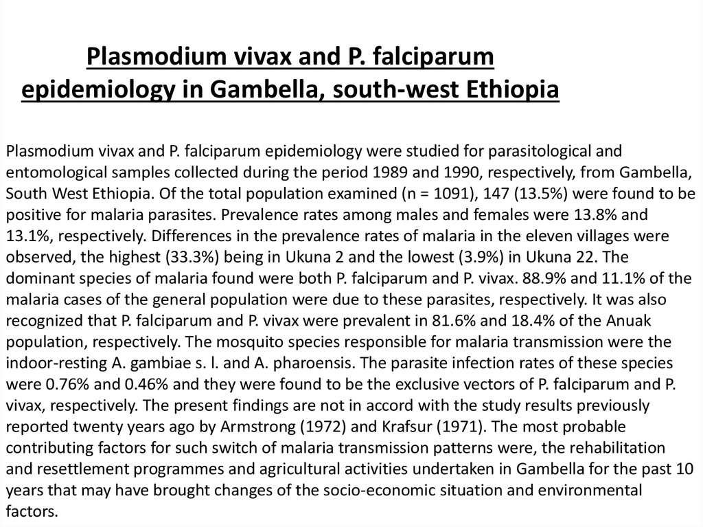
Plasmodium vivax and P. falciparumepidemiology in Gambella, south-west Ethiopia
Plasmodium vivax and P. falciparum epidemiology were studied for parasitological and
entomological samples collected during the period 1989 and 1990, respectively, from Gambella,
South West Ethiopia. Of the total population examined (n = 1091), 147 (13.5%) were found to be
positive for malaria parasites. Prevalence rates among males and females were 13.8% and
13.1%, respectively. Differences in the prevalence rates of malaria in the eleven villages were
observed, the highest (33.3%) being in Ukuna 2 and the lowest (3.9%) in Ukuna 22. The
dominant species of malaria found were both P. falciparum and P. vivax. 88.9% and 11.1% of the
malaria cases of the general population were due to these parasites, respectively. It was also
recognized that P. falciparum and P. vivax were prevalent in 81.6% and 18.4% of the Anuak
population, respectively. The mosquito species responsible for malaria transmission were the
indoor-resting A. gambiae s. l. and A. pharoensis. The parasite infection rates of these species
were 0.76% and 0.46% and they were found to be the exclusive vectors of P. falciparum and P.
vivax, respectively. The present findings are not in accord with the study results previously
reported twenty years ago by Armstrong (1972) and Krafsur (1971). The most probable
contributing factors for such switch of malaria transmission patterns were, the rehabilitation
and resettlement programmes and agricultural activities undertaken in Gambella for the past 10
years that may have brought changes of the socio-economic situation and environmental
factors.
29.
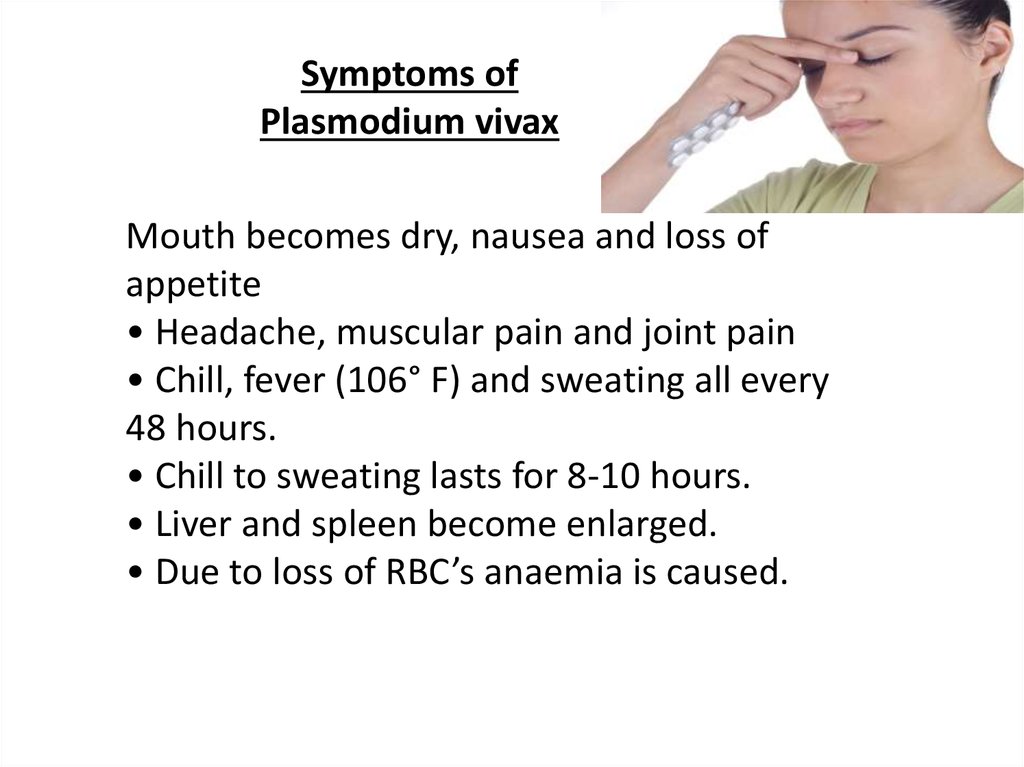
Symptoms ofPlasmodium vivax
Mouth becomes dry, nausea and loss of
appetite
• Headache, muscular pain and joint pain
• Chill, fever (106° F) and sweating all every
48 hours.
• Chill to sweating lasts for 8-10 hours.
• Liver and spleen become enlarged.
• Due to loss of RBC’s anaemia is caused.
30.
Life Cycle of Plasmodium vivax31.
• Hosts:• Plasmodium completes its lifecycle in two hosts (digenetic): Manand female Anopheles mosquito.
• 1. Primary or definitive host:
• Female Anopheles mosquito is the primary host of Plasmodium in
which it completes its sexual life cycle.
• 2. Secondary or Intermediate host:
• Man is the secondary host of plasmodium in which it completes its
asexual life cycle.
• The lifecycle of Plasmodium can be divided into three phases:
• 1. Asexual sehizogony
• 2. Sexual gamogony
• 3. Asexual sporogony
32.
• ASEXUAL CYCLE OF Plasmodium, IN MAN• Infective form of Plasmodium is known as
sporozoites. Sporozoites are 11-12µ long slender,
uni-nucleated. Sickle-shaped structure present in
the salivary glands of infected mosquito. When
an infected female Anopheles mosquito bites a
healthy man, a large number of sporozoites enter
into the blood stream of man. Within half an
hour, sporozoites enter the liver cells and
undergo asexual multiplication called schizogony.
33.
1. Asexual Schizogony:Schizogony is the asexual phase of reproduction of Plasmodium. It takes place in livercells and RBC’s of man. Schizogony can be divided into following phases:
a) Pre-erythrocytic schizogony
b) Exo-erythrocytic schizogony
c) Erythrocytic schizogony
d) Post- erythrocytic schizogony
a. Pre-erythrocytic schizogony:
In the liver cells, sporozoites grow to form a large and spherical schizont. Schizont
divides by multiple fission and forms a large number of cryptozoites. They may either
pass into the blood circulation to start erythrocytic schizogony or enter fresh liver cells
to start Exo-erythrocytic schizogony. Pre-erythrocytic schizogony takes 8 days to
complete.
34.
b. Exo-erythrocytic schizogony:
After re-entering fresh liver cell each cryptozoites divides to form a large number
of metacryptozoites similar to pre-erythrocytic schizogony.
Meta-cryptozoites are two types:
Smaller micro-metacryptozoites and larger macro-metacryptozoites. The micrometacryptozoites enter the RBC’s to start erythrocytic schizogony, while the
macro-metacryptozoites invade fresh liver cells to continue exo-erythrocytic
schizogony. It takes normally 4 days to complete.
c. Erythrocytic schizogony:As stated above, the erythrocytic schizogony begins when the RBC’s of blood are
attacked either by pre-erythrocytic cryptozoites or by exo-erythrocytic micrometacryptozoites. It takes normally in 8 to 12 days after above 2 phases. Stages of
erythrocytic schizogony are:
i. Trophozoite Stage:The merozoites (cryptozoites and micro- metacryptozoites) after entering into the
blood stream, feed on erythrocytes, become rounded and modify into trophozoite
35.
ii. Signet Ring Stage:As the merozoites grow a vacuole appears in the center and the nucleus is pushed to one
side. It gives a ring like appearance and known as signet ring stage.
The parasite ingests haemoglobin and decomposes it into protein and haematin. Protein is
use as food whereas unused haematin forms toxic. Yellowish brown malarial pigment,
haemozoin.
iii. Amoeboid Stage: As the signet ring parasite grows, vacuole disappears and the parasite becomes amoeboid in
appearance, thrusting out pseudopodial processes. This stage is called amoeboid stage. At
this stage RBC develops numerous granules, the Schuffner’s granules.
iv. Schizont Stage:Parasite grows in size, becomes rounded and almost completely fills the RBC called Schizont.
v. Rosette Stage:The nucleus of schizont divides by multiple fission to form 6 to 24 daughter nuclei. These
nuclei arrange at the periphery, while the toxic haemozoin granules accumulate at the center
of RBC. It appears as a flower rose, so called rosette stage.
Nuclei of rosette stage are surrounded by a little cytoplasm and are develop into merozoites.
With the rupture of the RBC, these merozoites are liberated into the blood plasma along with
toxic haemozoin. These normally attack fresh RBC’s to repeat the erythrocytic cycle or may
change into gametocytes. One complete erythrocytic cycle takes 48 hours in Plasmodium
vivax.
36.
d. Post-erythrocytic schizogony:Sometimes, some merozoites produced in erythrocytic schizogony reach the livercells and undergo schizogony development in liver cells. This is called posterythrocytic schizogony.
SEXUAL CYCLE OF Plasmodium in MAN
2. Sexual Gamogony:Formulation of gametocytes:
After many generations in about 4-5 is the blood some merozoites increase in size to
form two types of gametocytes; larger macro (9-10µ), less numerous and contain
large nucleus. Macro gametocytes are larger (10-12µ), more numerous and contain
smaller nucleus.
37.
SEXUAL CYCLE OF Plasmodium IN MOSQUITOWhen a female Anopheles sucks the blood of a malaria patient, the gametocytes reach
the stomach of mosquito and formation of gametes take palace as follows:
a. Gametogenesis (gemetogony) :
Process of formulation of gametes (male and female gametes).
i. Formulation of male gametes:
The nucleus of microgametocyte divides to form 6-8 daughter nuclei. The cytoplasm
gives out same number of flagella like projections and daughter nuclei enter in each
projection. These projections separate from the cytoplasm and form 6-8 haploid
microgamete or male gametes. This process of formation of microgamete is called
exflagellation.
ii. Formation of female gamete:The mega gametocyte undergoes some reorganization to form a single haploid mega
gamete or female gamete which is ready for fertilization.
38.
b. Fertilization:
The male gamete enters the female gamete through the fertilization cone formed at
female gamete and form diploid zygote or synkaryon. Fusion is anisogamous.
c. Ookinete stage:
The zygote remains inactive for sometimes and then elongates into a worm like
Ookinete or vermicule, which is motile. The Ookinete penetrates the stomach wall
and comes to lie below its outer epithelial layer.
d. Oocyst stage:
The Ookinete gets enclosed in a cyst. The encysted zygote is called Oocyst. The
Oocyet absorbs nourishment and grows in size.
3. Asexual Sporogony
The nucleus of Oocyet divides repeatedly to form a large number of haploid
daughter nuclei. At the same time, the cytoplasm develops vacuoles and gives
numerous cytoplasmic masses. The daughter nuclei pass into each cytoplasmic mass
and develop into slender sickle-shaped sporozoites are formed in each Oocyet. This
phase of asexual multiplication is known as sporogony.
Lastly, the Oocyet brusts and sporozoites are liberated into the haemolymph of the
mosquito. They spread throughout the haemolymph and eventually reach the
salivary glands and enter the duct of the hypopharyx. The mosquito is now becomes
infective and sporozoites get inoculated or injected the human blood when the
mosquito bites. The cycle is repeated.
In mosquito whole sexual cycle is completed in 10-12 days.
39.
Incubation period:The period between infection and the appearance of first symptoms is called incubation period.
It is about 10-14 days in Plasmodium vivax.
Pre-patent period:
The duration between the initial sporozoites infection and the first appearance of parasites in
the blood is called as pre-patient period. It is about 8 days in Plasmodium vivax.
40. Control
Controlcontrolled by three ways
1. Destruction of vector
2. Prevention of infection(prophylaxis)
3. Treatment of patient
4. Public awareness
41.
1. Destruction of vector (Anopheles mosquito)• Mosquito can be killed by spraying DDT, BHC, Dieldrin, Malathion etc.
• Filling up ditches, gutters and pits where the mosquito breeds.
• Water surface can be poisoned by spreading kerosene oil, petroleum etc.
• A speedly flow of water prevents the mosquito larva and pupa flourishing.
• Biological control: Certain fishes (trouts, minnows, stickle back), ducks, dragon flies etc feed on
larva and pupa of mosquito.
2. Prevention of infection (Prophylaxis)
• Use of mosquito nets.
• Screening doors, windows and ventilators.
• Using mosquito repellent creams (e.g. odomus), anti mosquito mat (e.g. Supermat) etc.
3. Treatment of patient:
There are several drugs that kill different stages of parasite in patient. The oldest drug is
Quinine; Paludrine kills almost all stages of parasite. Daraprism (single dose of 25 mg) is the
most effective drug.
42.
Plasmodium ovalePlasmodium ovale is a species of parasitic protozoa that causes
tertian malaria in humans. It is one of several species
of Plasmodiumparasites that infect humans
including Plasmodium falciparum and Plasmodium vivax which
are responsible for most malarial infection. It is rare compared
to these two parasites, and substantially less dangerous than P.
falciparum.
P. ovale has recently been shown by genetic methods to consist
of two subspecies, P. ovale curtisi and P. ovale wallikeri
43.
• Prepatent period.Humans are the only natural hosts for P. ovale. Much of what isknown about this parasite was obtained during malaria therapy of naïve patients
over 60 years ago. The prepatent period is the interval between sporozoite
inoculation and the first detection of parasites in the peripheral blood. Sinton et al.
reported a mean prepatent period of about 15 days, whereas James et al. working
with six different strains of the parasite, reported a mean of 13.6 days. The
Donaldson strain exhibited prepatent periods of 12 to 20 days, with a mean of 15.3
days; for the Liberian strain, prepatent periods of 13.5 to 15 days have been
reported . A retrospective examination of induced infections with P. ovalewas made
by Collins and Jeffery . These data were extracted from the records of patients that
were given malaria therapy for the treatment of neurosyphilis between 1940 and
1963.
• Prior to the introduction of penicillin for the treatment of syphilis, malaria was one
of the most effective treatments for the disease . The range in prepatent periods
following sporozoite injection was 14 to 20 days. A listing of prepatent periods for
30 patients infected via sporozoites with the Donaldson and Liberian strains
indicated prepatent periods of 12 to 20 days, with a median of 14.5 days.
44.
EpidemiologyWhile it is frequently said that P. ovale is very limited in its
range being limited to West Africa,the Philippines,
eastern Indonesia, and Papua New Guinea, it has been
reported
from Bangladesh , Cambodia, India, Thailand and Vietnam
The reported prevalence is low (<5%) with the exception of
West Africa, where prevalences above 10% have been
observed.
The epidemiology of this parasite is in need of updating
because the most recent global map of its distribution was
produced in 1969.
It has been estimated that there are about 15 million cases
of infection each year with this parasite.
45.
Clinical featuresThe prepatent period in the human ranges from 12 to 20
days. Some forms in the liver have delayed development and
relapse may occur after periods of up to 4 years after
infection.
The developmental cycle in the blood lasts approximately 49
h. An examination of records from induced infections
indicated that there were an average of 10.3 fever episodes of
> or = 101 °F (38,3 °C) and 4.5 fever episodes of > or = 104 °F
(40,0 °C). Mean maximum parasite levels were 6,944/microl
for sporozoite-induced infections and 7,310/microl for
trophozoite-induced infections.
46.
DiagnosisThe microscopic appearance of P. ovale is very similar to that of P.
vivax and if there are only a small number of parasites seen, it may be
impossible to distinguish the two species on morphological grounds
alone. There is no difference between the medical treatment of P.
ovale and P. vivax, and therefore some laboratory diagnoses report
"P. vivax/ovale", which is perfectly acceptable as treatment for the two
are very similar. Schüffner's dots are seen on the surface of the
parasitised red blood cell, but these are larger and darker than in P.
vivax and are sometimes called James' dots or James' stippling. About
twenty percent of the parasitised cells are oval in shape (hence the
species name) and some of the oval cells also have fimbriated edges
(the so-called "comet cell"). The mature schizonts of P. ovale never have
more than twelve nuclei within them and this is the only reliable way of
distinguishing between the two species.
47.
P. vivax and P. ovale that has been sitting in EDTA for morethan half-an-hour before the blood film is made will look
very similar in appearance to P. malariae, which is an
important reason to warn the laboratory immediately when
the blood sample is drawn so they can process the sample
as soon as it arrives.
Molecular tests (tests that look for DNA material from P.
ovale in blood) must take into account the fact that there
are two subspecies of ovale and tests designed for one
subspecies may not necessarily detect the other
48.
TreatmentStandard treatment is concurrent treatment with chloroquine
and primaquine . The combination atovaquone-proguanil may
be used in those patients who are unable to take chloroquine
for whatever reason
49.
Plasmodium malariaePlasmodium malariae is a parasitic protozoa that
causes malaria in humans. It is one of several species
of Plasmodium parasites that infect humans
including Plasmodium falciparum and Plasmodium
vivax which are responsible for most malarial infection. While
found worldwide, it is a so-called "benign malaria" and is not
nearly as dangerous as that produced by P. falciparum or P.
vivax. It causes feversthat recur at approximately three-day
intervals (a quartan fever), longer than the two-day (tertian)
intervals of the other malarial parasites, hence its alternate
names quartan fever and quartan malaria
50.
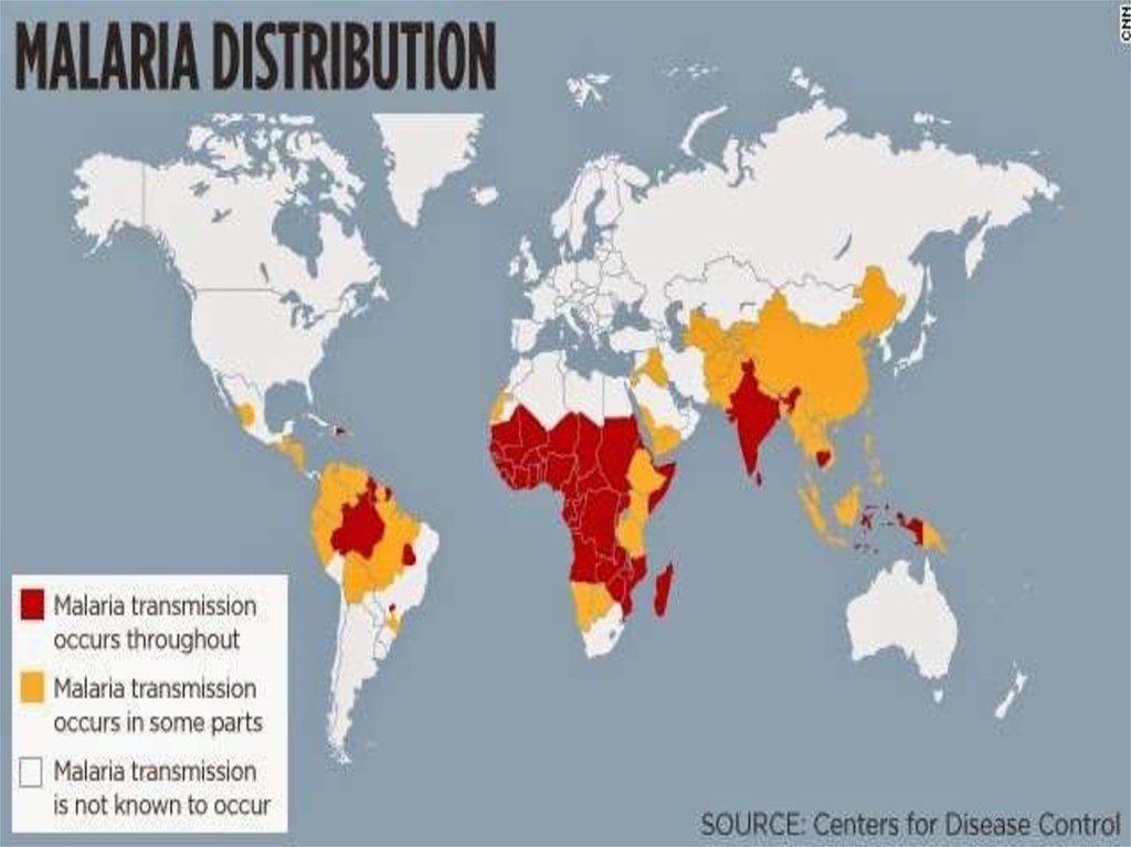
EpidemiologyEach year, approximately 500 million people will be infected with malaria worldwide Of those
infected, roughly two million will die from the disease. Malaria is caused by
six Plasmodium species: Plasmodium falciparum, Plasmodium vivax, Plasmodium ovale
curtisi,Plasmodium ovale wallikeri, Plasmodium malariae and Plasmodium knowlesi. At any one
time, an estimated 300 million people are said to be infected with at least one of
thesePlasmodium species and so there is a great need for the development of effective
treatments for decreasing the yearly mortality and morbidity rates.
P. malariae is the one of the least studied of the six species that infect
humans, in part because of its low prevalence and milder clinical
manifestations compared to the other species. It is widespread
throughout sub-Saharan Africa, much of southeast Asia, Indonesia, on
many of the islands of the western Pacific and in areas of the Amazon
Basin of South America. In endemic regions, prevalence ranges from less
than 4% to more than 20%, but there is evidence that P.
malariae infections are vastly underreported
51.
TransmissionP. malariae can be maintained at very low infection rates among a sparse and mobile
population because unlike the other Plasmodium parasites, it can remain in a human
host for an extended period of time and still remain infectious to mosquitoes
Vector
The vector of transmission of the parasite is the female Anopheles mosquito, but
many different species have been shown to transmit the parasite at least
experimentally.Collins and Jeffrey report over thirty different types of species,
which vary by geographic region.However, there are no animal reservoirs
for Plasmodium malariae.
52.
Incubation periodInformation about the prepatent period, or the period of time
between the infection of the parasite and demonstration of that
parasite within the body, of P. malariae associated malaria is
limited, but the data suggests that there is great variation, often
the length of time depending on the strain of P.
malariae parasite.[2] Usually, the prepatent period ranges from
16 to 59 days.
53.
MorphologyThe ring stages that are formed by the invasion of merozoites released by rupturing
liver stage schizonts are the first stages that appear in the blood.The ring stages grow
slowly but soon fill one-fourth to one-third of the parasitized cell.Pigment increases
rapidly and the half-grown parasite may have from 30 to 50 jet-black granules. The
parasite changes various shapes as it grows and stretches across the host cell to form
the band form.
Clinical presentation in humans
Plasmodium malariae causes a chronic infection that in some cases can last a lifetime. The P.
malariae parasite has several differences between it and the other Plasmodiumparasites, one
being that maximum parasite counts are usually low compared to those in patients infected
with P. falciparum or P. vivax.The reason for this can be accounted for by the lower number of
merozoites produced per erythrocytic cycle, the longer 72-hour developmental cycle (compared
to the 48-hour cycle of P. vivax and P. falciparum), the preference for development in older
erythrocytes and the resulting earlier development of immunity by the human host.Another
defining feature of P. malariae is that the fever manifestations of the parasite are more
moderate relative to those of P. falciparum and P. vivax and fevers show quartan periodicity.
54.
Along with bouts of fever and more general clinical symptoms such as chills andnausea, the presence of edema and the nephrotic syndrome has been documented
with some P. malariae infections. It has been suggested that immune complexes may
cause structural glomerular damage and that renal disease may also occur. Although P.
malariae alone has a low morbidity rate, it does contribute to the total morbidity
caused by all Plasmodium species, as manifested in the incidences of anemia, low birth
rate and reduced resistance to other infections.
Due to a similarity in the appearances of the pathogens, P. knowlesi infections are
often misdiagnosed as P. malariae infections. Molecular analysis is usually required for
an accurate diagnosis.
55.
DiagnosticsThe preferable method for diagnosis of P. malariae is through
the examination of peripheral blood films stained with
Giemsa stain.PCR techniques are also commonly used for
diagnoses confirmation as well as to separate
mixed Plasmodium infections. Even with these techniques,
however, it may still be impossible to differentiate infections,
as is the case in areas of South America where humans and
monkeys coexist and P. malariae and P. brasilianum are not
easily distinguishable
56.
Life cycleP. malariae is the only human malaria parasite
that causes fevers that recur at approximately
three-day intervals (therefore occurring evey
fourth day, a quartan fever), longer than the
two-day (tertian) intervals of the other malarial
parasites.
57.
Laboratory considerationsP. vivax and P. ovale sitting in EDTA for more than 30 minutes before the
blood film is made will look very similar in appearance to P. malariae,
which is an important reason to warn the laboratory immediately when
the blood sample is drawn so they can process the sample as soon as it
arrives.
Microscopically, the parasitised red blood cell (erythrocyte) is never
enlarged and may even appear smaller than that of normal red blood
cells. The cytoplasm is not decolorized and no dots are visible on the cell
surface. The food vacuole is small and the parasite is compact. Cells
seldom host more than one parasite. Band forms, where the parasite
forms a thick band across the width of the infected cell, are characteristic
of this species (and some would say is diagnostic). Large grains of malarial
pigment are often seen in these parasites: more so than any
other Plasmodium species, 8 merozoites
58.
Management and therapyFailure to detect some P. malariae infections has led to modifications of the species-specific
primers and to efforts towards the development of real-time PCR assays .The development of
such an assay has included the use of generic primers that target a highly conserved region of
the 18S rRNA genes of the four human-infecting species ofPlasmodium. This assay was found to
be highly specific and sensitive. Although serologic tests are not specific enough for diagnostic
purposes, they can be used as basic epidemiologic tools.The immunofluorescent-antibody (IFA)
technique can be used to measure the presence of antibodies to P. malariae.. A pattern has
emerged in which an infection of short duration causes a rapidly declining immune response,
but upon re-infection or recrudescence, the IFA level rises significantly and remains present for
many months or years.
The increasing need to correctly identify P. malariae infection is underscored by its possible antimalarial resistance. In a study by Müller-Stöver et al., the researchers presented three patients
who were found to be infected with the parasite after taking anti-malarial medications.[11] Given
the slower pre-erythrocytic development and longer incubation period compared to the other
malaria causing Plasmodium species, the researchers hypothesized that the anti-malarials may
not be effective enough against the pre-erythrocytic stages of P. malariae.[11] They thought that
further development of P. malariae can occur when plasma concentrations of the anti-malarials
gradually decrease after the anti-malarial medications are taken. According to Dr. William E.
Collins from the Center of Disease Control (CDC), chloroquines most commonly used for
treatment and no evidence of resistance to this drug has been found.In that event, it is possible
that the results from Müller-Stöver et al. provided isolated incidences.
59.
Public health, preventionstrategies and vaccines
The food vacuole is the specialized compartment that degrades hemoglobin during the asexual
erythrocytic stage of the parasite.It is implied that effective drug treatments can be developed
by targeting the proteolytic enzymes of the food vacuole. In a paper published in 1997,
Westling et al. focused their attention on the aspartic endopeptidase class of enzymes, simply
called plasmepsins. They sought to characterize the specificity for the enzymes cloned from P.
vivax and P. malariae. Using substrate specificity studies and inhibitor analysis, it was found that
the plasmepsins for P. malariae and P. vivax showed less specificity than that for P.
falciparum. Unfortunately, this means that the development of a selective inhibitor for P.
malariae may prove more challenging than the development of one for P. falciparum.Another
study by Bruce et al.presented evidence that there may be regular genetic exchange within P.
malariae populations. Six polymorphic genetic markers from P. malariae were isolated and
analyzed in 70 samples of naturally acquired P. malariae infections from different parts of the
world. The data showed a high level of multi-genotypic carriage in humans.
Both of these experiments illustrate that development of vaccine options will prove challenging,
if not impossible. Dr. William Collins doubts that anyone is currently looking for possible
vaccines for P. malariae and given the complexity of the parasite it can be inferred why. He
states that very few studies are conducted with this parasite, perhaps as a result of its perceived
low morbidity and prevalence. Collins sights the great restrictions of studies with chimpanzees
and monkeys as a sizeable barrier.Since thePlasmodium brasilianium parasite that infects South
American monkeys is thought to be an adapted form of P. malariae, more research with P.
brasilianium may hold valuable insight into P. malariae.