 Медицина
Похожие презентации:
")


Diabetes Mellitus
1.
Diabetes MellitusDiabetes is not a single disease. Rather, it is a
heterogeneous group of syndromes characterized by an
elevation of blood glucose level caused by a relative or
absolute deficiency of insulin.
Etiology of diabetes mellitus:
Diabetes can be divided into two main groups based on
their requirement for insulin:
A. Type 1 diabetes (IDDM): Insulin–dependent
diabetes mellitus most commonly occurs in individual
around the time of puberty.
Causes: Massive ß-cell destruction due to autoimmuneprocesses or an invasion of viruses or by the action of
chemical toxins. As results of the ß-cell destruction, the
pancreas fails to respond to glucose and the classic
symptoms of insulin deficiency appear (polydipsia,
polyuria, and polyphagia and weight loss).
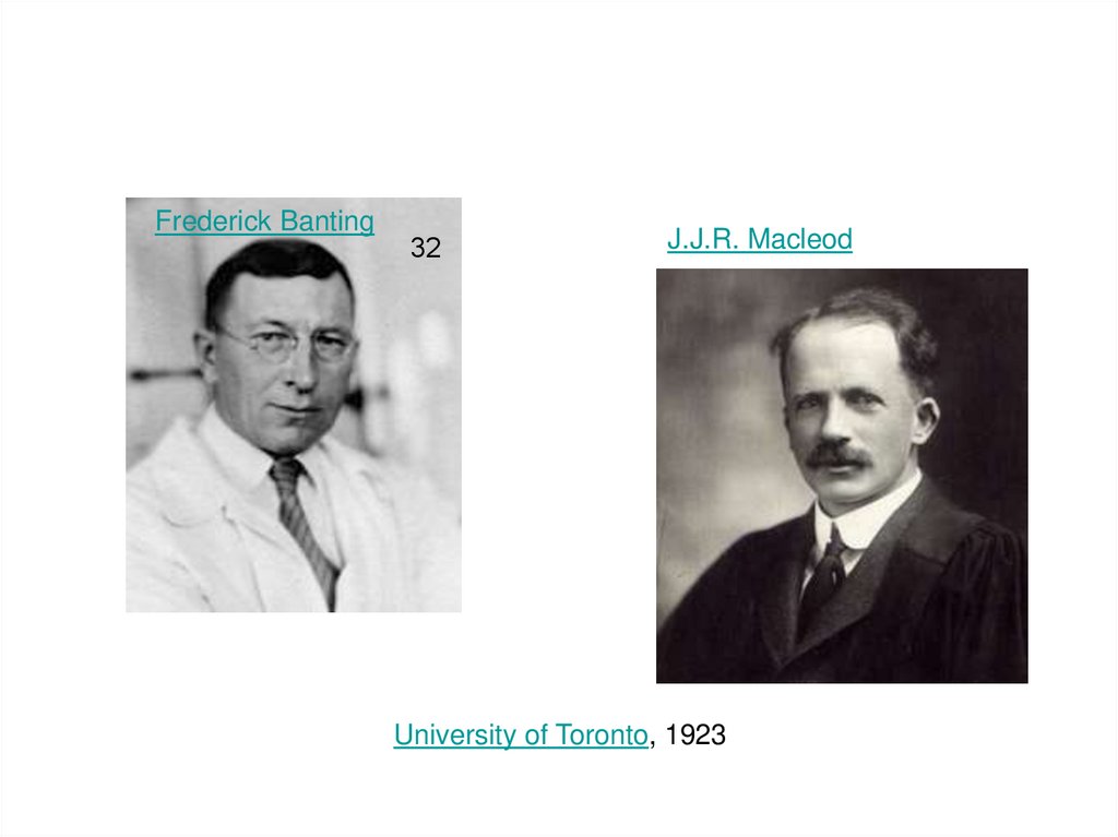
2.
Frederick Banting32
J.J.R. Macleod
University of Toronto, 1923
3.
Treatment: Type 1 diabetic must depend on exogenous(injected) insulin to control hyperglycemia avoid
ketoacidosis and maintain acceptable levels of
glycosylated hemoglobin (HbA1c). The rate of formation
of HbA1c is proportional to the average blood glucose
concentration over the previous several months; thus
HbA1c provides a measure of how well treatment
has normalized blood glucose in diabetics. The goal in
administrating insulin is to maintain blood glucose conc.
close to normal to avoid long-term complications.
4.
ما بعد األكلNormal ß-cell function: Before ingesting a meal, low,
basal levels of circulating insulin are maintained through
constant ß-cell secretion. This is suppresses lipolysis,
proteolysis and glycogenolysis. A burst of insulin
secretion occurs within two minutes after ingesting a
meal, in response to transient increases in the levels of
circulating glucose and amino acids. This lasts up to 15
minutes and is followed by postprandial insulin secretion.
However in type 1 diabetics, the ß-cell of pancreas can
neither maintain a basal secretion level of insulin nor
respond in variation in circulating fuels.
5.
B-Type 2 diabetes (NIDDM) (maturity-onset): Mostdiabetics are type 2 (80-90 %). The disease is influenced by
genetic factors, aging, obesity, and peripheral insulin
resistance.
Causes: The pancreas in NIDDM retains some ß-cell
function, but insulin secretion is insufficient to maintain
glucose homeostasis. The ß-cell mass may become gradually
reduced in type 2 diabetes. In contrast with type 1 diabetes,
those with type 2 are often obese. Type 2 diabetes is
frequently accompanied by the lack of sensitivity of target
organs to endogenous or exogenous insulin. The resistance
to insulin is considered the major cause of this type of
diabetes (sometimes referred to as ''metabolic syndrome").
Treatment: the goal of treatment of type 2 diabetes is to
maintain blood glucose concentration within normal limits;
most are dependent on administration of oral hypoglycemic
agents. Weight reduction, exercise, and dietary
modification may decrease insulin resistance and correct
hyperglycemia of type 2 diabetics.
6.
3-Type 3 (maturity-onset diabetes of the young(MODY):
Due to mutation of particular genes, resulting in
deregulation of glucose levels and insulin secretion. It
occurs before 25 years of age. Patients with type 3
are not obese and insulin resistance is absent
4- Type 4 (Gestational diabetes):
It is a glucose intolerance associated with
pregnancy. Tight glycemic control must be maintained
close to normal range during pregnancy. Hyperglycemia
can lead to congenital abnormalities.
Diet, exercise, and/ or insulin administration are
effective in this case.
7.
Clinical picture of diabetes in general:1. Polyurea (frequent urination especially
during night).
2. Polydepsia (excessive thirst)
3. Polyphagia (increase appetite) with loss of
weight
4. General weakness and easy fatigue.
5. May present with symptoms of complications
8.
Possible Complications in Diabetics:1-CVS complications:
-Microangiopathy: which is the thickening of the
basement membrane of endothelium of capillaries,
arterioles, and venules due to deposition of
mucopolysaccharide materials causing narrowing of
blood vessel (it is more pronounced in retina
(retinopathy), glomeruli (nephropathy), vasa nervosa
(neuropathy).
-Atherosclerosis of large vessel: Around 50% of
people with diabetes have disorders of lipid
metabolism that is marked by high triglyceride levels
or low High Density Lipoprotein (HDL) levels. If it
deposited in cerebral blood vessel it produces
thrombosis with hemiplegia. In coronary blood
vessel (angina with infarction).
9.
2- Cerebral complications (diabetic coma):- Diabetic ketoacidosis (DKA):
DKA progresses from hyperglycemia to ketosis, which
is a build-up of ketones in the body. Ketosis can lead
to acidosis, which is a condition in which the blood has
too much acid. When this happens it is known as
diabetic ketoacidosis. DKA is a potentially lifethreatening complication of diabetes. If left
untreated the electrolyte and the acid-base
disturbances can result in coma or death. Although
DKA is generally seen in people with type-1 diabetes,
it also has been described in patients with type-2
diabetes. DKA is identified by 3 clinical features:
Hyperglycemia, ketonuria or ketonemia, and
acidosis. By definition, the following laboratory values
are present with DKA: serum blood glucose greater
than 250 mg/dl, moderate or large ketonuria or
ketonemia and an arterial blood pH below 7.3 and/or
serum bicarbonate level below 15 mEq/L.
10.
Signs and symptoms: Feeling tired, excessivethirst and/or excessive urination, signs of
dehydration such as dry mouth, confusion,
rapid deep breathing, breathe that smells
fruity, fever, unconsciousness.
Treatment: It's important to treat dehydration
by replacing fluids that have been lost, so
most likely IV therapy will be used.
Electrolyte imbalances need to be corrected
and insulin therapy started to control
hyperglycemia. All of this must be done
under careful medical supervision.
11.
- Hypoglycemic Coma:It results from missing a meal or insulin overdose.
Clinical picture include hunger, sweating (moist tongue),
dizziness, headache, irritability, shakiness, clammy skin,
loss of coordinator, blurred vision, nausea, confusion,
nightmares, heart palpitations or rapid heart rate, and
numbness in the lips or tongue, dilated pupil, convulsion,
coma. If one doesn’t take action as mild hypoglycemia
develops, the lack of glucose may seriously impair brain
function, causing delirium, seizures or loss of consciousness
(hypoglycemic coma).
Treatment: If one becomes hypoglycemic, he should take 10 to
15 grams of carbohydrate as quickly as possible to boost
blood glucose level and avoid falling into a hypoglycemic
coma. All of the following contain 10 to 15 grams of
carbohydrate: Two to three 5-gram glucose tablets. Four to
six ounces of orange juice. Half a can of a cola or other soft
drink, Two teaspoons of sugar. Two teaspoons of honey.
12.
3- Diabetic Retinopathy (ocular complications)The elevated blood sugar levels are the main factor in the
development of damage and sclerosis to the endothelium of
blood vessels. This is particularly marked in the retina where
the vessels are very thin. If the management of DM is poor
(no improvement in blood sugar levels or uncontrolled
hypertension) significantly reduced vision or blindness may
result from hemorrhage or retinal detachment In addition to
managing the DM laser treatment can be given.
4-Diabetic nephropathy (renal complications)
This is caused by thickening of the basement membrane of
tubules, inter-intra-capillaries causing damage to the kidneys.
An early sign of this disorder is a gradual loss of protein
through the excretion of tiny protein particles in urine, a
condition known as “microalbuminuria”. This early indication
of diabetic. People diagnosed with diabetic nephropathy have a
high risk of suffering further kidney damage and edema,
possibly leading to kidney failure requiring dialysis or
transplant.
13.
Diabetic foot syndrome:This may lead to ischemia (cyanosis-coldness), neuropathy
(painless ulcer), infections (fungus infection):
combination of diabetic neuropathy (damage to the nerves)
with resulting pain and insensitivity, plus a circulatory
disorder is the reason for the high number of amputations
that still have to be performed on people with diabetes. In
most cases a minor injury to a neuropathic foot results in
damage to the skin. Because the person feels no pain, they
do not take the important step of relieving pressure on or
immobilizing the foot so the lesion cannot heal. If a
circulatory disorder such as occlusive arterial disease is an
added factor, treatment of the wound can be a long process
and there is an increased risk of amputation.
6-Genital complications: genital tract infection (puerperal
sepsis), impotence, menorrhagia (abnormally heavy bleeding
at menstruation), may be abortion, premature labor.
14.
Diagnosis:1- Urine analysis:
*Urine tests for detection of glucose in the urine using test-strips. These strips
impregnated in the urine to detect glucose by specific color reaction.
* Urine tests for detection of ketone bodies by ketostix or ketodiastix.
2- Blood glucose tests:
A-Fasting plasma glucose test: Overnight fasting then measuring plasma
glucose level in the morning it should be (80-120 mg/dl) above 140 is
considered abnormal.
B-Glucose tolerance test: Used in border-line case (i.e. fasting plasma glucose
120-140). Fasting blood glucose level is determined and urine samples are
collected, then 75 gm /100 ml glucose solution is taken orally, then samples
from venous blood and urine are tested for glucose after 30, 60, 90, 120,
150 minutes of administration. Normal person blood glucose reach the peak
level below 160 mg/dl in 30-60 min then return to fasting level again after
120-150 min. For diabetic person blood glucose reach the peak level above
180 mg/dl in 30-60 min then fails to return to its fasting level again after 120150 min. Renal threshold for glucose is 180 mg/dl).
C- Two-hours postprandial blood glucose: fasting plasma glucose level was
determined then a meal or 75 gm /100 ml glucose solution is taken orally.
After 2 hours plasma glucose level is detected. It should return to normal
fasting level after 2 hours in normal subject. If it is above 130 mg/dl so its
suggestive. If it is above 180 mg/dl so it is diagnostic.
15.
3- Glycosylated hemoglobin (HbA1c): (normal level3.9-6.9%)
This glycosylated hemoglobin is formed by nonenzymatic glycosylation reaction between glucose and
N-terminal amino acid of β-chain of the hemoglobin
molecule. It becomes stable for 6-8 weeks throughout
the life span of RBCs (120 days). It is level is high in
diabetics, reflecting the state of hyperglycemia over
the preceding 8 weeks, so it is useful to asses the
efficiency of diabetic control but it is not diagnostic. All
red blood cells have some glucose bound to them.
With normal blood glucose levels, glycated
hemoglobin is expected to be 3.9 % to 6.9 %. As
blood glucose concentration rise, however, more
binding occurs. Poor blood sugar control over time is
suggested when the glycated hemoglobin measure
exceeds 8.0%.
16.
Management of DiabetesI. Treatment with insulin
Chemistry of insulin: Insulin hormone is protein in nature
consists of two polypeptide chains, A and B. chain A is
composed of 21 amino acids while chain B consists of
30 amino acids. The chains are connected by two
disulphide linkages (S-S), which is essential for the
biological activity of insulin.
Source of insulin secretion: Insulin is the hormone
secreted by the ß (beta) cells of the islets of Langerhans.
Glucagon hormone secreted from α (alpha) cell of
pancreas and somatostatin is secreted from δ (delta) cells
of pancreas.
17.
Synthesis of insulin: The beta cells of the pancreatic isletssynthesize insulin from a single chain precursor termed
proinsulin. In the process of conversion of human proinsulin
to insulin, 4 amino acids and the remaining connector or C
peptide are removed by proteolysis. Insulin and C peptide
are secreted in equimolar amounts in response to any
stimulant.
Regulation of insulin secretion: The beta cells receive a
dual autonomic nerve supply:
1-The parasympathetic: which reaches the beta cells as
postganglionic vagal nerve endings upon stimulation; it
enhances the release of insulin, an effect which can be
blocked by atropine.
2-The sympathetic: which feeds both α and ß2-receptors:
stimulation of α-receptors inhibits the release of insulin,
whereas stimulation of ß2-receptors promotes its release.
Adrenaline had a predominant effect on the α-receptors of
the islets. It therefore inhibits release of insulin.
However, if the α-receptors are blocked by drugs e.g.
phentolamine, adrenaline would act mainly on the ß2receptors to enhance release of the hormone.
18.
Stimulants of insulin secretion: Normally, the release ofinsulin is controlled by the blood glucose level, which
directly stimulate insulin release, as well as its synthesis.
Insulin secretion is also increased by certain amino acids
(e.g. arginine, and leucine) and by GIT hormones such as
secretin, gastrin, pancreozymin gastric-inhibitory peptide
(GIP). On the other hand adrenalin is a potent inhibitor of
insulin secretion.
Mechanism of insulin secretion: Secretion is most commonly
triggered by high blood glucose which is taken up by the
glucose transporter into beta-cells of pancreas. There, it is
phosphorylated by glucokinase, which acts as a glucose
sensor. The products of glucose metabolism enter the
mitochondrial respiratory chain and generate adenosine
triphosphate (ATP). The rise in ATP levels causes a block of
k channels, leading to membrane polarization and an influx of
Ca++, which results in pulsatile insulin exocytosis.
Glucose-induced insulin secretion appears to occur in two
phases:
1. An initial-burst phase, which peaks in minutes then
rapidly declines.
2. A slow phase, which takes an hour to reach a peak.
19.
Insulin receptors:They are highly specific glycoprotein complexes,
consisting of two α subunits (on the external surface
of the cells) and two ß subunits (across the cell
membrane) linked together by disulphide bonds.
When insulin binds to the α subunits a tyrosine
residue on the inner ß subunits undergoes
autophosphorylation, leading to activation of kinase
which become capable of phosphorylation of other
proteins and enzymes. This initiates a cascade of
events, facilitating glucose entry into the cells as
well as transporting of amino acids and certain
ions. Insulin receptors vary in number inversely with
insulin concentration to which they are exposed. Thus
with low insulin concentration, the number of receptors
increases (up regulation) and with high insulin
concentration, the number of receptors
decreases.(down regulation).
20.
Types of insulin preparations:1) Regular insulin:
It is a short acting, clear aqueous soluble, crystalline zinc insulin. It is
rapid in action but short in duration. Therefore, it should frequently
administered daily to control DM. It is usually injected subcutaneously
30 minutes before meals but can be also given intravenously in
emergency, e.g., diabetic acidosis. The ultrashort acting insulins, e.g.,
Lispro, aspart and glulisine have more rapid absorption than regular
insulin, so it is usually injected 15 minutes prior to meals. Peaks
after 30-90 minutes of its injection with shorter duration of activity.
Injected
subcutaneously and intravenously in emergency usually in combination
with long acting insulin to assure proper glucose control.
21.
2) Protamine Zinc Insulin (PZI): (Long-acting insulin)The combination of crystalline zinc insulin and excess
protamine causes the formation of large crystals. Therefore
this preparation is sparingly soluble. When injected this
formulation serves as a tissue depot, producing a slow
absorption and longer duration of action lasts up to 36
hours. Because it contains excess protamine it should not
be combined in the same syringe with soluble insulin to
avoid its binding with excess protamine.
3) Isophane insulin [Neutral Protamine Hagedorn NPH)]
It is a suspension of crystalline zinc insulin combined at
neutral pH with just enough protamine (but no excess).
This intermediate acting insulin due to delayed absorption
of insulin because of its conjugation with protamine. It
should be given subcutaneously (never IV). It can be
administered in the same syringe with soluble insulin without
fear of binding with excess protamine.
22.
4) Lente Insulin:Lent insulin formulations do not contain protamine; their insolubility
results from the addition of excess zinc in an acetate buffer
rather than a phosphate buffer. The onset of action depend on the physical
state, the ambient zinc concentration, and the pH.
a) Semi-lente insulin: a microamorphous crystalline form known as
prompt insulin zinc suspension. Its onset after 1 hour and has duration
of action of 12-16 hours. It can considered as fast acing insulin.
b) Ultra-lente insulin: A large crystalline form with high zinc content,
known as extended insulin zinc suspension. It is long acting insulin
with an onset of 4-6 hours and a duration of 20-36 hours.
c) Lente insulin: Combining 7 parts of ultra-lente and 3 parts of semilente produces insulin zinc suspension. It is intermediate acting insulin,
similar to NPH in its onset (1-2 hrs) and in its duration (18-28 hrs).
5) Insulin glargine: The isoelectric point of insulin glargine is lower than that of human
insulin, leading to precipitation at the injection site, so extending its action. It has a
flat prolonged hypoglycemic effects, that is, it has no peak.
Insulin Combination:
Various premixed combinations of human insulins, such as 70% NPH and 30%
regular insulin. 50% of each of these is also available.
23.
Insulin Combination:Various premixed combinations of human insulins,
such as 70% NPH and 30% regular insulin. 50% of
each of these is also available.
Sources of insulin:
Recently human insulin has been produced either by
enzymatic modification of pork or bacterial synthesis
involving recombinant DNA technique. Human
insulin produced by recombinant DNA technique
(Humulin) is available in several formulations:
regular, NPH, Lente, Ultra-lente. It is largely
replaced most of the clinically used insulin which is
derived from either beef (cow) (differs by 3 AA from
human) and pork (differs by 1 AA from human). The
beef insulin is slightly more antigenic than pork in
humans.
24.
Adverse effects of insulin:1. Hypoglycemia:
The worst sequela of hypoglycemia is insulin shock. The
early symptoms of the hypoglycemia is the sympathetic
overactivity such as sweating, tachycardia, tremors,
palpitations, restlessness and hunger are thought to be
occurred by the compensatory secretion of epinephrine.
Then hypoglycemia affects the CNS causing mental
confusion, motor incoordination, loss of consciousness
with or without convulsion. Hypoglycemia is best treated
by administrating glucose (5% IV) or glucagon (1 mg
vial, IV, IM, SC.) or by giving oral fruit juice, or
soluble carbohydrate.
25.
2. Local reactions: Irritation at the injection site canleads to lipoatrophy or lipodystrophy. Site of
injection should be rotated. Subcutaneous infusion
can results in infection and local allergic
reactions.
3. Antigenic response (insulin resistance):
With the development of new, more highly purified
animal insulins and the advent of human insulin,
the production of insulin antibodies and
hypersensitivity reactions are less of a problem.
4. Weight gain: Is an undesirable effect of intensive
insulin therapy.
26.
Oral Hypoglycemic DrugsA- Insulin Secretagogues: These agents are
useful in:
1-Patients with Type 2 diabetes that can not
managed by diet alone.
2- Patients who develop diabetes after the age
of forty, and had diabetes
less than five years.
27.
1. Sulfonylureas1st generation: Tolbutamide (8 hr)
2nd generation:
Glibenclamide (Daonil) (18 hr), Gliclazide (Diamicron) (20 hr)
Glipizide (Minidiab) (20 hr),
Glimepiride (Amaryl) (24 hr)
Mechanisms of action
1) Stimulation of insulin secretion from ß-cells of pancreas (by blocking ATP-sensitive
K+ channels resulting in depolarization and Ca++ influx)
2) Reduction of serum glucagon level.
3) Increase binding of insulin to target tissues and receptors.
Pharmacokinetic
Given orally, metabolized by liver, excreted by kidney.
Adverse Effects
Hyperinsulinemia and hypoglycemia
Weight gain- GIT disturbance.
Contraindicated in renal and hepatic insufficiency as accumulation may occur.
Cross placenta and cause insulin depletion.
28.
2. MeglitinidNateglinide (Starlix) (2 hr)
Repaglinide (NovoNorm) (2 hr)
Mechanisms of action
1) Like sulfonylurea blocking ATP-sensitive K+ channels
2) In contrast to sulfonylurea they have rapid onset and
short duration.
3) Particularly effective in the early insulin release that occur
after a meal (postprandial glucose regulator)
Pharmacokinetic
Effective orally and inactivated by liver CYP3A4, excreted in
bile
Adverse Effects
Low incidence of hypoglycemia compared to sulfonylurea.
Drugs that inhibit CYP3A4 (erythromycin, ketoconazole) may
cause hypoglycemia whereas drugs that increase level of
this enzyme (barbiturate, carbamazepine, rifampin) may
have the opposite effect.
29.
B- Insulin Sensitizer:These agents lower blood sugar by
improving target cell response to insulin
without increasing pancreatic insulin
secretion. This group includes two
classes, Biguanides and
Thiazolidinediones.
30.
1. Biguanides(Metformin (6 hr)(Cidophage, Diaphage)
Mechanisms of action
) Reduction of hepatic gluconeogenesis
2) Slow intestinal glucose absorption.
3) Reduction of hyperlipidemia (LDL,
VLDL
cholesterol conc., fall and HDL cholesterol
concentration rise.
4) Metformin is DOC for newly diagnosed Type 2
diabetics as it reduce cardiovascular mortality.
5) Metformin requires insulin for its action, but it dos not
promote insulin secretion (hyperinsulinemia is not a
problem).
6) Metformin is effective in treatment of polycystic ovary
diseases, by lowering insulin resistance in these
women so can results in ovulation and pregnancy.
31.
PharmacokineticGiven orally, not bound to plasma proteins and not
metabolized by liver, excreted mainly in the urine.
Adverse Effects
Contraindicated in sever infection, pregnancy, renal
and hepatic insufficiency as accumulation may occur.
2) Long-term use may interfere with vitamin B12
absorption.
3) The drug should be discontinued in patients requiring
IV radiographic contrast agents. Fatal lactic acidosis
may occur.
4) Increased risk of lactic acidosis in patients treated
with heart failure medications.
32.
Thiazolidinediones (TZDs) (Glitazones)Pioglitazone (Glustin) (>24 hr) Rosiglitazone
(Rosizone) >24 hr)
Mechanisms of action
Regulate adipocyte production and secretion of fatty
acids as well as glucose metabolism, resulting in
increased insulin sensitivity in adipose tissues, liver,
skeletal ms.
2) LDL levels have increased with rosiglitazone.
3) HDL levels increase with both drugs.
4) TZDs lead to an expansion in the subcutaneous
tissues.
5) They also effective in treatment of polycystic ovary
diseases, by lowering insulin resistance in these
women so can results in ovulation and pregnancy.
33.
PharmacokineticEffective orally, bound to serum albumin and
inactivated by liver CYP450, excreted in urine and
bile.
Adverse Effects
1) Hepatotoxicity with troglitzone
2) Weight gain (due to increase in subcutaneous fat or
due to fluid retention).
3) The latter may lead to or worsen heart failure.
4) Headache and anemia.
5) Women taking oral contraceptives and TZDs may
become pregnant ( reduce plasma conc., of
estrogen-containing contraceptives
34.
Alpha 2 inhibits insulin release frompancrease but it stimulate glucagon
release