")


 Медицина
Похожие презентации:


Molar pregnancy
1. Molar pregnancy (Hydatidiform Mole)
Prepared by:Dr. Mohannad Shalaldeh
Dr.Yazan Zatari
Supervisor :
Dr.Anan Amro
Hebron Governmental Hospital
2. Outline
Gestational trophoblastic disease.Molar pregnancy.
Classification.
Pathogenesis.
Risk factors.
Presentation.
Treatment .
Follow up.
3. GTD
Gestational trophoblastic disease (GTD) is a diverse group ofinterrelated diseases resulting in the abnormal proliferation of
trophoblastic (placental) tissue.
These tumors results from abnormal fetal tissue rather than
maternal tissue.
Produce human chorionic gonadotropin (hCG).
Extremely sensitive to chemotherapy.
The most curable gynecologic malignancy.
4. GTD classification;
GTDBenign GTD ( molar
pregnancy)
80%
Malignant GTD
20%
Complete (classical) mole 90%
Choriocarcenoma
Incomplete (partial ) mole 10%
Placental site trophoblastic tumors
Persistent/invasive mole
5. Molar pregnancy
The incidence of molar pregnancy is about 1 in 1,000pregnancies
highest among Asian women occur in 1 in 500
pregnancies.
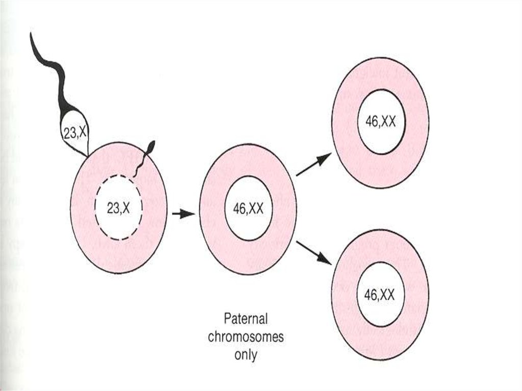
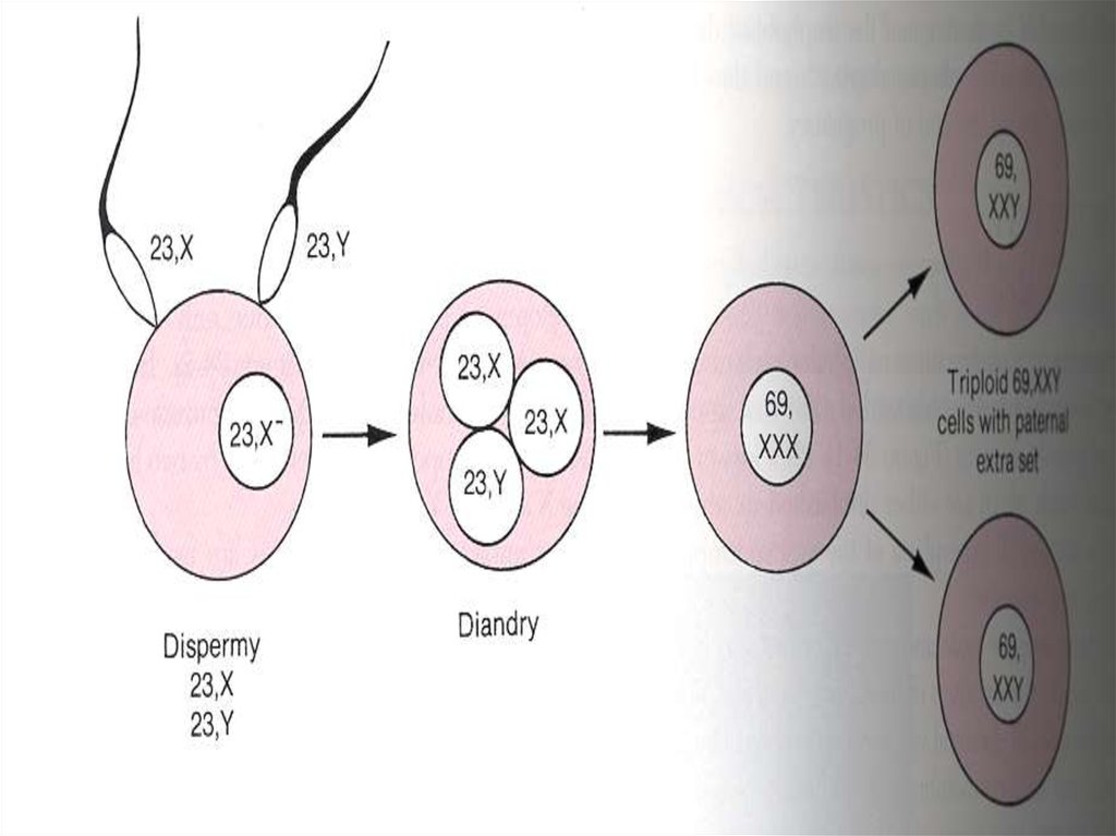
6. Molar Pregnancy
Complete moleIncomplete mole
- Fertilization an empty egg by one
sperm.
-All placental villa swollen.
-Fetus, cord, amniotic membrane
are absent.
-Paternal chromosomes only. 46
XX.
-diploidy
-fertilization of an egg by two
sperms
-some placental villa swollen
- Fetus, cord, amniotic
membrane are present
- Paternal and maternal
69XXY
-Triploid
7.
8.
9. Clinical risk factors for molar pregnancy
Age (extremes of reproductive years)<15
Clinical risk
factors
for
molar
pregnancy
>40
Reproductive history
prior hydatidiform mole
prior spontaneous abortion
Nullparity (70%)
Diet
Vitamin A deficiency
Birthplace
Outside North America( occasionally has
this disease)
10. Complete hydatidiform mole demonstrating enlarged villi of various size
11. A large amount of villi in the uterus.
12.
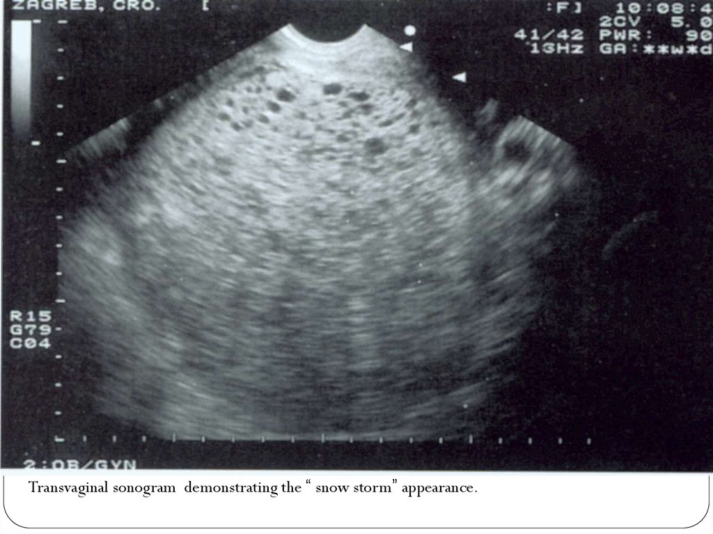
Transvaginal sonogram demonstrating the “ snow storm” appearance.13. Molar Pregnancy
14. Molar Pregnancy
Diagnosis:-Ultrasound shows snowstorm-like appearance, no
fetus, theca lutein cyst
-Beta hCG in normal pregnancy the level is at it peak at
around 14 weeks (100,000 mIU/ml)
15. Management
Baseline hCG level.Rh(D) status.
Suction curettage (D&C).
(RhoGAM) should be given to all Rhnegative
Women
hysterectomy
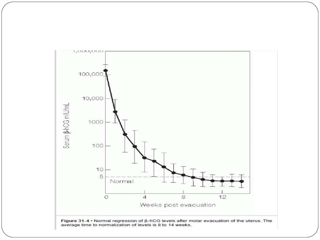
16. Follow up
95% to 100% cure rates after suction curettagePersistent disease will
develop in 15% to 25% of patients with complete moles and in 4% of
patients with partial moles
Levels should be measured within 48 hours of uterine evacuation and
then weekly until negative for 3
consecutive weeks
followed monthly for 6 months
A plateau or rise in hCG levels during
monitoring or the presence of hCG greater than 6 months after the
D&C is indicative of persistent/invasive disease.
prevent pregnancy
The risk of developing recurrent GTD
is approximately 1% to 2% after one molar pregnancy
(compared to 0.1% in the general population) but as high as
16% to
17.
18. Follow up
HCG weekly until normal for two values then monthly forone year.
Repeat x- ray if HCG rises or plateau.
Contraception for one year.
Pelvic examination every 3 weeks for 3 months.
19. Follow up
Initiate chemotherapy if:-HCG level is increasing or plateaus
-Metastasis disease is present
-HCG level is still elevated after 6 months of evacuation
-HCG starts to rise after being undetectable