


 Биология
Похожие презентации:


Seminar B5 The cell: Organelles
1. Seminar B5 The cell: Organelles
Foundation Year ProgramSeminar B5
The cell: Organelles
Introduction to Biology
2019-20
2. What is a Cell?
Foundation Year ProgramWhat is a Cell?
• A cell is the basic unit of life
• All organisms are made of cells
• In the hierarchy of biological organization, the cell
is the simplest collection of matter that can be
alive
• Many forms of life exist as single-celled organisms
• More complex organisms, including plants and
animals, are multicellular
• Multicellular organisms are cooperatives of many
kinds of specialized cells that could not survive for
long on their own. However, the cell remains the
organism’s basic unit of structure and function
Introduction to Biology
2019-20
3. What do we call the study of cells?
Foundation Year ProgramWhat do we call the study of cells?
• Cytology
• But… How can we study
cells?
• The most obvious way is by
observing them
• How can we observe cells?
Introduction to Biology
2019-20
4. Microscopy!!! What types of microscopy do you know?
Foundation Year ProgramMicroscopy!!!
What types of microscopy do you
know?
Introduction to Biology
2019-20
5. Microscopy Terms
Foundation Year ProgramMicroscopy Terms
• Magnification is the increase in an object’s
image size compared with its actual size.
• Resolution is a measure of the clarity of an
image. It is the ability of an instrument to
show two nearby objects as separate.
Introduction to Biology
2019-20
6. The Discovery
Foundation Year ProgramThe Discovery
• The progress in cell
discovery is associated
with the evolution of
microscopy.
• Larger cells were spotted
first while smaller
organisms had to wait for
the technology to be
developed.
Introduction to Biology
2019-20
7. Major events in cell biology & imaging
Foundation Year ProgramMajor events in cell biology & imaging
Introduction to Biology
2019-20
8.
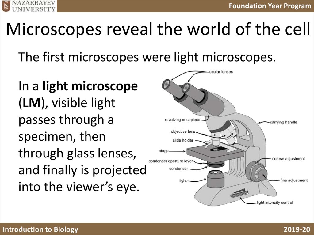
Foundation Year ProgramMicroscopes reveal the world of the cell
The first microscopes were light microscopes.
In a light microscope
(LM), visible light
passes through a
specimen, then
through glass lenses,
and finally is projected
into the viewer’s eye.
Introduction to Biology
2019-20
9. Microscopes reveal the world of the cell
Foundation Year ProgramWhat can we see with an LM?
Plant cell (non-leaf)
Introduction to Biology
Red blood cells
Plant cell (leaf)
2019-20
10. What can we see with an LM?
Foundation Year ProgramElectron microscopes
• Beginning in the 1930s, scientists started using a
very powerful microscope called the electron
microscope (EM) to view the ultrastructure of cells.
• Instead of light, EM uses a beam of electrons.
• Electron microscopes can
– resolve biological structures as small as 0.2 nm
– magnify up to 1,000,000 times.
Introduction to Biology
2019-20
11. Electron microscopes
Foundation Year ProgramWhat do they look like?
Scanning Electron
Microscope
Introduction to Biology
Transmission Electron
Microscope
2019-20
12. What do they look like?
Foundation Year ProgramElectron Microscopy
Electron microscopes use a beam of electrons to view
very small objects. The beam acts in different ways:
It will either scan the surface or pass through the
samples.
Introduction to Biology
2019-20
13. Electron Microscopy
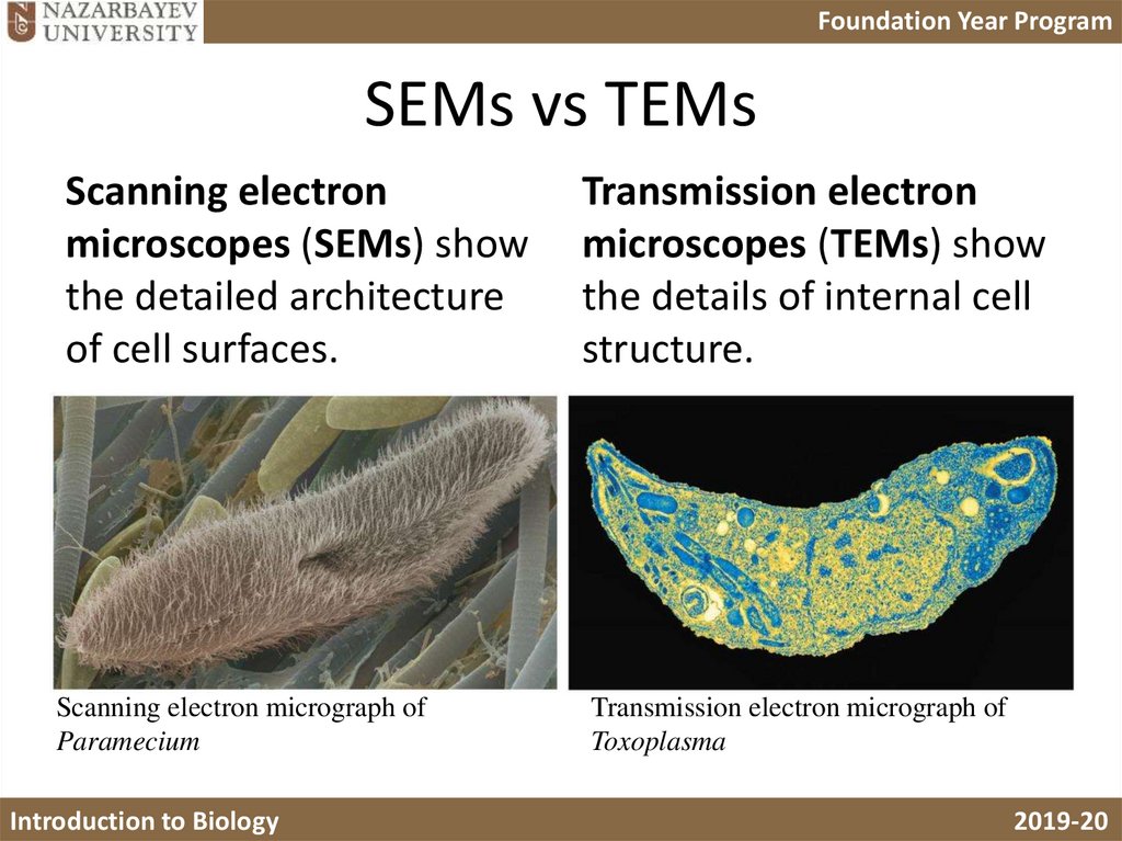
Foundation Year ProgramSEMs vs TEMs
Scanning Electron
Microscope
Introduction to Biology
Transmission Electron
Microscope
2019-20
14.
Foundation Year ProgramSEMs vs TEMs
Scanning electron
microscopes (SEMs) show
the detailed architecture
of cell surfaces.
Scanning electron micrograph of
Paramecium
Introduction to Biology
Transmission electron
microscopes (TEMs) show
the details of internal cell
structure.
Transmission electron micrograph of
Toxoplasma
2019-20
15. SEMs vs TEMs
Foundation Year ProgramYour Task
• You are going to be given a set of cards with
pictures of 8 organelles.
• There are 6-8 pictures for each organelle.
• For each organelle:
– Collect the micrographs showing that organelle
– Identify the drawing that shows the organelle
– Choose the correct description
• Hint: you can spy on other groups to confirm
your ideas.
Introduction to Biology
2019-20
16. Your Task
Foundation Year ProgramLet’s check your conclusions!!
Introduction to Biology
2019-20
17. Let’s check your conclusions!!
Foundation Year ProgramNucleus
Introduction to Biology
2019-20
18. Nucleus
Foundation Year ProgramRER
Introduction to Biology
2019-20
19. RER
Foundation Year ProgramSER
Introduction to Biology
2019-20
20. SER
Foundation Year ProgramGolgi Body
Introduction to Biology
2019-20
21. Golgi Body
Foundation Year ProgramMitochondria
Introduction to Biology
2019-20
22. Mitochondria
Foundation Year ProgramChloroplast
Introduction to Biology
2019-20
23. Chloroplast
Foundation Year ProgramLysosomes
Introduction to Biology
2019-20
24. Lysosomes
Foundation Year ProgramCentrioles/Flagella
Introduction to Biology
2019-20
25. Centrioles/Flagella
Foundation Year ProgramWould you be able to tell which
photomicrographs are from…
• Light microscope?
Introduction to Biology
2019-20
26. Would you be able to tell which photomicrographs are from…
Foundation Year ProgramWhich photomicrographs are SEM?
Introduction to Biology
2019-20
27. Which photomicrographs are SEM?
Foundation Year ProgramTEM?
• ALL THE REST!!!
• Thanks For Your Attention!!
Introduction to Biology
2019-20