


")


 Биология
Похожие презентации:


Skeletal tissues
1. SKELETAL TISSUES
BONE TISSUEAssociate Professor Kharchenko S.V.
Department of Histology and Embryology
Medical Academy named after S.I. Georgievsky
1
2. BONE TISSUE
This is a specialized type of connectivetissue with high mineralization of the
intercellular substance.
It contains 67-70% of inorganic salts
represented by salts of calcium
phosphates.
Organic matter of bone is represented by
proteins and lipids.
2
3.
BONE TISSUEPRYMARY
RETICULOFIBROSIS
SECONDARY LAMELLAR
3
4. RETICULARFIBROUS BONE TISSUE
It is found in skeleton offetus, in adults - in the
places of attachment of
tendons to bones, in
places of cranial sutures,
in dental alveoli, in the
bony labyrinth of the inner
ear. May appear during
regeneration in places of
bone damage.
4
5. LAMELLAR BONE
t is characterized by an ordered arrangement of collagenfibers in the composition of bone lamella.
Bone lamella form parallel concentric layers - osteons structural and functional units of the compact bone
5
6. CELLS OF BONE
OSTEOGENIC DIFFERONOsteogenic cell - osteoblast osteocyte
HEMATOGENOUS DIFFERON
PHSC - Promonocyte - Monocyte Osteoclast
6
7. OSTEOBLASTS - cells building bone tissue
They are located on thesurface of bone spicules in
the developing bone.
After the final formation of
the bone - in the deep layers
of the periosteum or in
places of bone damage.
They have a cuboidal or
poligonal in shape.
7
8. FUNCTION OF OSTEOBLAST
Create a bone in two stages:1. Actively synthesize the organic bone
matrix (osteoid). For this, the cell
contains a well-developed synthetic
apparatus.
2. Provide mineralization of osteoid due
to the enzyme alkaline phosphatase.
9. OSTEOCYTE
Highlydifferentiated cells
They have cell body
and process.
with a large nucleus
and basophilic
cytoplasm.
Body located in
bone cavities –
lacunae, process in
the canaliculi.
Provide the exchange
of water, proteins and 9
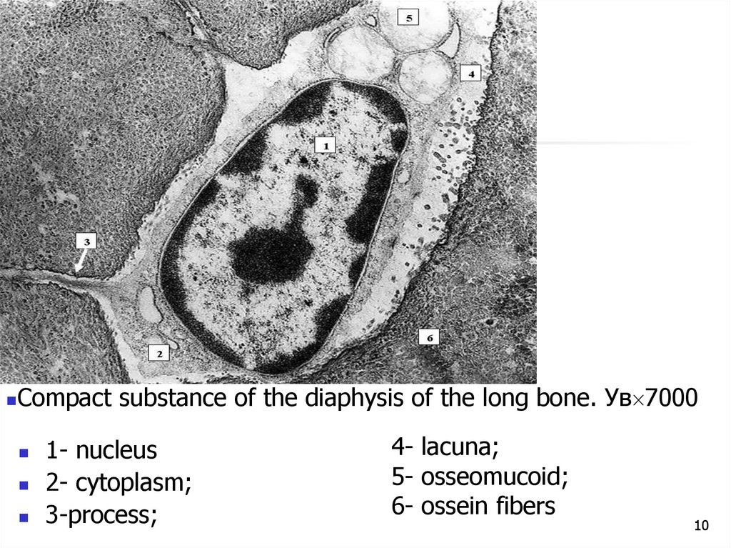
10.
Compact substance of the diaphysis of the long bone. Ув 70001- nucleus
2- cytoplasm;
3-process;
4- lacuna;
5- osseomucoid;
6- ossein fibers
10
11. OSTEOCLAST
Polynuclear macrophages of bone tissue are formed fromblood monocytes. They are located on the surface of bone
plates.
The osteoclast cytoplasm zone adjacent to the bone plate
forms a ruffled border (cytoplasmic outgrowths that contain
many lysosomes)
11
12. Osteoclast
On the periphery of the osteoclast there is a zone oftight fit, which seals the area of action of enzymes
Functions - destruction of calcified cartilage and
bone
12
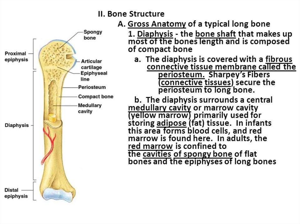
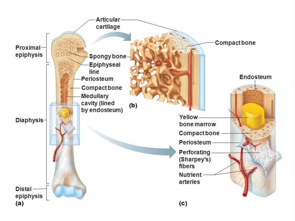
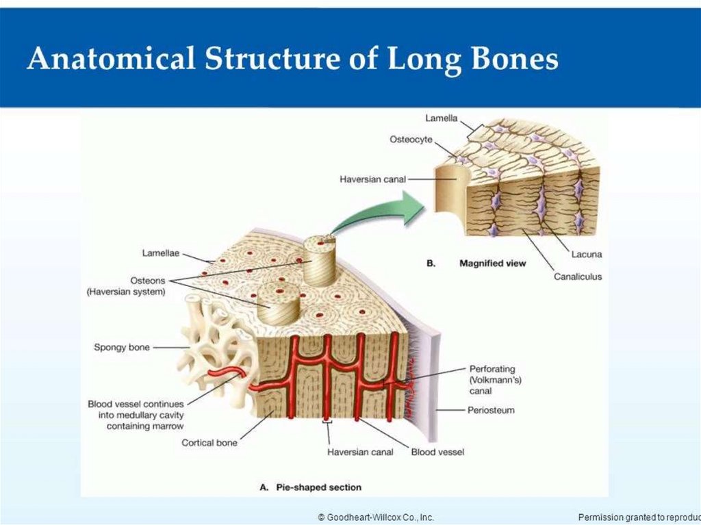
13. Long bone as an organ
Consists of:- head of the long bone - epiphysis
- long bone bodies - diaphysis
- bone marrow cavity
The epiphysis is formed by a spongy substance
and contains red bone marrow
The diaphysis is formed by several layers. It
basically has a compact substance.
The bone marrow cavity is filled with yellow bone
marrow
13
14.
1415.
1516. The structure of the diaphysis of the long bone
histologically consists of three layers:1.
The periosteum
2. Compact bone
3. Endostium
16
17.
1718.
1819. PERIOSTEUM
PERIOSTIUM consists of 2layers:
1. external - fibrous
(contains collagen fibers and
blood vessels enter the bone
through it)
2. internal - osteogenic
(contains osteoblasts that
provide bone growth in
thickness and bone
regeneration in case of injury)
19
20. ENDOSTIUM
Is lining the medullarycanal
It is formed by loose
fibrous connective tissue,
where there are
osteoblasts, osteoclasts
and cells of loose
connective tissue
20
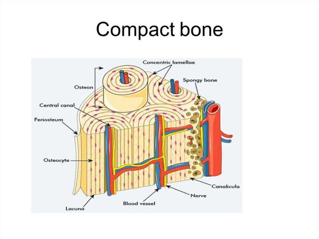
21. COMPACT BONE
Consists of three layersof bone lamella
1. External
circumferential (general)
bone lamella
2. Osteon layer
3. Internal circumferential
(general) bone lamella
21
22. OSTEON (haversian system)
Is structural andfunctional unit of
bone consists of
bone lamella
concentrically
stacked on top of
each other in the
form of cylinders
inserted one into the
other.
Osteocytes lie
between the bony
plates in the gaps
22
23. ЛАКУАНАРНО-КАНАЛЬЦЕВАЯ СИСТЕМА
2324. ГАВЕРСОВА СИСТЕМА
A blood vessel passesthrough the osteon
canal or Haversian
canal.
Between the osteons
are inserted bone
lamella (remnants of
decaying osteons).
24
25. OSTEOGENESIS PRENATAL
BONE FORMATION BEGINS ON 1 MONTH OF PRENATALDEVELOPMENT
CONTINUES UNTIL 25 YEARS
1. INTRAMEMRANOUS BONE FORMATION (DIRECT OSTEOGENESIS
FROM Mesenchyma)
Characteristic for coarse fibrous bone tissue - flat bones of the skull,
collarbone, phalanx of the fingers
2. ENDOCHONDRAL BONE FORMATION (INDIRECT OSTEOGENESIS
from the cartilage model to the long bone)
25
26. INTRAMEMRANOUS BONE FORMATION
1. osteogenic islet formation -mesenchymalcells in places of future flat bones condense and form
dense clusters - skeleton islets! Mesenchymal cells later
differentiate into preosteoblasts-osteoblasts
2. osteoid stage - osteoblasts begin to form the
intercellular substance of the bone (due to secretion of
ECM components - collagen)
3. mineralization of the intercellular
substance (impregnation with calcium salts due to
enzyme alkaline phosphatase of osteoblasts ).
4. bone remodeling and growth - old sections of
primary reticulofibrous bone are gradually destroyed
(due to osteoclasts) and new sections of lamellar bone
are formed in their place (due to osteoblasts)
26
27. INTRAMEMRANOUS BONE FORMATION
FORMATION OF OSTEOGENIC ISLANDSCELL DIFFERENTIATION AND OSTEOID FORMATION
ORIGINAL OSTEOID
27
28. ПРЯМОINTRAMEMRANOUS BONE FORMATIONЙ ОСТЕОГЕНЕЗ
The development of bone spicules, trabeculae occurs withthe appositional growth of bone tissue.
SUCH BONE - PRIMARY SPONGE
THEN primary bone IS REPLACED BY A - SECONDARY28
SPONGY bone
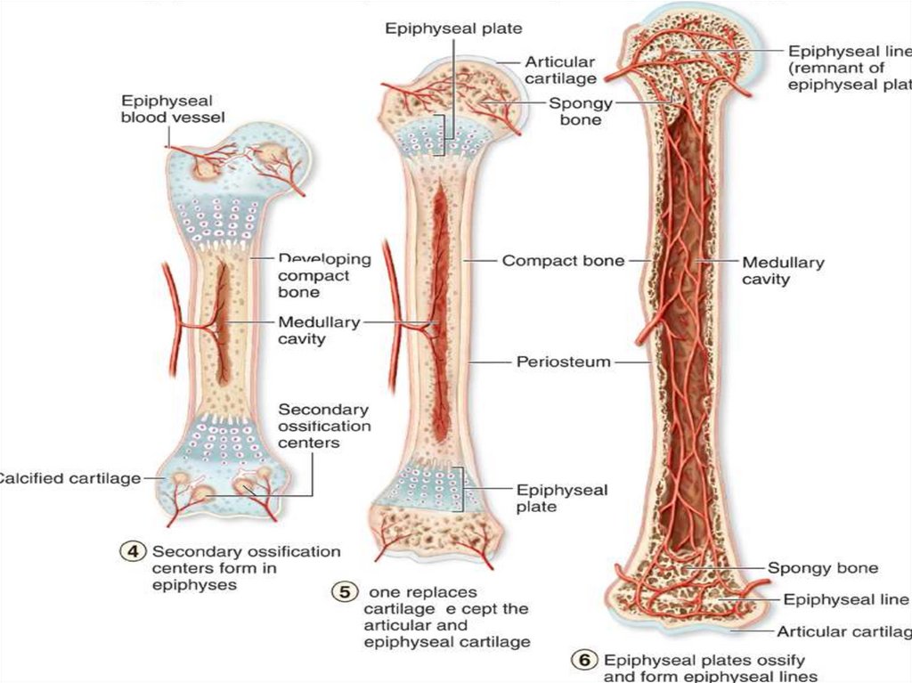
29. ENDOCHONDRAL BONE FORMATION
BEGINS ON THE SECOND MONTHFORMATION OF THE CARTILAGE MODEL
FORMATION OF THE PERIOSTEAL BONE COLLAR
(PERICONDRAL OSSIFICATION)
ENDOCHONDRAL OSSIFICATION IN THE DIAPHYSIS
ENDOCHONDRAL OSSIFICATION IN THE EPIPHYSIS
FORMATION OF EPIPHYSICAL PLATES OF GROWTH
29
30. ENDOCHONDRAL BONE FORMATION
1. the formation of a cartilage model (hyaling) ofthe future bone;
2. in the area of the diaphysis of the
cartilaginous model, perichondral ossification
occurs
while the perichondrium turns into the
periosteum, in which stem (osteogenic) cells
differentiate into osteoblasts;
osteoblasts begin to form bone tissue in the form
of common plates forming a PERIOSTEAL
BONE COLLAR
30
31.
3132. endochondral ossification
3. In parallel with this, endochondral ossificationis also observed, which occurs both in the
diaphysis and in the epiphysis; ossification of the
epiphysis is carried out only by endochondral
ossification; blood vessels grow into the
cartilage, in the adventitia of which there are
osteogenic cells that turn into osteoblasts.
Osteoblasts, producing intercellular substance,
form bone plates around the vessels in the form
of osteons; cartilage destruction occurs
simultaneously with bone formation
32