


 Биология
Похожие презентации:


Общая гистология. Ткань
1. ОБЩАЯ ГИСТОЛОГИЯ. GENERAL HISTOLOGY.
ТКАНЬ - это исторически сложившаяся системаклеток и неклеточных структур, обладающая
общностью строения и специализированная на
выполнении определенной функции.
The TISSUE is historically developed system of cells
and not cellular structures, possessing a generality of a
structure and specialized on performance of the certain
function.
2. DEVELOPMENT of TISSUES - HISTOGENESIS
DEVELOPMENT of TISSUES HISTOGENESISIt is a formation of an embryonal tissue rudiment and
its transformation into a mature tissue.
Tissues form in the end of gastrulation of embryogenesis
as a result of a differentiation of a germinal material.
EMBRYOGENESIS consists of some stages:
• fertilisation,
• cleavage,
• gastrulation,
• histogenesis and organogenesis.
3.
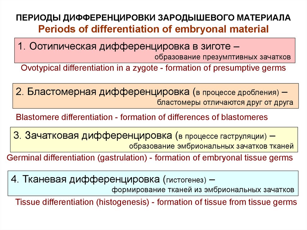
ПЕРИОДЫ ДИФФЕРЕНЦИРОВКИ ЗАРОДЫШЕВОГО МАТЕРИАЛАPeriods of differentiation of embryonal material
1. Оотипическая дифференцировка в зиготе –
образование презумптивных зачатков
Ovotypical differentiation in a zygote - formation of presumptive germs
2. Бластомерная дифференцировка (в процессе дробления) –
бластомеры отличаются друг от друга
Blastomere differentiation - formation of differences of blastomeres
3. Зачатковая дифференцировка (в процессе гаструляции) –
образование эмбриональных зачатков тканей
Germinal differentiation (gastrulation) - formation of embryonal tissue germs
4. Тканевая дифференцировка (гистогенез) –
формирование тканей из эмбриональных зачатков
Tissue differentiation (histogenesis) - formation of tissue from tissue germs
4. Сперматозоид
Яйцеклетка млекопитающих(вторично изолецитальная)
Animal’s ovum
Bird’s ovum
Ядро
Вторичные
оболочки
Ядро
Гранулы
желтка
Сперматозоид
Гранулы
желтка
жгутик
Spermatozoon
Яйцеклетка курицы
(резко телолецитальная)
ядро
акросома
5.
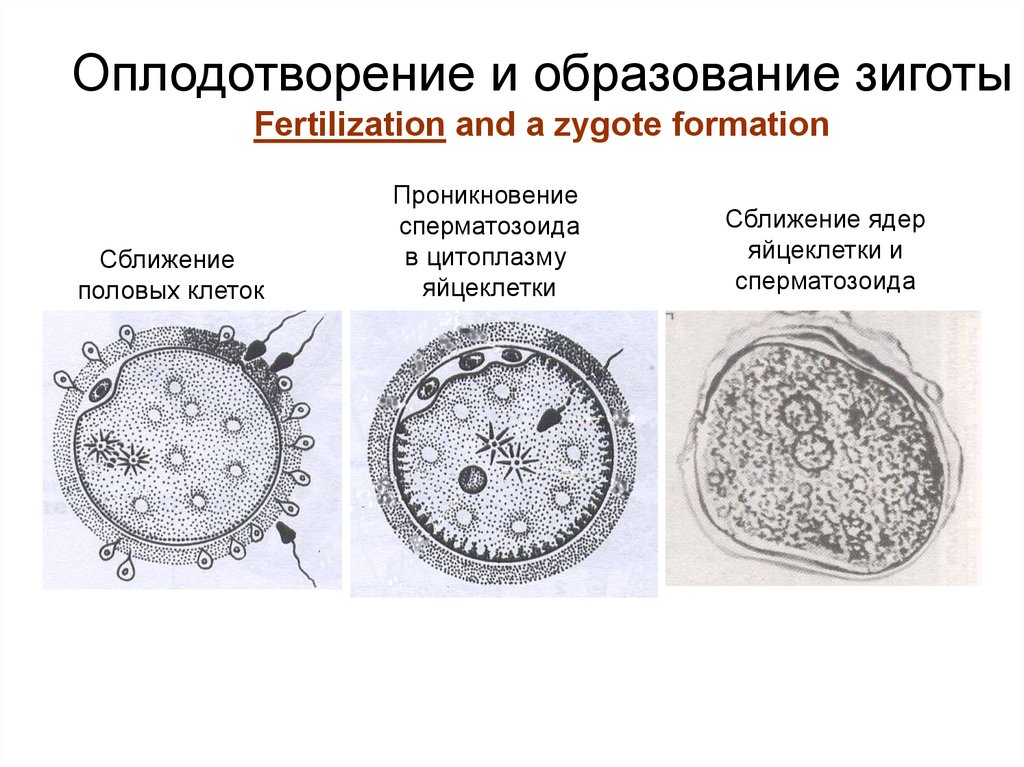
Оплодотворение и образование зиготыFertilization and a zygote formation
Сближение
половых клеток
Проникновение
сперматозоида
в цитоплазму
яйцеклетки
Сближение ядер
яйцеклетки и
сперматозоида
6.
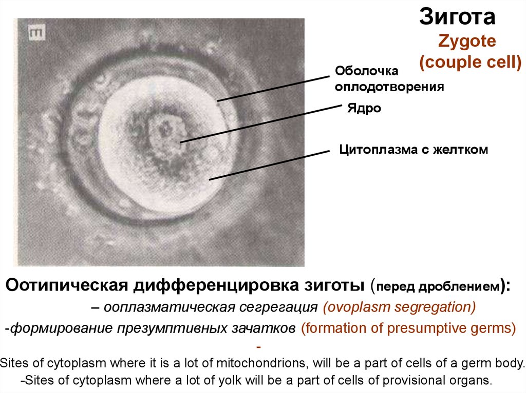
ЗиготаZygote
(couple cell)
Оболочка
оплодотворения
Ядро
Цитоплазма с желтком
Оотипическая дифференцировка зиготы (перед дроблением):
– ооплазматическая сегрегация (ovoplasm segregation)
-формирование презумптивных зачатков (formation of presumptive germs)
Sites of cytoplasm where it is a lot of mitochondrions, will be a part of cells of a germ body.
-Sites of cytoplasm where a lot of yolk will be a part of cells of provisional organs.
7. Бластомерная дифференцировка в процессе дробления Blastomere differentiation during cleavage
• Дробление - это митотическое деление зиготына бластомеры, которые образуют многоклеточный
однослойный зародыш.
• Cleavage is mitotic division of a zygote on blastomeres
which form a multicellular single-layered germ.
Отличия дробления от митоза:
1) дочерние клетки не растут;
2) дробление идет под оболочкой
оплодотворения,
3) бластомеры не расходятся;
4) происходит бластомерная
дифференцировка,
бластомеры отличаются
друг от друга содержимым
цитоплазмы.
Differences of cleavage from mitosis:
1) daughter cells do not grow;
2) cleavage goes under
a membrane of fertilization,
3) blastomeres do not miss;
4) there is blastomere differentiation,
blastomeres differ from each other
by contents of cytoplasm.
8.
The cleavage is ended by formation of the multicellular germ,but its size does not increase and = to the size of a zygote.
1. Blastomeres form the dense cellular ball – MORULA.
2. Cells of morula allocate a liquid inside morula,
are moved apart .
Formed single-layered germinal vesicle
is called BLASTULA.
9.
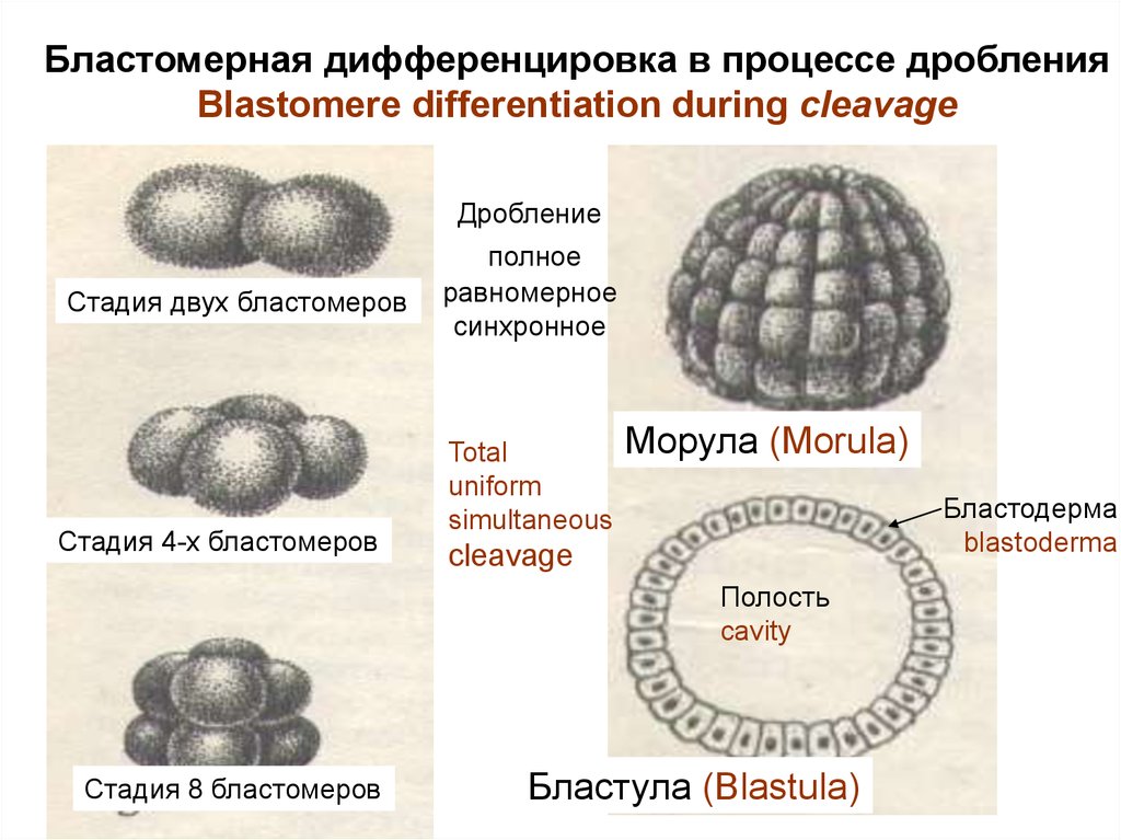
Бластомерная дифференцировка в процессе дробленияBlastomere differentiation during cleavage
Дробление
Стадия двух бластомеров
Стадия 4-х бластомеров
полное
равномерное
синхронное
Total
uniform
simultaneous
Морула (Morula)
Бластодерма
blastoderma
cleavage
Полость
cavity
Стадия 8 бластомеров
Бластула (Blastula)
10.
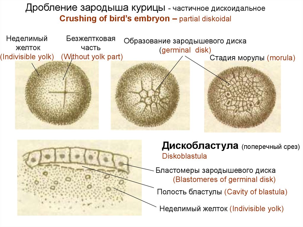
Дробление зародыша курицы - частичное дискоидальноеCrushing of bird’s embryon – partial diskoidal
Неделимый
Безжелтковая Образование зародышевого диска
желток
часть
(germinal disk)
(Indivisible yolk) (Without yolk part)
Стадия морулы (morula)
Дискобластула
(поперечный срез)
Diskoblastula
Бластомеры зародышевого диска
(Blastomeres of germinal disk)
Полость бластулы (Cavity of blastula)
Неделимый желток (Indivisible yolk)
11. Зачатковая дифференцировка Germinal differentiation
Germinal differentiation - formation of tissue germs –happens during gastrulation
Gastrulation – formation of the three-layers embryo.
Cells are made multiple copies by mitosis, are differentiated and move.
Gastrulation happens to 2 stages:
1 stage – formation of germ layers ectoderm and entoderm.
2 stage - formation of germ layer mesoderm.
12.
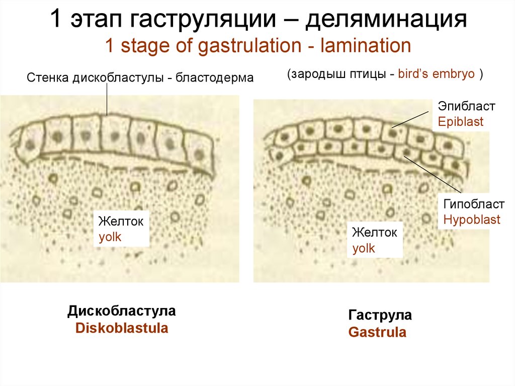
1 этап гаструляции – деляминация1 stage of gastrulation - lamination
Стенка дискобластулы - бластодерма
(зародыш птицы - bird’s embryo )
Эпибласт
Epiblast
Желток
yolk
Дискобластула
Diskoblastula
Желток
yolk
Гаструла
Gastrula
Гипобласт
Hypoblast
13.
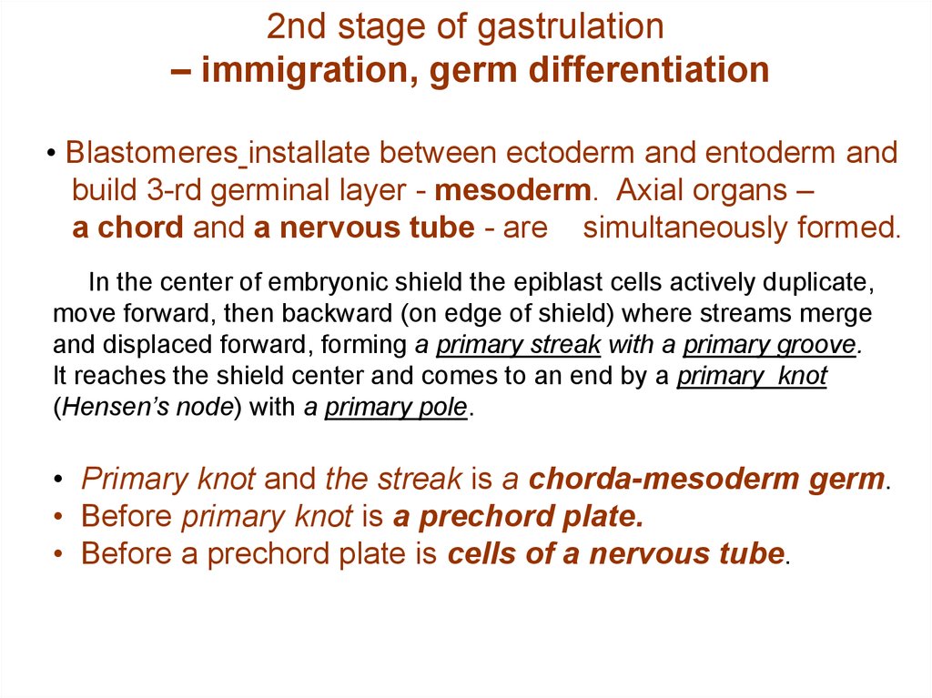
2nd stage of gastrulation– immigration, germ differentiation
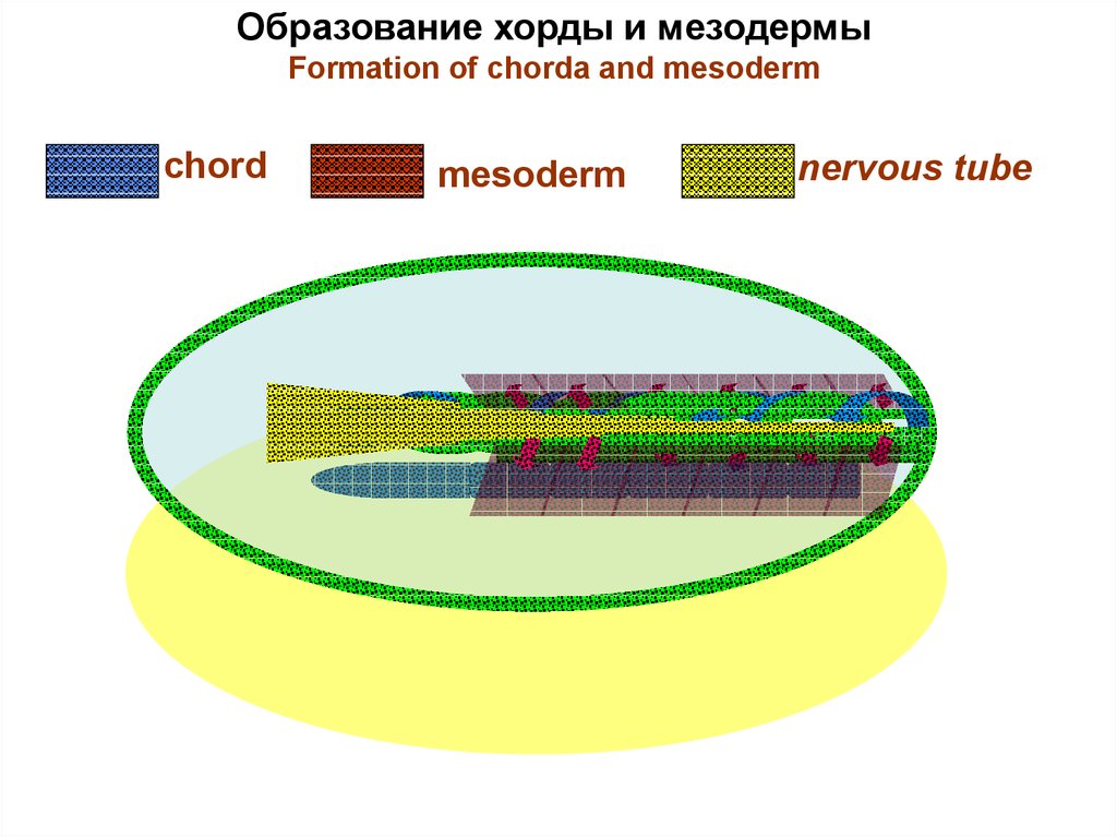
• Blastomeres installate between ectoderm and entoderm and
build 3-rd germinal layer - mesoderm. Axial organs –
a chord and a nervous tube - are simultaneously formed.
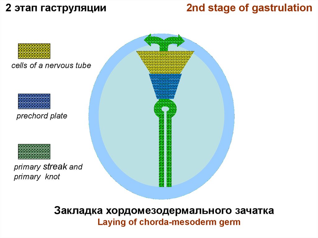
In the center of embryonic shield the epiblast cells actively duplicate,
move forward, then backward (on edge of shield) where streams merge
and displaced forward, forming a primary streak with a primary groove.
It reaches the shield center and comes to an end by a primary knot
(Hensen’s node) with a primary pole.
• Primary knot and the streak is a chorda-mesoderm germ.
• Before primary knot is a prechord plate.
• Before a prechord plate is cells of a nervous tube.
14.
2 этап гаструляции2nd stage of gastrulation
cells of a nervous tube
prechord plate
primary streak and
primary knot
Закладка хордомезодермального зачатка
Laying of chorda-mesoderm germ
15.
• A chord and mesoderm are pawnedfrom a primary strip.
• A head chord shoot is pawned
from a prechord plate.
• Cells of a nervous tube are placed
above a chord.
16.
Образование хорды и мезодермыFormation of chorda and mesoderm
chord
mesoderm
nervous tube
17. Neurulation
• Above a chord an ectoderm cells becomehigh and form a nervous plate.
• Then it is guttering and form a nervous
crest.
• Its edges are closed, and a nervous tube is
formed.
Above a nervous tube the epiblast is closed and forms to
skin ectoderm.
18.
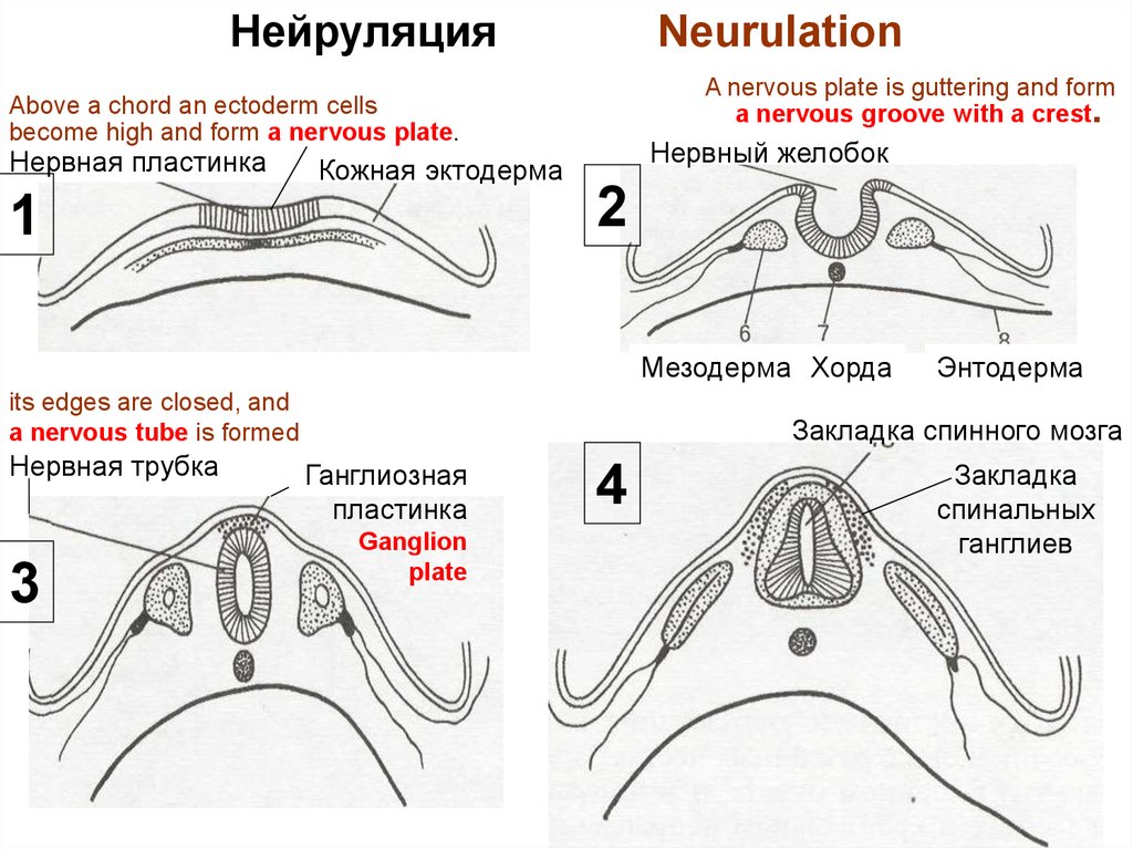
НейруляцияNeurulation
A nervous plate is guttering and form
a nervous groove with a crest.
Above a chord an ectoderm cells
become high and form a nervous plate.
Нервная пластинка
Кожная эктодерма
1
Нервный желобок
2
Мезодерма Хорда
its edges are closed, and
a nervous tube is formed
Нервная трубка
3
Энтодерма
Закладка спинного мозга
Ганглиозная
пластинка
Ganglion
plate
4
Закладка
спинальных
ганглиев
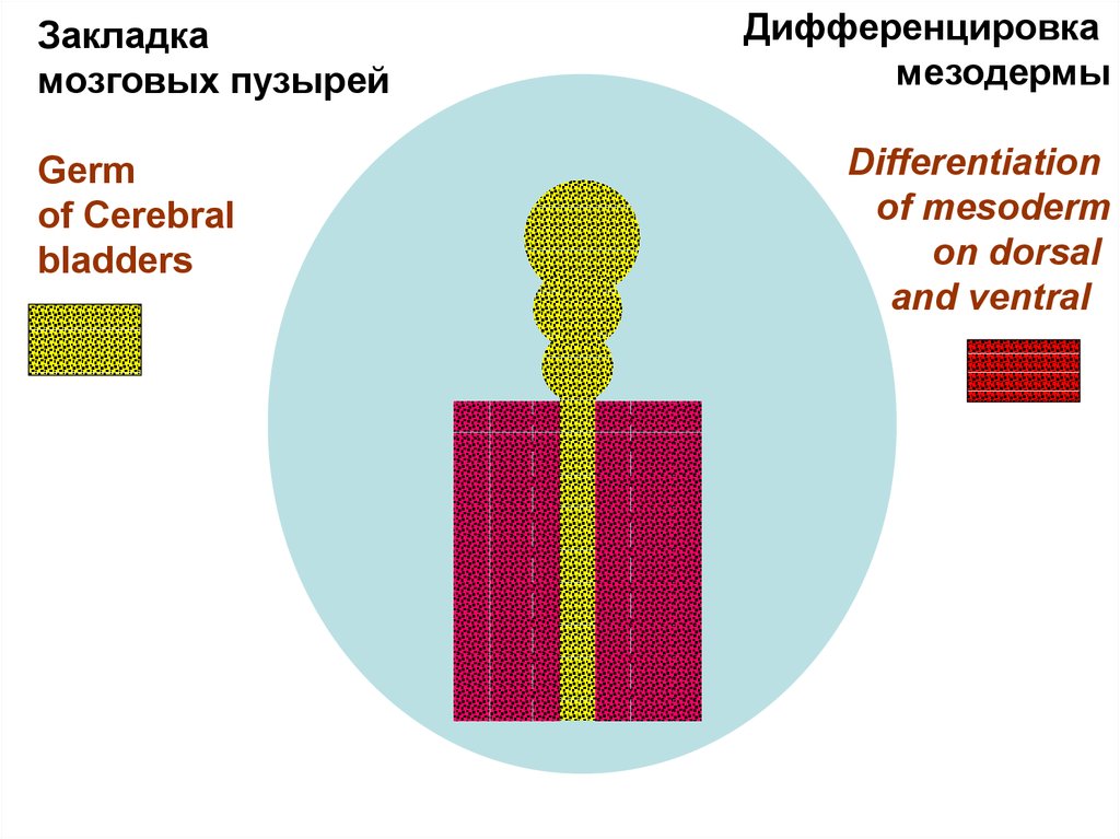
19.
Закладкамозговых пузырей
Germ
of Cerebral
bladders
Дифференцировка
мезодермы
Differentiation
of mesoderm
on dorsal
and ventral
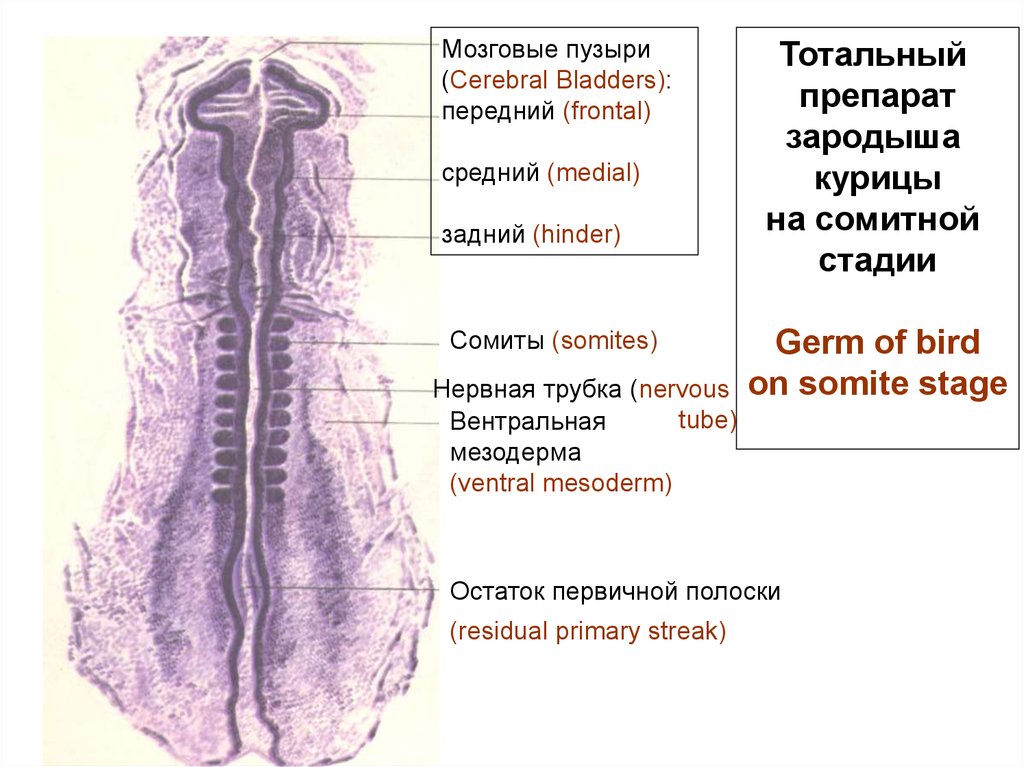
20.
Мозговые пузыри(Cerebral Bladders):
передний (frontal)
средний (medial)
задний (hinder)
Сомиты (somites)
Нервная трубка (nervous
tube)
Вентральная
мезодерма
(ventral mesoderm)
Тотальный
препарат
зародыша
курицы
на сомитной
стадии
Germ of bird
on somite stage
Остаток первичной полоски
(residual primary streak)
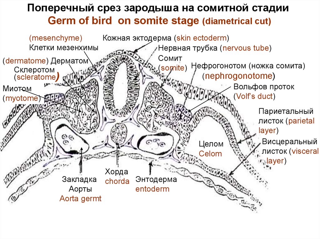
21. Differentiation of dorsal and ventral mesoderm
• Dorsal mesoderm is segmented on somites.• Ventral mesoderm is split on 2 layers - parietal (near ectoderm) and
visceral (near entoderm). Between them the celom (secondary cavity of a
body) is formed. Ventral mesoderm and the celom form splanchnotome.
(Layers of ventral mesoderm form mucosal layer - peritoneum. The internal
peritoneum layer grows together with a wall of internal organs, external layer forms a
wall of a peritoneum cavity).
• Splanchnotome is connected with somites by means of segment legs
which form nephrogonotome - a germ of urogenital system.
• Somites are differentiated on 3 germs:
- dermatome (a germ of a connective tissue of a skin),
- scleratome (a germ of skeletal tissues - cartilages, bones),
- myotome (a germ of a skeletal muscular tissue).
Cells with shoots (are evicted from all germinal layeres, but basically from
mesoderm, fill all intervals between germinal layeres) form an embryonal tissue -
mesenchyme.
It is a development source of connective tissue, vessels, blood, a
lymph and smooth muscle tissue.
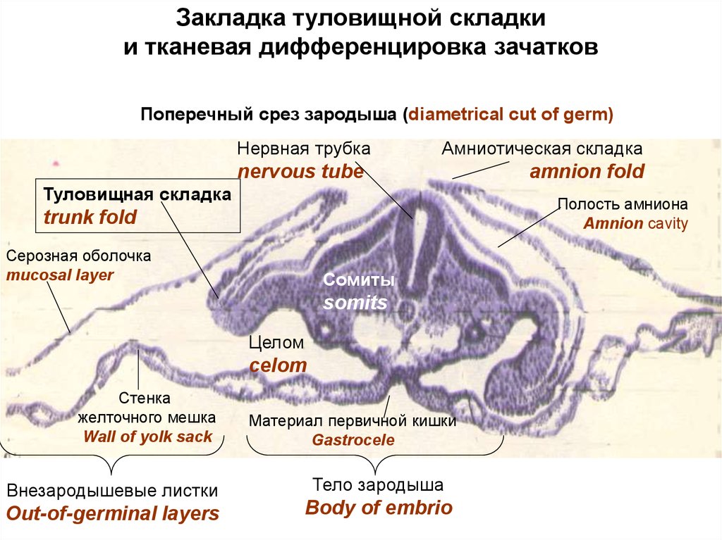
A germ body formation begins by means of trunk folds which are
formed from ectoderm and parietal layer of mesoderm. These folds bend
under a germ and separate a germ body from not germ organs.
22.
Поперечный срез зародыша на сомитной стадииGerm of bird on somite stage (diametrical cut)
(mesenchyme)
Кожная эктодерма (skin ectoderm)
Клетки мезенхимы
Нервная трубка (nervous tube)
Сомит
(dermatome) Дерматом
(somite) Нефрогонотом (ножка сомита)
Склеротом
(nephrogonotome)
(scleratome)
Вольфов проток
Миотом
(Volf’s duct)
(myotome)
Целом
Celom
Закладка
Аорты
Aorta germt
Хорда
chorda Энтодерма
entoderm
Париетальный
листок (parietal
layer)
Висцеральный
листок (visceral
layer)
23.
Закладка туловищной складкии тканевая дифференцировка зачатков
Поперечный срез зародыша (diametrical cut of germ)
Нервная трубка
Амниотическая складка
nervous tube
Туловищная складка
Полость амниона
Amnion cavity
trunk fold
Серозная оболочка
mucosal layer
Сомиты
somits
Целом
celom
Стенка
желточного мешка
Wall of yolk sack
Внезародышевые листки
Out-of-germinal layers
amnion fold
Материал первичной кишки
Gastrocele
Тело зародыша
Body of embrio
24.
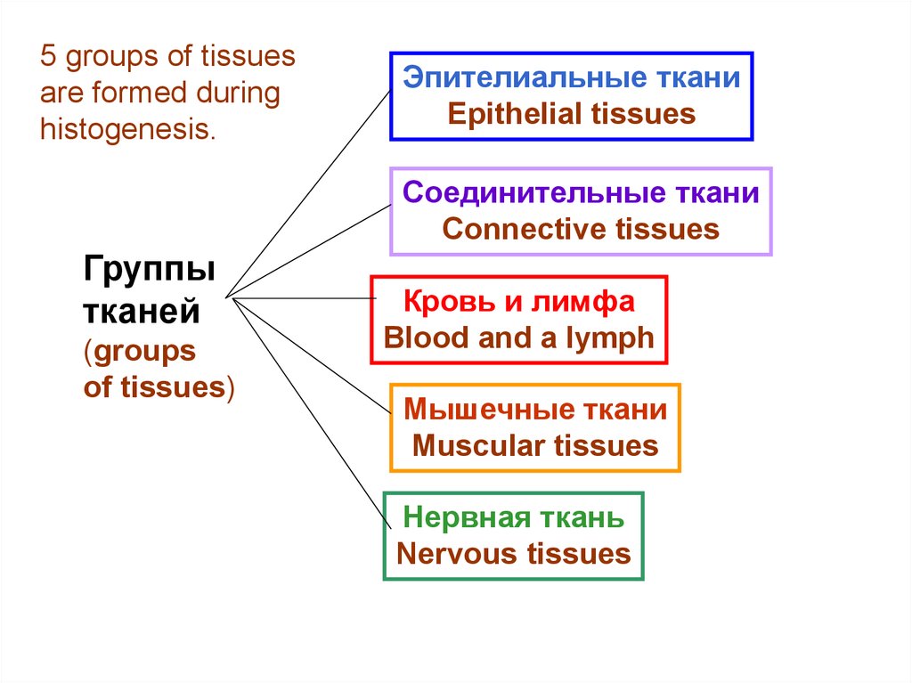
5 groups of tissuesare formed during
histogenesis.
Эпителиальные ткани
Epithelial tissues
Соединительные ткани
Connective tissues
Группы
тканей
(groups
of tissues)
Кровь и лимфа
Blood and a lymph
Мышечные ткани
Muscular tissues
Нервная ткань
Nervous tissues
25.
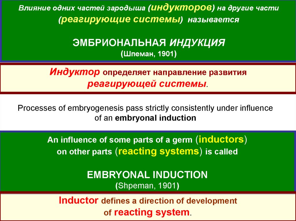
Влияние одних частей зародыша (индукторов) на другие части(реагирующие системы) называется
ЭМБРИОНАЛЬНАЯ ИНДУКЦИЯ
(Шпеман, 1901)
Индуктор определяет направление развития
реагирующей системы.
Processes of embryogenesis pass strictly consistently under influence
of an embryonal induction
An influence of some parts of a germ (inductors)
on other parts (reacting systems) is called
EMBRYONAL INDUCTION
(Shpeman, 1901)
Inductor defines a direction of development
of reacting system.
26.
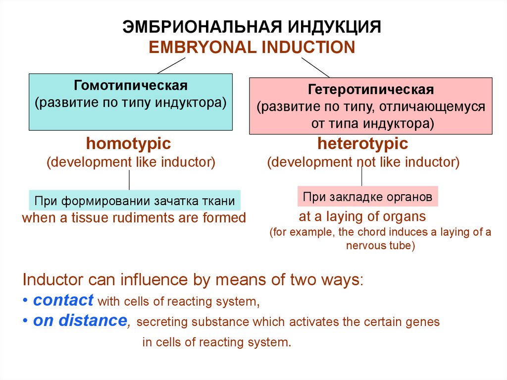
ЭМБРИОНАЛЬНАЯ ИНДУКЦИЯEMBRYONAL INDUCTION
Гомотипическая
(развитие по типу индуктора)
Гетеротипическая
(развитие по типу, отличающемуся
от типа индуктора)
homotypic
heterotypic
(development like inductor)
(development not like inductor)
При формировании зачатка ткани
when a tissue rudiments are formed
При закладке органов
at a laying of organs
(for example, the chord induces a laying of a
nervous tube)
Inductor can influence by means of two ways:
• contact with cells of reacting system,
• on distance, secreting substance which activates the certain genes
in cells of reacting system.
27.
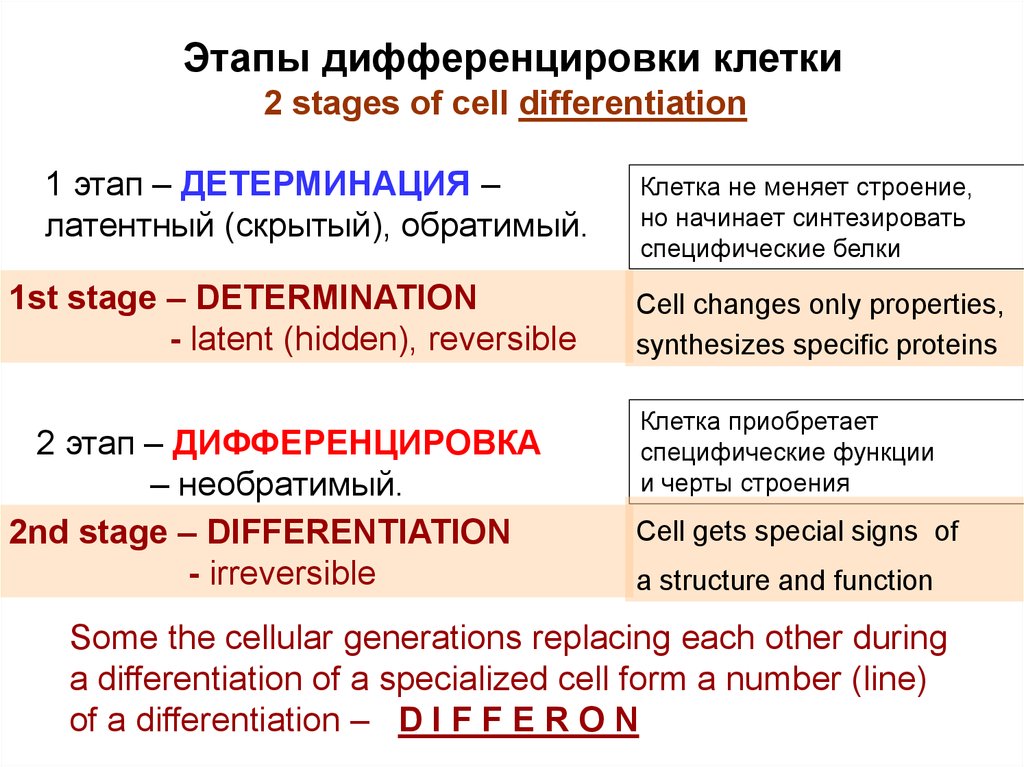
Этапы дифференцировки клетки2 stages of cell differentiation
1 этап – ДЕТЕРМИНАЦИЯ –
латентный (скрытый), обратимый.
1st stage – DETERMINATION
- latent (hidden), reversible
2 этап – ДИФФЕРЕНЦИРОВКА
– необратимый.
2nd stage – DIFFERENTIATION
- irreversible
Клетка не меняет строение,
но начинает синтезировать
специфические белки
Cell changes only properties,
synthesizes specific proteins
Клетка приобретает
специфические функции
и черты строения
Cell gets special signs of
a structure and function
Some the cellular generations replacing each other during
a differentiation of a specialized cell form a number (line)
of a differentiation – D I F F E R O N
28.
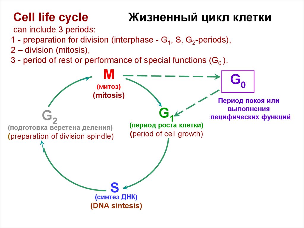
Жизненный цикл клеткиCell life cycle
can include 3 periods:
1 - preparation for division (interphase - G1, S, G2-periods),
2 – division (mitosis),
3 - period of rest or performance of special functions (G0 ).
М
G0
(митоз)
(mitosis)
G1
G2
(подготовка веретена деления)
(preparation of division spindle)
(период роста клетки)
(period of cell growth)
S
(синтез ДНК)
(DNA sintesis)
Период покоя или
выполнения
специфических функций
29.
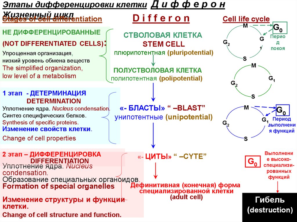
Этапы дифференцировки клетки Д и ф ф е р о нЖизненный
цикл
Stages of cell differentiation
Differon
Cell life cycle
М
НЕ ДИФФЕРЕНЦИРОВАННЫЕ
СТВОЛОВАЯ КЛЕТКА
STEM CELL
(NOT DIFFERENTIATED CELLS):
Упрощенная организация,
низкий уровень обмена веществ
The simplified organization,
low level of a metabolism
1 этап - ДЕТЕРМИНАЦИЯ
DETERMINATION
Уплотнение ядра. Nucleus condensation.
Синтез специфических белков.
Synthesis of specific proteins.
G0
G
G2
1
плюрипотентная (pluripotential)
Перио
д
покоя
S
М
ПОЛУСТВОЛОВАЯ КЛЕТКА
полипотентная (polipotential)
G1
G2
S
«- БЛАСТЫ» “ –BLAST”
унипотентные (unipotential)
Изменение свойств клетки.
.Change of cell properties
2 этап – ДИФФЕРЕНЦИРОВКА
DIFFERENTIATION
М
Change of cell structure and function.
G1 Период
G2
выполнени
я функций
S
«- ЦИТЫ» “ –CYTE”
Уплотнение ядра. Nucleus
condensation.
Образование специальных органоидов.
Дефинитивная (конечная) форма
Formation of special organelles
Изменение структуры и функции
клетки.
G0
специализированной клетки
(adult cell)
G0
Выполнени
е высокоспециализированных
функций
Гибель
(destruction)