


 Медицина
Похожие презентации:


Peptic Ulcer Disease Complications
1.
Medical Academy named afterS.I. Georgievsky of Vernadsky CFU
Department of Surgery №2
Lecturer Baranovskiy Yu.G., PhD
Peptic Ulcer Disease
Complications
2.
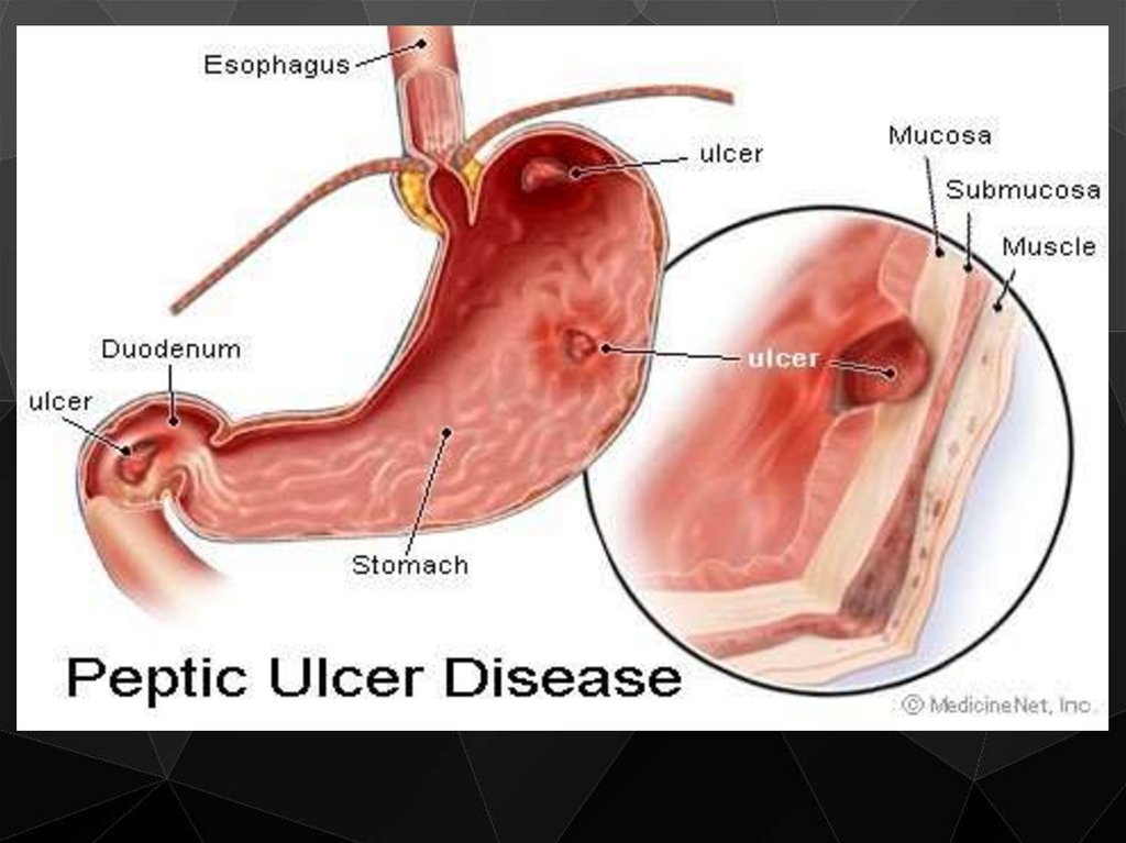
Ais an open sore in
the upper digestive tract. There are
two types of peptic ulcers, a gastric
ulcer, which forms in the lining of the
stomach, and a duodenal ulcer,
which forms in the upper part of the
small intestine.
3.
4. Classification
₪ Stomach (called gastric ulcer)₪ Duodenum (called duodenal ulcer)
₪ Oesophagus (called Oesophageal ulcer)
₪ Types of peptic ulcers:
₪ Type I: Ulcer along the lesser curve of stomach
₪ Type II: Two ulcers present - one gastric, one
duodenal
₪ Type III: Prepyloric ulcer
₪ Type IV: Proximal gastroesophageal ulcer
₪ Type V: Anywhere
5. SYMPTOMS
6.
SYMPTOMSBurning pain
Bloating
Nausea
Water brash
Unexplained weight loss
Hematemesis (vomiting of blood)
Appetite changes
Melina
Vomiting
Blood in the stools
Low blood cell count (anemia)
Frequent burping or hiccupping
Stomach pain wakes you up at night
An early sense of fullness with
eating
7.
CAUSES8.
CAUSES• Helicobacter pylori, a bacteria that is frequently found
in the stomach
• Nonsteroidal anti-inflammatory drugs (NSAIDS) such
as ibuprofen
• In addition, smoking and certain other genetic and
environmental factors (such as medications) may
influence the course of peptic ulcer disease.
• Psychological stress and dietary factors were once
thought to be the cause of ulcers, although these factors
are no longer thought have a major role.
9.
Helicobacter pylori infection• H. pylori is a helix-shaped
• Gram-negative, slow-growing organism
10.
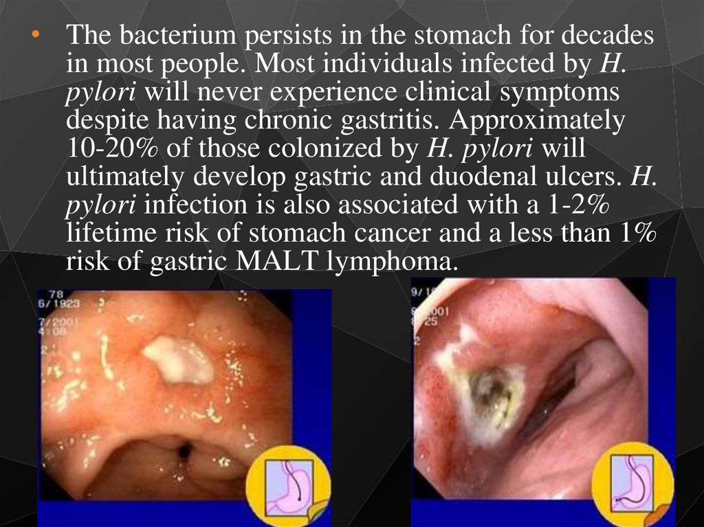
• The bacterium persists in the stomach for decadesin most people. Most individuals infected by H.
pylori will never experience clinical symptoms
despite having chronic gastritis. Approximately
10-20% of those colonized by H. pylori will
ultimately develop gastric and duodenal ulcers. H.
pylori infection is also associated with a 1-2%
lifetime risk of stomach cancer and a less than 1%
risk of gastric MALT lymphoma.
11.
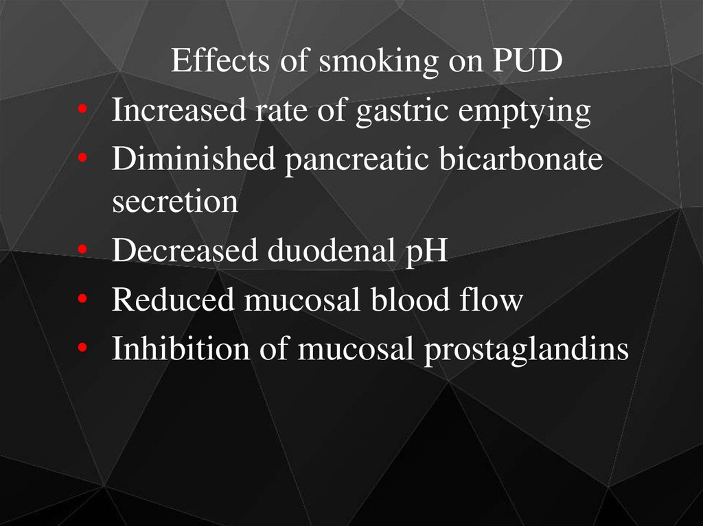
Effects of smoking on PUD
Increased rate of gastric emptying
Diminished pancreatic bicarbonate
secretion
Decreased duodenal pH
Reduced mucosal blood flow
Inhibition of mucosal prostaglandins
12.
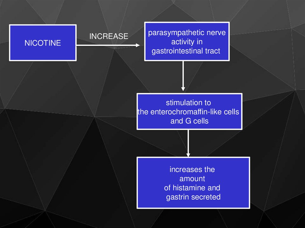
NICOTINEINCREASE
parasympathetic nerve
activity in
gastrointestinal tract
stimulation to
the enterochromaffin-like cells
and G cells
increases the
amount
of histamine and
gastrin secreted
13.
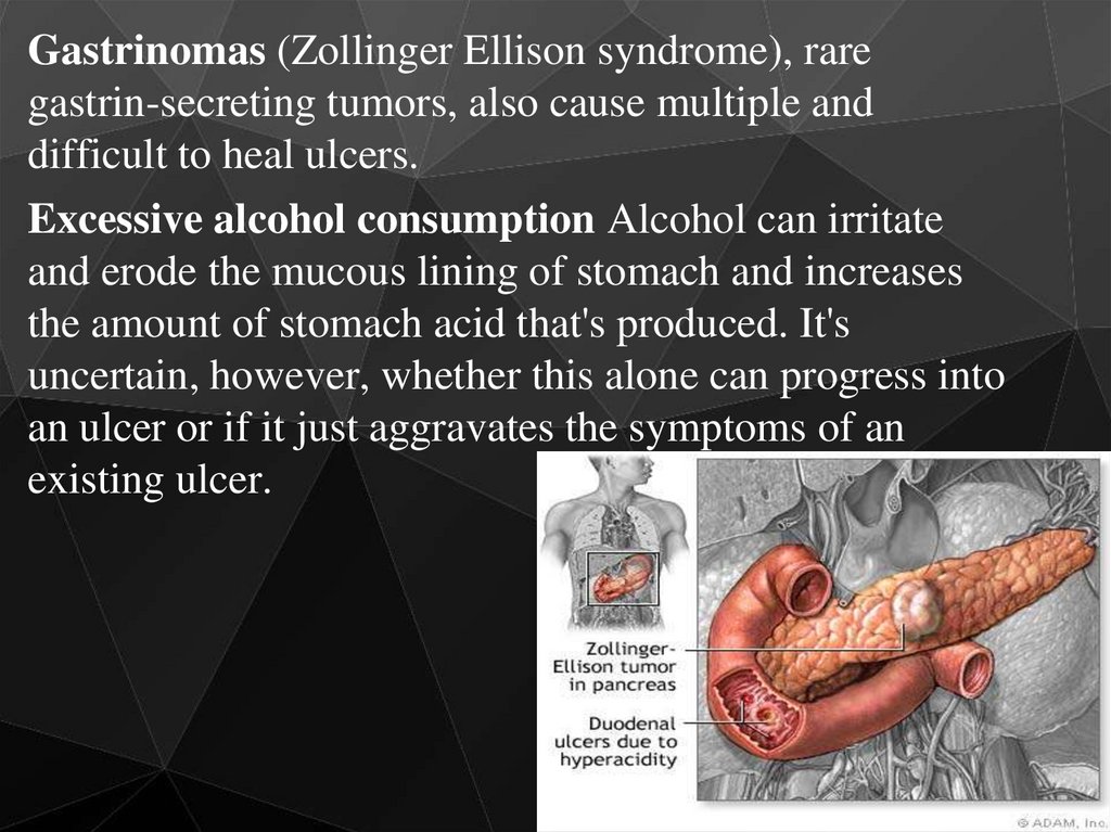
Gastrinomas (Zollinger Ellison syndrome), raregastrin-secreting tumors, also cause multiple and
difficult to heal ulcers.
Excessive alcohol consumption Alcohol can irritate
and erode the mucous lining of stomach and increases
the amount of stomach acid that's produced. It's
uncertain, however, whether this alone can progress into
an ulcer or if it just aggravates the symptoms of an
existing ulcer.
14.
CaffeineBeverages and foods that contain caffeine can
stimulate acid secretion in the stomach. This
can aggravate an existing ulcer, but the
stimulation of stomach acid can't be
attributed solely to caffeine.
15.
The complications of PepticUlceration
The common complications are:
Perforation
Penetration
Bleeding
Stenosis
16.
Perforation (a hole in the wall) often leads tocatastrophic consequences. Erosion of the
gastro-intestinal wall by the ulcer leads to
spillage of stomach or intestinal content into the
abdominal cavity. Perforation at the anterior
surface of the stomach leads to acute peritonitis,
initially chemical and later bacterial peritonitis.
The first sign is often sudden intense abdominal
pain. Posterior wall perforation leads to
pancreatitis; pain in this situation often radiates
to the back
17.
PerforationClinical Features
History of peptic ulcer
Sudden onset, severe, generalized
abdominal pain
Starts as chemical peritonitis, then bacterial
peritonitis which will be accompanied by
deterioration of the patient’s condition
18.
PerforationClinical symptoms
Tachycardia, pyrexia
Shock
Board like rigidity of abdomen
Abdominal splinting
19.
PerforationClinical Features
In elderly, the classical presentation of PPU
may be absent
Use of NSAID
Board like abdominal rigidity may be not
present
Epigastric tenderness
20.
PerforationClinical Features
The most frequent place for perforation
is the anterior wall of duodenum
Anterior or incisural part of gastric ulcer
may perforate
Gastric ulcer may perforate in gland
bag (difficult to diagnose)
21.
PerforationInvestigations
Observe chest X-ray will reveal free gas
under the diaphragm in more than 50% of
the cases
Amylase level to R/O pancreatitis
CT scan of the abdomen
Endoscopy
22.
On X-ray is crescent-shaped illumination under the diaphragm23.
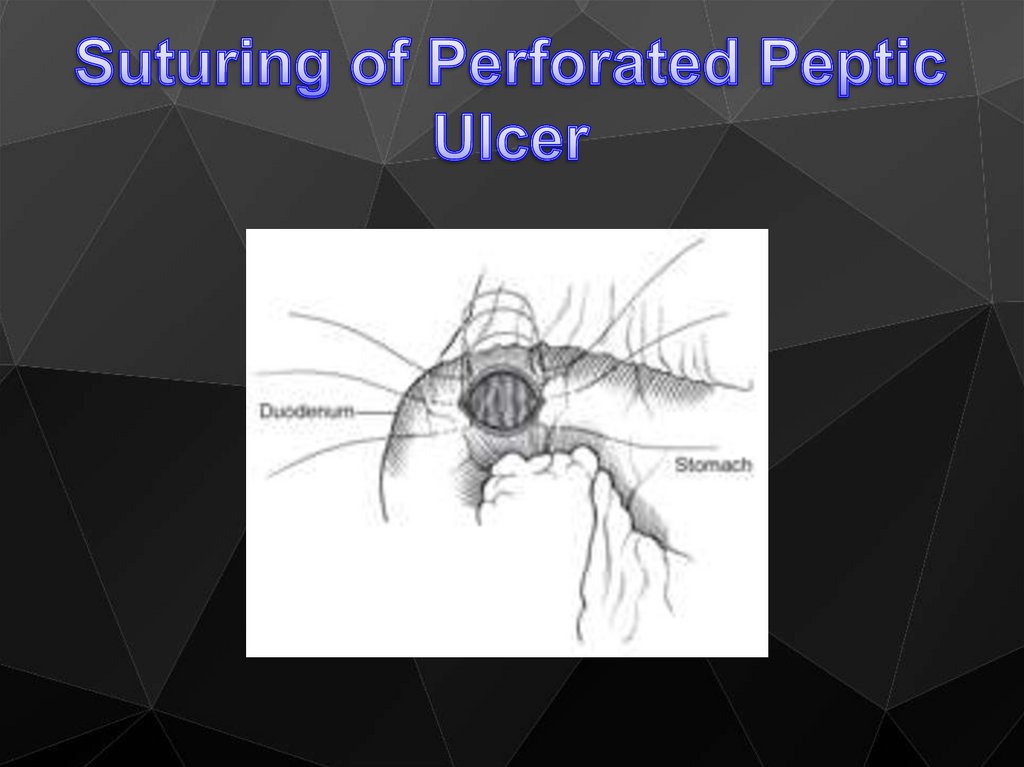
PerforationTreatment
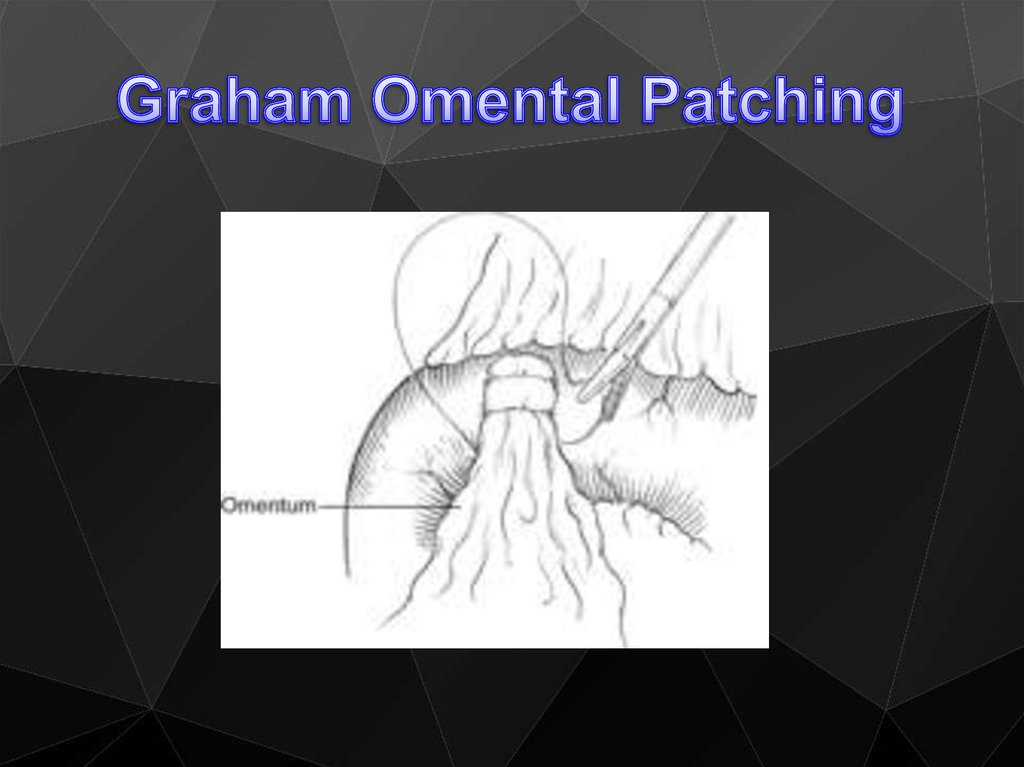
Hospitalisation and analgesia
The treatment is principally surgical
Midline laparotomy
Thorough peritoneal toilet
Duodenal ulcer, close and patch with omentum
Gastric ulcer, should if possible, excised and
closed
If suturing is not possible, Billroth resection.
24.
25.
26.
27.
28.
29.
PerforationTreatment
Systemic antibiotics
Vagotomy, highly selective vagotomy
Minimally invasive
Conservative treatment
- Small leak
- Mild peritoneal contamination
- I.V fluid, N/G tube
Proton pump inhibitors lifelong especially if to
continue on NSAID & H pylori eradication
therapy
30.
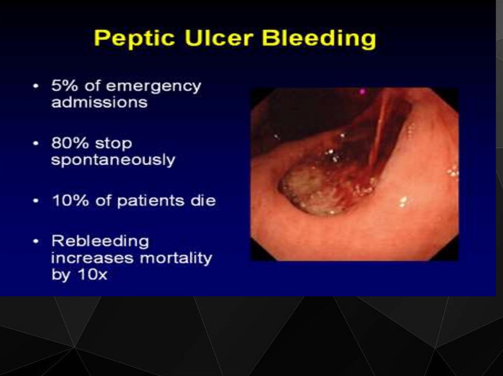
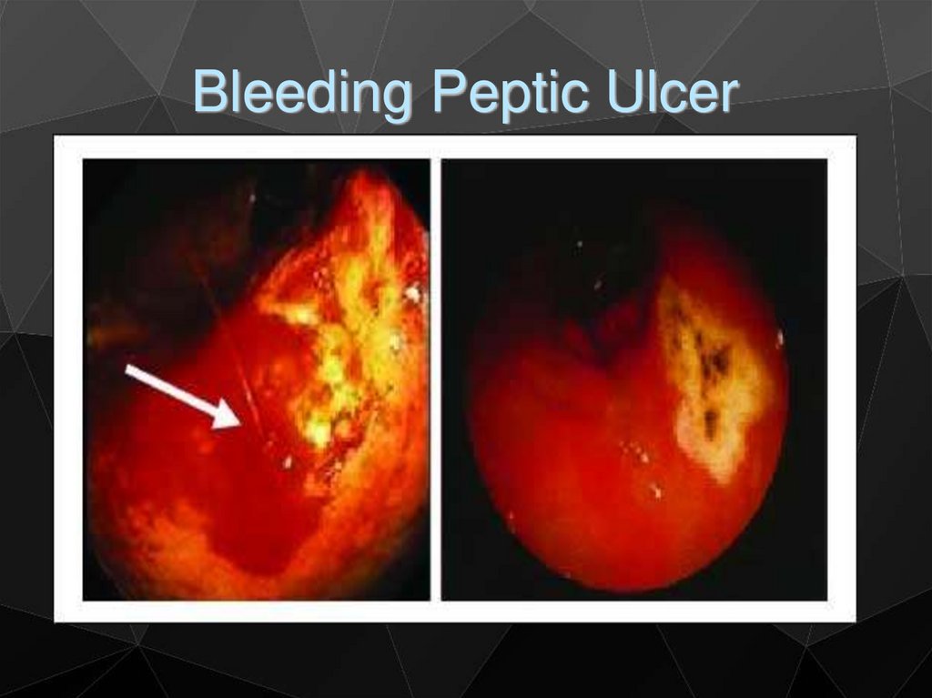
BleedingGastrointestinal bleeding is the most
common complication. Sudden large bleeding
can be life-threatening. It occurs when the
ulcer erodes one of the blood vessels.
Bleeding can occur as slow blood loss that
leads to anemia or as severe blood loss that
may require hospitalization or a blood
transfusion.
31.
Penetration is when the ulcercontinues into adjacent organs such as
the liver and pancreas
32.
Penetration is a form of perforation inwhich the perforating ulcers erode the
whole thickness of the stomach or
duodenal wall, into adjacent abdominal
organs such as liver, pancreas, bile duct or
intestines. Pancreas is the most typical site
of penetration. A combination of serious
ulcer symptoms including abnormal pain
distribution and decreased response to
conventional treatment are signs of ulcer
penetration.
33.
BleedingEpidemiology
Mirror that of PPU
NSAID
34.
Bleeding35.
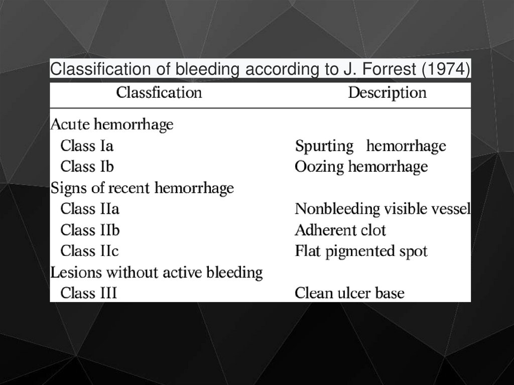
Classification of bleeding according to J. Forrest (1974)36.
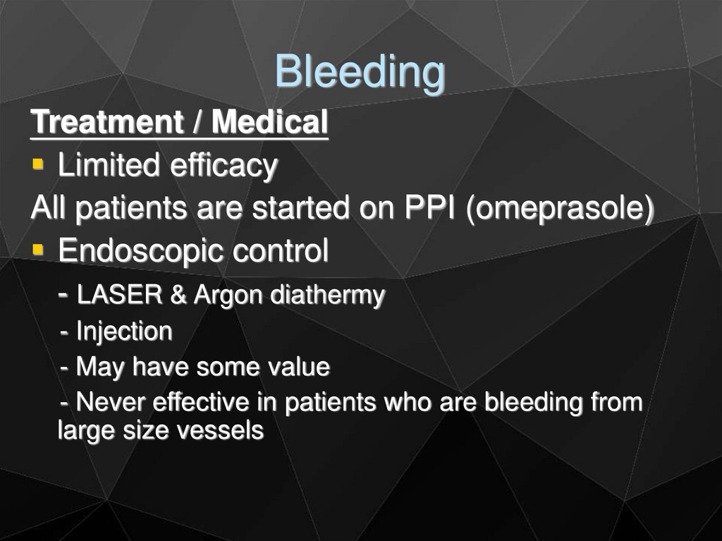
BleedingTreatment / Medical
Limited efficacy
All patients are started on PPI (omeprasole)
Endoscopic control
- LASER & Argon diathermy
- Injection
- May have some value
- Never effective in patients who are bleeding from
large size vessels
37.
Bleeding Peptic Ulcer38.
39.
BleedingTreatment / Surgical
Indications
Patient continue to bleed
Visible vessel in ulcer base
Spurting vessel
Ulcer with a clot
Elderly
Patient who has required more than 6 units of
blood
40.
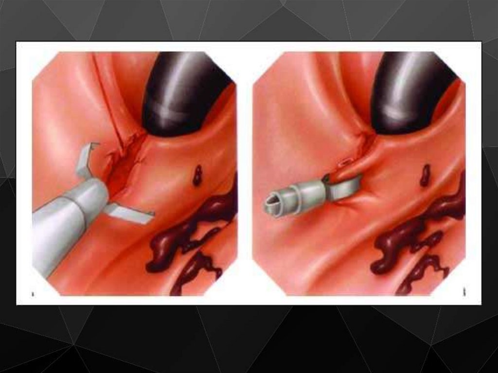
BleedingTreatment / Surgical
Aim to stop bleeding
Upper midline incision
Site usually localized by prior Endoscopy
Duodenal mobilization
Pyloro-duodenotomy
Suture that under-run the bleeding vessel
Gastric ulcer, excise ulcer if possible, if not,
under-run bleeding vessel and take biopsies
41.
BleedingTreatment / Surgical
Definitive acid lowering surgery is not
required
PPI (omeprasole)
Anti H pylori
42.
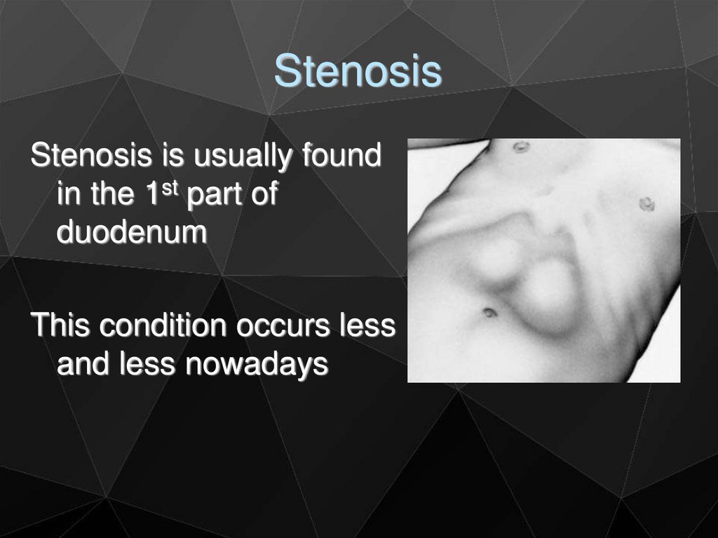
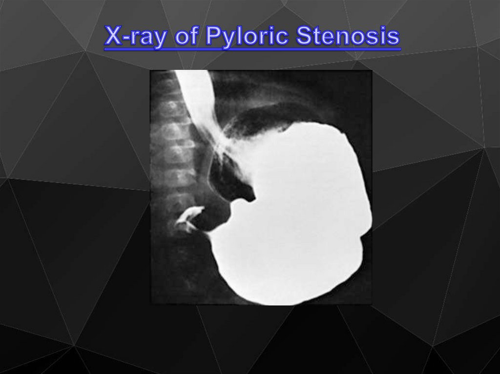
StenosisStenosis is usually found
in the 1st part of
duodenum
This condition occurs less
and less nowadays
43.
Scar tissue Scarring and swelling due toulcers causes narrowing in the duodenum
and gastric outlet obstruction. Patient
often presents with severe vomiting. Peptic
ulcers can also produce scar tissue that can
obstruct passage of food through the
digestive tract, causing you to become full
easily, to vomit and to lose weight.
44.
45.
StenosisClinical Features
Long history of peptic ulcer disease
Vomiting, unpleasant in nature, totally lacking in
bile, containing foodstuff taken several days
previously
Weight loss
Patient looks unwell and dehydrated
On examination you can see distended
stomach, succussion splash may be audible on
shaking the patient’s abdomen
46.
47.
StenosisMetabolic effects
Vomiting of HCl results in hypochloremic
acidosis
Initially Na+ & K+ levels are normal
With dehydration, more profound metabolic
abnormalities arise
Renal dysfunction
Initially urine has low chloride and high HCO3
content , HCO3 is excreted with Na+, so patient
become hyponatremic and more dehydrated
48.
StenosisMetabolic effects
Then because of dehydration, a phase of
Na+ retention follows and K+ and Hydrogen
are excreted in preference
Paradoxical aciduria
Hypokalemia
Alkalosis leads to lowering of circulating
ionized calcium and tetany may occur
49.
StenosisManagement
1) Correct metabolic abnormality
Rehydration with isotonic saline with K +
supplementation
Replacing NaCl and water allows kidney to
correct the acid-base abnormality
Correct anemia which may appear after
rehydration
50.
StenosisManagement
2) Empty the stomach with wide-bore N/G
tube, may need lavage
3) Endoscopy and contrast radiology to
confirm and R/O malignancy
4) Parenteral anti-secretory agent
51.
StenosisManagement
Early cases may settle with conservative
measurement, presumably as the edema
around the ulcer diminishes as the ulcer is
healed
Gastroenterostomy
Endoscopic balloon dilatation
- Effective in early cases
- Risk of perforation
- Dilatation may have to be performed several times
52.
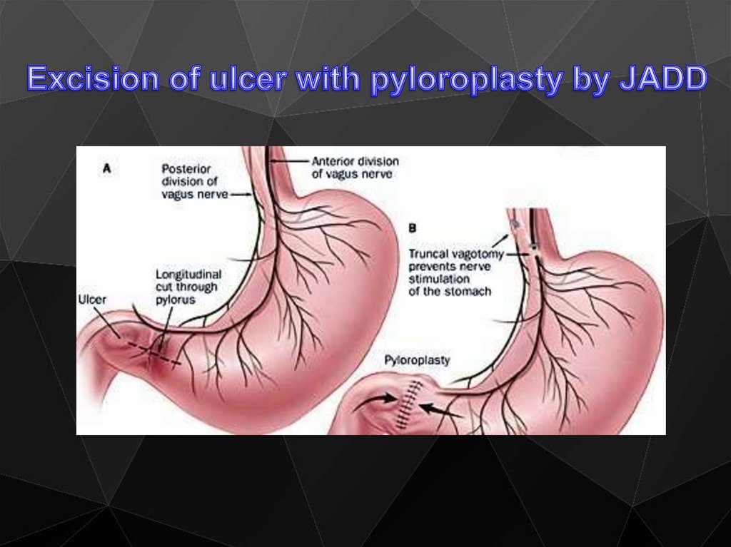
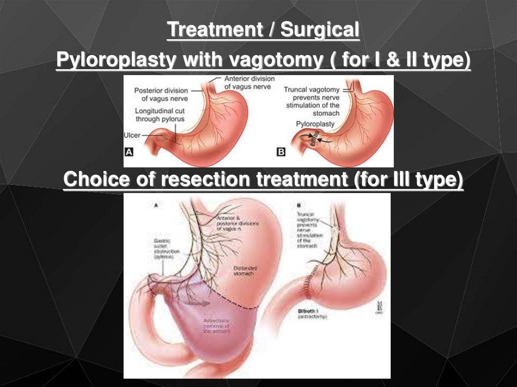
Treatment / SurgicalPyloroplasty with vagotomy ( for I & II type)
Choice of resection treatment (for III type)