


 platel")


, 1 neutrophilic myelocyte, 1 segmented neutrophil, 1 late NRBC. Normal marrow - 100X")
, 1 eosinophilic metamyelocyte (lower right), 1 eosinophilic band (top right), 1 intermediate (myelocyte) eosinophil in mitosis, 1 neutrophilic myelocyte. Normal")
, 2 neutrophilic myelocytes. Normal marrow - 100X")


, 1 eosinophilic metamyelocyte (lower right), 1 eosinophilic band (top right), 1 intermediate (myelocyte) eosinophil in mitosis, 1 neutrophilic myelocyte. Normal")
 Note differences in chromatin and cytoplasmic color - 100X")


 and 2 segmented neutrophils (lower frame). Normal blood - 100X")


; Normal blood - 100X")
; Normal blood - 100X")
, 1 large lymphocyte. Normal blood - 100X")


. Imprint of marrow biopsy - 100X")

 Медицина
 Биология
Похожие презентации:


Клеточное строение костного мозга
1. Клеточное строение костного мозга
2. Все клетки, которые можно встретить в костном мозге в норме и при патологии можно разделить на следующие основные группы:
1. Клетки ретикулярной стромы (не принимаютнепосредственного участия в кроветворении,
но имеют большое значение, т. к. создают
необходимое микроокружение для
кроветворных клеток). К ним относятся:
• ретикулярные клетки;
• клетки эндотелия;
• остеобласты;
• жировые клетки.
3. Ретикулярные клетки
Reticular cells, polychromatic NRBCs, normal marrowTwo reticular cells (center), 4 lymphocytes, 5 polychromatic NRBCs, 1 segmented
neutrophil, 1 smudge cell. Normal marrow - 100X
4. Ретикулярные клетки
Reticular cell, normal marrowOne reticular cell. Normal marrow - 100X
5. Ретикулярные клетки
Reticular cell, neutrophilic myelocyte, normal marrowOne reticular cell, 1 neutrophilic myelocyte. Normal marrow - 100X
6. Клетки эндотелия
Endothelial cells.Strand of endothelial cells. Normal marrow -20X
7. Клетки эндотелия
Endothelial cellsHigher magnification of strand of endothelial cells. Normal marrow - 100X
8. Остеобласты
OsteoblastsClump of osteoblasts. Normal marrow particle preparation -50X Osteoblast are
large in size, often are oval in shape, have an eccentrically located nucleus which
appears to be falling out of the cell, blue-gray cytoplasm with a clear area away
from the nucleus.
9. Остеобласты
Osteoblasts, Normal marrowSame clump of osteoblasts. Normal marrow particle preparation -100X
10. Остеобласты
Рlasma cell, osteoblastsOne small and 1 large plasma cell in top left frame. 2 osteoblasts in top right
frame, 1 osteoblast in lower frame. Normal marrow -100X
11. Жировые клетки
Fat cells. Aplastic anemia marrowMultiple fat cells. Aplastic anemia marrow -20X
12. Жировые клетки
Fat cells, Aplastic anemia marrowTwo very large fat cells. Aplastic anemia marrow -50X
13. 2. Миелокариоциты - клетки кроветворной ткани костного мозга с их производными - зрелыми клетками крови. К ним относятся:
• недифференцированные бласты;• клетки миелоцитарного ряда;
• клетки моноцитарного ряда;
• клетки лимфоцитарного ряда;
• клетки эритроцитарного ряда;
• клетки мегакариоцитарного ряда.
14. Недифференцированные бласты
15. Миелобласт
Myeloblast, late neutrophil, normal marrowOne myeloblast, 1 late neutrophil. Normal marrow - 100X
16. Миелобласт
Myeloblast, neutrophilic metamyelocyte, normal marrowOne myeloblast, 1 neutrophilic metamyelocyte. Normal marrow - 100X
17. Миелобласт
Myeloblast, late NRBC, normal marrowOne myeloblast / monoblast, 1 late NRBC. Normal marrow - 100X
18. Progranulocyte, band neutrophils One late progranulocyte, 2 band neutrophils. Normal marrow - 100X
19. Progranulocyte, band neutrophils, normal marrow One progranulocyte, 2 band neutrophils. Normal marrow - 100X
20. Progranulocyte, myelocyte, normal marrow One progranulocyte, 1 myelocyte. Normal marrow - 100X
21. Late neutrophilic myelocyte, normal marrow One late neutrophilic myelocyte. Normal marrow - 100X
22. Neutrophilic myelocyte, normal marrow One neutrophilic myelocyte. Normal marrow - 100X
Neutrophilic myelocyte, normal marrowOne neutrophilic myelocyte. Normal marrow 100X
23. Myelocytes, abnormal platelets, AML blood Two myelocytes, one with and the other without primary or coarse azurophilic granules. Both contain vacuoles. Their nuclear chromatin is showing some heterochromatin deveopment. Several abnormal (agranular) platel
Myelocytes, abnormal platelets, AML bloodTwo myelocytes, one with and the other without primary or
coarse azurophilic granules. Both contain vacuoles. Their
nuclear chromatin is showing some heterochromatin
deveopment. Several abnormal (agranular) platelets are
present. Acute Megakaryocytic Leukemia (M-7). Blood - 100
24. Myelocyte, disrupted band neutrophil One early myelocyte with many azure granules, 1 disrupted band neutrophil. Normal marrow - 100X
25. Eosinophilic myelocyte, Normal marrow Left frame: 1 normal eosinophilic myelocyte. Center frame: 1 eosinophilic myelocyte with large blue and large eosinophilic granules. Right frame: 1 eosinophilic myelocyte with large blue granules and large eosinophili
Eosinophilic myelocyte, Normal marrowLeft frame: 1 normal eosinophilic myelocyte. Center frame:
1 eosinophilic myelocyte with large blue and large
eosinophilic granules. Right frame: 1 eosinophilic
myelocyte with large blue granules and large eosinophilic
granules. Normal marrow - 100X
26. Eosinophilic myelocyte, neutrophilic myelocyte One eosinophilic myelocyte (lower left), 1 neutrophilic myelocyte, 1 segmented neutrophil, 1 late NRBC. Normal marrow - 100X
27. Eosinophilic myelocyte, eosinophilic metamyelocyte, mitosis One eosinophilic myelocyte (left), 1 eosinophilic metamyelocyte (lower right), 1 eosinophilic band (top right), 1 intermediate (myelocyte) eosinophil in mitosis, 1 neutrophilic myelocyte. Normal
Eosinophilic myelocyte, eosinophilic metamyelocyte,mitosis
One eosinophilic myelocyte (left), 1 eosinophilic metamyelocyte (lower right),
1 eosinophilic band (top right), 1 intermediate (myelocyte) eosinophil in
mitosis, 1 neutrophilic myelocyte. Normal marrow - 100X
28. Eosinophilic myelocyte, neutrophilic myelocytes One eosinophilic myelocyte (left), 2 neutrophilic myelocytes. Normal marrow - 100X
29. Metamyelocyte, normal marrow One metamyelocyte - late. Normal marrow - 100X
30. Metamyelocyte, smudge cell, normal marrow One metamyelocyte, 1 smudge cell. Normal marrow - 100X
31. Metamyelocyte, smudge cell, normal marrow One metamyelocyte - early, 1 smudge cell. Normal marrow - 100X
32. Metamyelocyte, neutrophilic myelocyte, normal marrow One metamyelocyte - early, 1 neutrophilic myelocyte. Normal marrow - 100X
33. Eosinophilic myelocyte, eosinophilic metamyelocyte, mitosis One eosinophilic myelocyte (left), 1 eosinophilic metamyelocyte (lower right), 1 eosinophilic band (top right), 1 intermediate (myelocyte) eosinophil in mitosis, 1 neutrophilic myelocyte. Normal
Eosinophilic myelocyte, eosinophilic metamyelocyte,mitosis
One eosinophilic myelocyte (left), 1 eosinophilic
metamyelocyte (lower right), 1 eosinophilic band (top right),
1 intermediate (myelocyte) eosinophil in mitosis, 1
neutrophilic myelocyte. Normal marrow - 100X
34. Band neutrophils, monocyte Three band neutrophils, 1 monocyte (top left center) Note differences in chromatin and cytoplasmic color - 100X
35. Band neutrophils Three band neutrophils - 100X
36. Band eosinophil, mature neutrophil, Normal blood One band eosinophil, 1 mature neutrophil. Normal blood - 100X
Band eosinophil, mature neutrophil, Normal bloodOne band eosinophil, 1 mature neutrophil. Normal blood 100X
37. Mature Neutrophils, Platelet Satellitism One huge clump of nine Mature Neutrophils, each of which is surrounded by platelets. It is probably the stickiness of the platelets for each other that enhances this clumping. The white cell count may be erroneousl
Mature Neutrophils, Platelet SatellitismOne huge clump of nine Mature Neutrophils, each of which is
surrounded by platelets. It is probably the stickiness of the platelets for
each other that enhances this clumping. The white cell count may be
erroneously low in such cases. Platelet Satellitism. EDTA blood - 100X
38. Young megakaryocyte, neutrophilic myelocytes, normal marrow Young megakaryocyte showing early granulation adjacent to lobulated nuclei. 1 progranulocyte, 2 neutrophilic myelocytes, 1 eosinophilic myelocyte, 1 neutrophilic metamyelocyte, 2 band and 2 segme
Young megakaryocyte, neutrophilic myelocytes, normal marrowYoung megakaryocyte showing early granulation adjacent to lobulated
nuclei. 1 progranulocyte, 2 neutrophilic myelocytes, 1 eosinophilic
myelocyte, 1 neutrophilic metamyelocyte, 2 band and 2 segmented
neutrophils and 3 late NRBC surround it. Normal marrow - 100X
39. Segmented neutrophils, band neutrophils, normal blood Segmented neutrophils and band neutrophils composite; 2 band neutrophils (top frames) and 2 segmented neutrophils (lower frame). Normal blood - 100X
40. Eosinophil, Normal blood One 3-lobed eosinophil, 1 2-lobed eosinophil, 2 mature neutrophils; composite. Normal blood - 100X
41. Basophils, mature neutrophils, CML blood Three mature basophils, 2 mature neutrophils. Chronic myeloid leukemia blood - 100X
42. Basophil One mature basophil. Normal blood - 100X
43. Монобласт
44. Промоноцит
45. Young monocyte, normal blood One young monocyte with immature chromatin and nucleoli. Normal blood - 100X
46. Immature Monocytes, cytoplasmic fragmentation, M-5 Two Immature Monocytes, one of which shows cytoplasmic fragmentation. These fragments resemble Platelets and may be erroneously counted as such. The two fragments to the right of the monocytes may be real
platelets. AcuteMonocytic Leukemia ( M-5). Blood - 100X
47. Young monocytes, normal blood Two young monocytes, 1 with many vacuoles. Normal blood - 100X
48. Macrophage, mature megakaryocyte, normal marrow A macrophage with an oval shaped nucleus and a highly vacuolated cytoplasm containing a NRBC and a few scattered azure granules is at left center. A mature megakaryocyte with a single large nucleus and a ful
Macrophage, mature megakaryocyte, normal marrowA macrophage with an oval shaped nucleus and a highly vacuolated
cytoplasm containing a NRBC and a few scattered azure granules is at
left center. A mature megakaryocyte with a single large nucleus and a
fully granulated cytoplasm lies to its right. Normal marrow - 100X
49. Osteoclast One osteoclast with 12 nuclei. Normal marrow -100X
50. Very small lymphocyte, monocyte, normal blood One very small lymphocyte, 1 monocyte. Normal blood - 50X
51. Lymphocytes, normal blood Two lymphocytes (1 with granules); Normal blood - 100X
52. Large reactive lymphocyte, normal blood One very large lymphocyte (reactive); Normal blood - 100X
53. Monocyte, large lymphocyte, normal blood One monocyte (left), 1 large lymphocyte. Normal blood - 100X
Monocyte, large lymphocyte, normal bloodOne monocyte (left), 1 large lymphocyte. Normal blood 100X
54. КЛЕТКИ ЛЕЙКОЛИЗА
55. Plasmacytoid lymphoid, infectious mononucleosis blood One small mature lymphocyte, 1 Plasmacytoid Lymphoid with a contorted nucleus. Infectious mononucleosis blood - 100X
56. Plasmacytoid lymphocyte, severe arthritis with osteoporosis Left frame: 1 Plasmacytoid Lymphocyte with a double nucleus, 1 mature neutrophil. Center frame: 1 mature lymphocyte and 1 small Plasmacytoid Lymphocyte. Right frame: 1 Plasmacytoid Lymphocyte wit
Plasmacytoid lymphocyte, severe arthritis with osteoporosisLeft frame: 1 Plasmacytoid Lymphocyte with a double nucleus, 1 mature
neutrophil.
Center frame: 1 mature lymphocyte and 1 small Plasmacytoid Lymphocyte.
Right frame: 1 Plasmacytoid Lymphocyte with a contorted nucleus and 1 late
smudged form.
Severe arthritis with osteoporosis blood - 100X
57. Monocyte, Plasmacytoid Lymphocyte, drug reaction buffy coat One monocyte, 1 Plasmacytoid Lymphocyte and 1 eosinophil. Drug reaction buffy coat preparation - 100X
58. Plasma cell, alcoholic, liver disease One monocyte at left edge and one Plasma Cell at right edge. The plasma cell is medium in size, has an eccentrically located nucleus which shows a dense chromatin with a few randomly located open areas. The abundant c
Plasma cell, alcoholic, liver diseaseOne monocyte at left edge and one Plasma Cell at right edge. The plasma cell
is medium in size, has an eccentrically located nucleus which shows a dense
chromatin with a few randomly located open areas. The abundant cytoplasm is
very basophilic blue, contains a few vacuoles and shows an almost colorless
area (Golgi) adjacent to the nucleus. Alcoholic with liver disease blood - 100X
59. Plasma Cell One large Plasma Cell, oval in shape, with an eccentrically located nucleus, a very low nuclear/cytoplasm ratio, a gray-blue cytoplasm containing several vacuoles of variable size. The nuclear chromatin is dense with several clear areas random
Plasma CellOne large Plasma Cell, oval in shape, with an eccentrically located
nucleus, a very low nuclear/cytoplasm ratio, a gray-blue cytoplasm
containing several vacuoles of variable size. The nuclear chromatin is
dense with several clear areas randomly distributed. Normal marrow 100X
60. ЭРИТРОБЛАСТ
61. Plasma cells, proerythroblast, NRBC Two Plasma Cells with low nuclear/cytoplasmic ratio, deep basophilic blue cytoplasm, distinct clear area adjacent to the nucleus and eccentrically located nucleus. At the center top edge is a Proerythroblast with a high
Two Plasma Cells with low nuclear/cytoplasmic ratio, deep basophilicblue cytoplasm, distinct clear area adjacent to the nucleus and
eccentrically located nucleus. At the center top edge is a
Proerythroblast with a high nuclear/cytoplasmic ratio but deep
basophlic blue cytoplasm and dense nuclear chromatin similar to the
plasma cells. Its clear area (Golgi) adjacent to the nucleus is not as
pronounced as in the plasma cells. 1 late NRBC is at left center as well
as a 5-lobed mature neutrophil. Normal marrow - 100X
62. Plasma cell, proerythroblast One Plasma Cell and 1 Proerythroblast. Early erythroblasts have a deep blue cytoplasm, and a coarse nuclear chromatin similar to plasma cells, but differ in having a high nuclear/cytoplasmic ratio and a centrally located nucle
Plasma cell, proerythroblastOne Plasma Cell and 1 Proerythroblast. Early erythroblasts have a
deep blue cytoplasm, and a coarse nuclear chromatin similar to plasma
cells, but differ in having a high nuclear/cytoplasmic ratio and a
centrally located nucleus in a round shaped cell. Also, they usually lack
cytoplasmic vacuoles. Normal marrow - 100X
63. Mature lymphocyte, proerythroblast, basophilic erythroblast
64. Late NRBC, Mature Lymphocyte, AML One Late NRBC at upper left and one Mature Lymphocyte at lower right. Many platelets are scattered throughout the field. An Abnormal Megathrombocyte, which is huge, irregular in shape and agranular, is at the left center.
Acute Megakaryocytic Leukemia (M-7) untreated.Blood - 50X
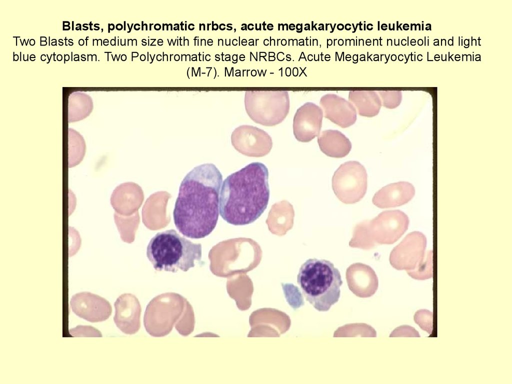
65.
Blasts, polychromatic nrbcs, acute megakaryocytic leukemiaTwo Blasts of medium size with fine nuclear chromatin, prominent nucleoli and light
blue cytoplasm. Two Polychromatic stage NRBCs. Acute Megakaryocytic Leukemia
(M-7). Marrow - 100X
66. Binucleated Plasma Cell, orthochromatic NRBC One Binucleated Plasma Cell. Compare its nuclear chromatin, which is typical for a plasma cell, with that in previous image. A smudge nuclear mass and an orthochromatic NRBC are in the field. Normal marrow - 10
Binucleated Plasma Cell, orthochromatic NRBCOne Binucleated Plasma Cell. Compare its nuclear chromatin, which is
typical for a plasma cell, with that in previous image. A smudge nuclear
mass and an orthochromatic NRBC are in the field. Normal marrow 100X
67. РЕТИКУЛОЦИТЫ
68. Эритроцит
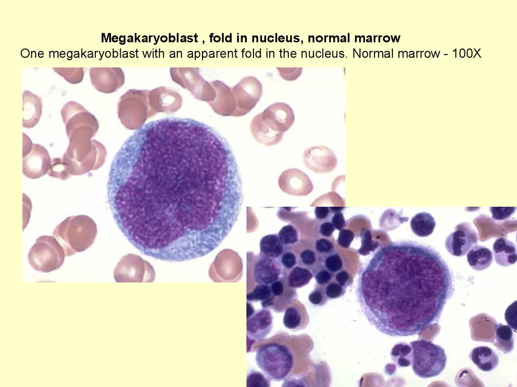
69.
Megakaryoblast , fold in nucleus, normal marrowOne megakaryoblast with an apparent fold in the nucleus. Normal marrow - 100X
70. Promegakaryocyte with nuclear separation, normal marrow One promegakaryocyte with nuclear separation and some nuclear lobulation. Normal marrow - 100X
71. Abnormal megakaryocyte, Wright's-Giemsa stain, AML marrow Mature Abnormal Megakaryocyte with two apparent nuclei; one is very large. Wright's-Giemsa stain. Acute Megakaryocytic Leukemia (M-7). Imprint of marrow biopsy - 100X
Abnormal megakaryocyte, Wright's-Giemsa stain, AML marrowMature Abnormal Megakaryocyte with two apparent nuclei; one is very
large. Wright's-Giemsa stain. Acute Megakaryocytic Leukemia (M-7).
Imprint of marrow biopsy - 100X
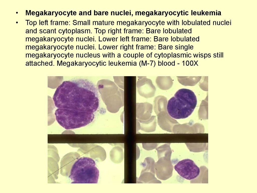
72.
Megakaryocyte and bare nuclei, megakaryocytic leukemia
Top left frame: Small mature megakaryocyte with lobulated nuclei
and scant cytoplasm. Top right frame: Bare lobulated
megakaryocyte nuclei. Lower left frame: Bare lobulated
megakaryocyte nuclei. Lower right frame: Bare single
megakaryocyte nucleus with a couple of cytoplasmic wisps still
attached. Megakaryocytic leukemia (M-7) blood - 100X
73. Normal Platelets , Proper Thickness, Field to Examine Normal Platelets scattered throughout the field. There are slightly fewer than in the previous fields, but within normal numbers. This is a Proper Thickness of a Field to Examine, where the red cells a
Normal Platelets , Proper Thickness, Field to ExamineNormal Platelets scattered throughout the field. There are
slightly fewer than in the previous fields, but within normal
numbers. This is a Proper Thickness of a Field to Examine,
where the red cells are just touching each other or barely
overlapping. Count the number of platelets in at least 10
similar fields and figure the average number per field to derive
a platelet estimate. The Red Cells are Normocytic,
Normochromic. The white cell is a Normal Mature Neutrophil.
Normal blood - 100X.