


 Медицина
Похожие презентации:
 of thebreast")


Fibroadenoma fibrocytic and mastitis
1.
Mastitis/Fibrocystic
change/
Fibroadenoma
2.
Mastitis1) Acute Mastitis :
•Occurs during the first month of
breastfeeding.
•Caused by a local bacterial infection
when breast is most vulnerable due
to cracks and fissures in nipples.
From this portal of entry, S. aureus
or streptococci invade breast tissue.
•One duct system or sector of breast
is involved.
3.
•Infection may spread to entirebreast.
•Staphylococcal abscesses- single or
multiple,
•Streptococci- spread infection in the
form of cellulitis.
• Breast- erythematous and painful.
• Fever is present.
4.
2) Duct Ectasia•Presents as a palpable periareolar
mass with thick, white nipple
secretions and occasionally with skin
retraction.
•Occurs in 5th or 6th decade of life in
multiparous women.
• Pain and erythema-uncommon.
5.
Morphology•Ectatic dilated ducts are filled
with inspissated secretions and
numerous lipid-laden macrophages.
•Whenruptured marked
periductal
and interstitial chronic
inflammatory reaction consisting of
lymphocytes, macrophages, and
plasma cells.
6.
• Granulomas may form aroundcholesterol deposits and
secretions.
•Subsequent fibrosis irregular
mass with skin and nipple
retraction.
7.
3) Granulomatous Mastitis:•Can be a manifestation of systemic
granulomatous diseases (e.g.
polyangiitis, sarcoidosis, TB) or of
disorders that are localized to breast
(granulomatous lobular mastitis, rare
infections).
8.
Granulomatous lobular mastitis:•Uncommon disease, occurs in
parous women.
•Granulomas are closely associated
with lobules, suggesting disease may
be caused by a hypersensitivity
reaction to antigens expressed
during lactation.
9.
•Localized infections are mostcommon in immunocompromised
patients or adjacent to foreign
objects such as breast prostheses or
nipple piercings.
10.
FIBROCYSTIC CHANGES• Changes in female breast that range
from innocuous to patterns
associated with increased risk of
breast carcinoma.
• Arise during reproductive period of
life, may persist after menopause.
• Small minority-forms of epithelial
hyperplasia.
11.
Alterations subdivided intononproliferative and proliferative
patterns.
1)Nonproliferative lesions- cysts
and/or fibrosis and adenosis focally.
2)Proliferative lesions-epithelial
cell hyperplasia.
12.
Nonproliferative Change• Most common type of alteration.
• Involved areas show ill-defined,
diffusely increased density and
discrete nodularities.
Morphology
Gross
• Cysts-multifocal and bilateral, may
be single large cyst.
13.
• Cysts:<1cm to 5cm in diameter.• Brown to blue cysts filled with
serous, turbid fluid.
• Secretory products may calcify,
appear as microcalcifications in
mammograms.
14.
Microscopy:Three principal morphologic changes:
cystic change often with apocrine
metaplasia, fibrosis, and focally
adenosis.
Cysts• In smaller cysts, epithelium-cuboidal
to columnar, sometimes multilayered
focally.
• In larger cysts, epithelium-flattened
or atrophic.
15.
• Mild epithelial proliferation- smallpapillary projections.
• Frequently, cysts are lined by large
polygonal cells that have an abundant
granular, eosinophilic cytoplasm, with
small, round, deeply chromatic nuclei,
called apocrine metaplasia.
16.
Stroma• Compressed fibrous tissue with lossof its normal delicate, myxomatous
appearance and lymphocytic
infiltrate.
Adenosis•Defined as an increase in the
number of acini per lobule.
• Focal adenosis
•Calcifications-occasionally within
lumens.
17.
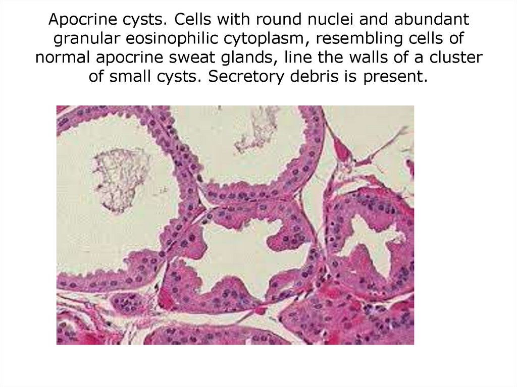
Apocrine cysts. Cells with round nuclei and abundantgranular eosinophilic cytoplasm, resembling cells of
normal apocrine sweat glands, line the walls of a cluster
of small cysts. Secretory debris is present.
18.
Proliferative ChangeDisease Without Atypia
• Lesions characterized by proliferation
of epithelial cells without atypia.
• Small increase in risk of subsequent
carcinoma in either breast.
Gross:
not distinctive, dominated by
coexisting fibrous or cystic changes.
19.
Microscopy• Wide spectrum• Ducts, ductules, or lobules may be
filled with orderly cuboidal cells,
within which small gland patterns
can be discerned (fenestrations) or
as papilloma or sclerosing
adenosis.
• No atypia.
20.
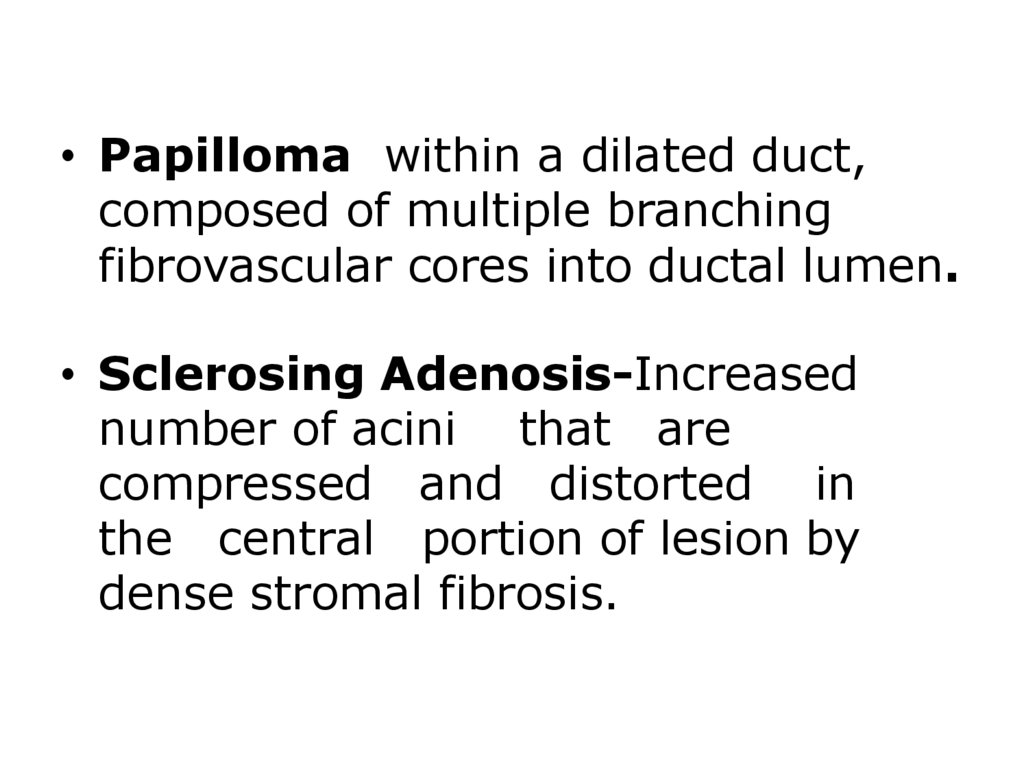
• Papilloma within a dilated duct,composed of multiple branching
fibrovascular cores into ductal lumen.
• Sclerosing Adenosis-Increased
number of acini that are
compressed and distorted in
the central portion of lesion by
dense stromal fibrosis.
21.
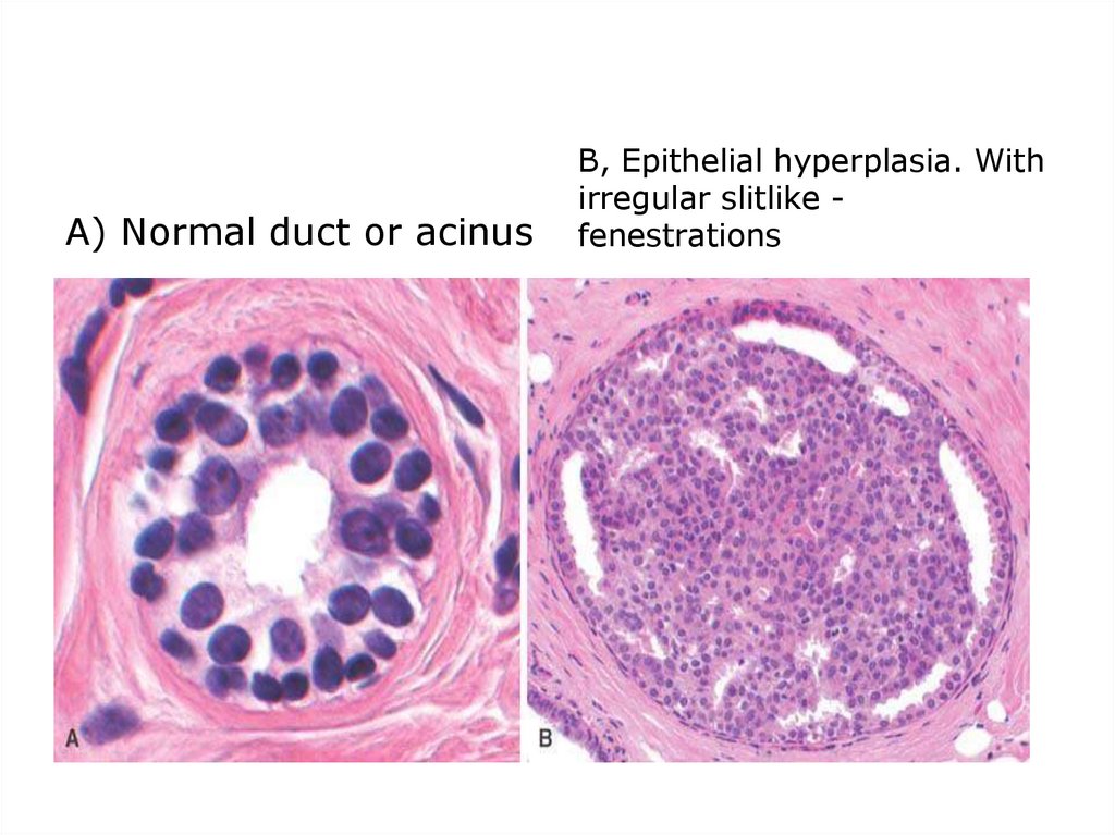
A) Normal duct or acinusB, Epithelial hyperplasia. With
irregular slitlike fenestrations
22.
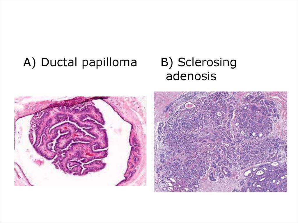
A) Ductal papillomaB) Sclerosing
adenosis
23.
Proliferative Breast Diseasewith Atypia
• Hyperplasia with atypia is present in
ducts or lobules.
• Moderately increased risk of
carcinoma.
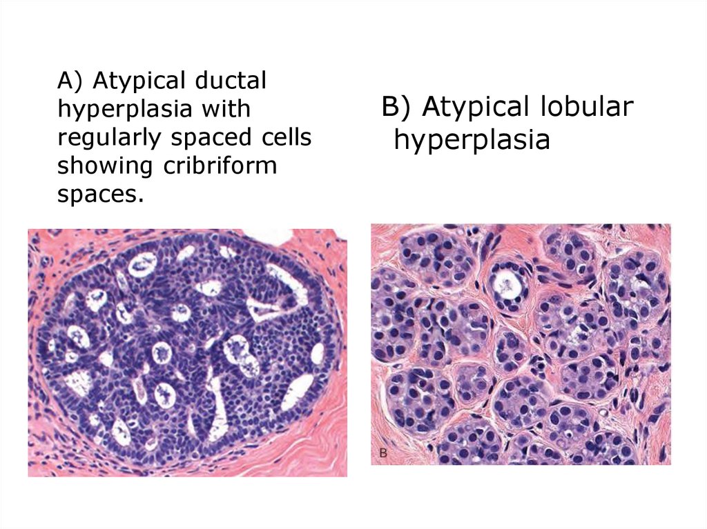
24.
A) Atypical ductalhyperplasia with
regularly spaced cells
showing cribriform
spaces.
B) Atypical lobular
hyperplasia
25.
Fibroadenoma•Most common benign fibroepithelial
tumor of female breast.
•Increase in estrogen activity
contributes to its development.
•Usually in young women; peak
incidence- 3rd decade of life.
26.
Morphology
Gross:
• Discrete, usually solitary, freely
movable nodule, 1-10 cm in diameter.
• Rarely multiple tumors and rarely may
exceed 10 cm in diameter (giant
fibroadenoma)
• Well-circumscribed, smooth, or mildly
lobulated masses.
• Cut surface- bulging, uniform gray
white, and gelatinous or mucoid.
27.
Microscopy:•Loose fibroblastic stroma containing
ductlike, epithelium-lined spaces of
various forms and sizes.
•Ductlike or glandular spaces are
lined with single or multiple layers of
cells that are regular and have welldefined, intact basement membrane.
28.
Two patterns:•Pericanalicular fibroadenoma- Ductal
spaces are open, round to oval, and
regular.
•Intracanalicular fibroadenoma- Duct
spaces are compressed by extensive
proliferation of stroma.
29.
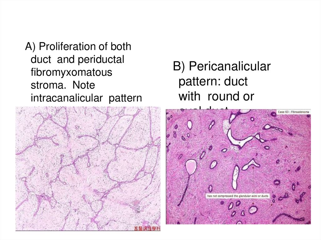
A) Proliferation of bothduct and periductal
fibromyxomatous
stroma. Note
intracanalicular pattern
of slit-like duct
B) Pericanalicular
pattern: duct
with round or
oval duct
30.
Clinical features•Present as solitary, discrete, movable
painless masses.
•May enlarge late in menstrual cycle
and during pregnancy.
•After menopause, may regress and
calcify.
•Almost never become malignant.
31.
THANKYOU