


 Медицина
 Биология
Похожие презентации:


The hormonal regulation of the body
1.
• Mechanisms of physiologicalregulation.
Humoral regulation of
physiological functions.
Interrelations of nervous and
humoral regulation
Snegir AG
2.
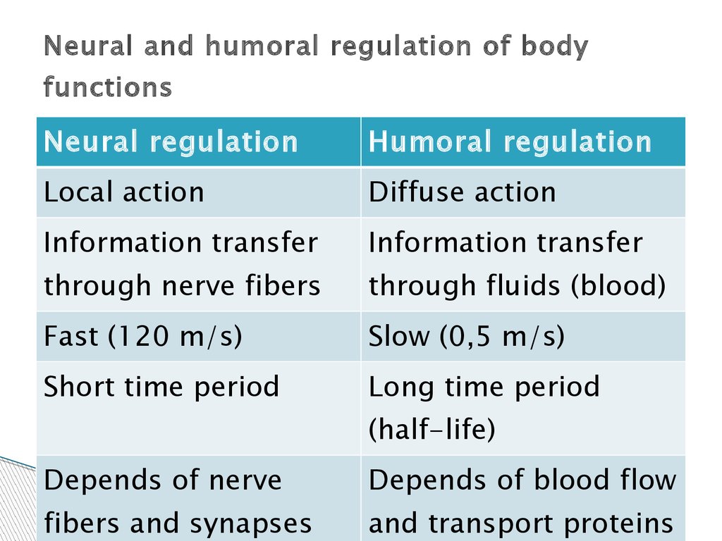
Neural and humoral regulation of bodyfunctions
Neural regulation
Humoral regulation
Local action
Diffuse action
Information transfer
Information transfer
through nerve fibers
through fluids (blood)
Fast (120 m/s)
Slow (0,5 m/s)
Short time period
Long time period
(half-life)
Depends of nerve
Depends of blood flow
fibers and synapses
and transport proteins
3.
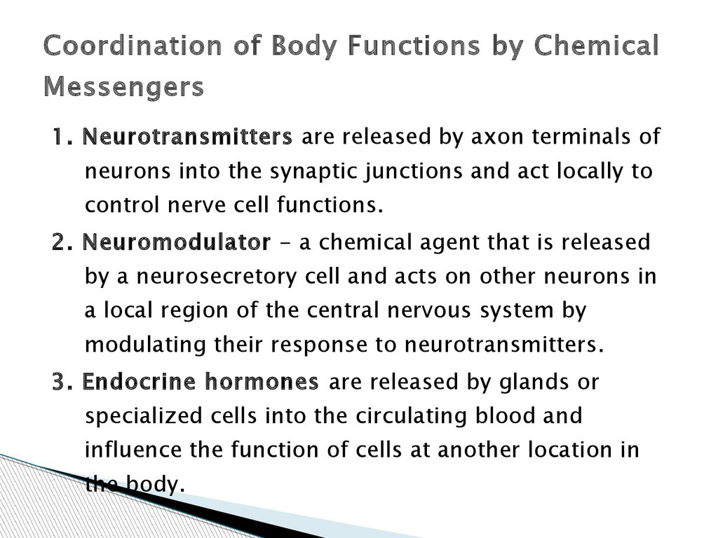
Coordination of Body Functions by ChemicalMessengers
1. Neurotransmitters are released by axon terminals of
neurons into the synaptic junctions and act locally to
control nerve cell functions.
2. Neuromodulator - a chemical agent that is released
by a neurosecretory cell and acts on other neurons in
a local region of the central nervous system by
modulating their response to neurotransmitters.
3. Endocrine hormones are released by glands or
specialized cells into the circulating blood and
influence the function of cells at another location in
the body.
4.
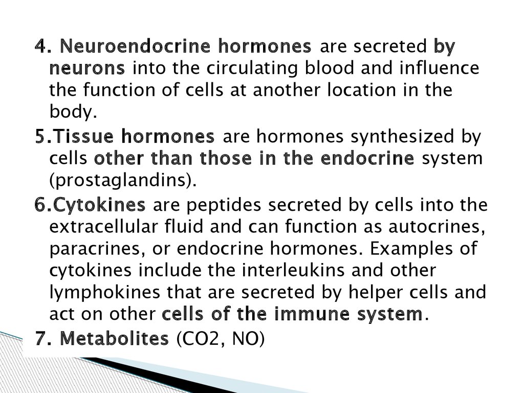
4. Neuroendocrine hormones are secreted byneurons into the circulating blood and influence
the function of cells at another location in the
body.
5.Tissue hormones are hormones synthesized by
cells other than those in the endocrine system
(prostaglandins).
6.Cytokines are peptides secreted by cells into the
extracellular fluid and can function as autocrines,
paracrines, or endocrine hormones. Examples of
cytokines include the interleukins and other
lymphokines that are secreted by helper cells and
act on other cells of the immune system.
7. Metabolites (CO2, NO)
5.
The action of humoralfactors
Endocrine hormones are released by glands or
specialized cells into the circulating blood and
influence the function of cells at another location in
the body.
Neuroendocrine hormones are secreted by neurons
into the circulating blood and influence the function
of cells at another location in the body.
Paracrines are secreted by cells into the extracellular
fluid and affect neighboring cells of a different type.
Autocrines are secreted by cells into the
extracellular fluid and affect the function of the same
cells that produced them by binding to cell surface
receptors
6.
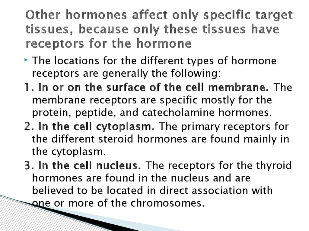
Other hormones affect only specific targettissues, because only these tissues have
receptors for the hormone
The locations for the different types of hormone
receptors are generally the following:
1. In or on the surface of the cell membrane. The
membrane receptors are specific mostly for the
protein, peptide, and catecholamine hormones.
2. In the cell cytoplasm. The primary receptors for
the different steroid hormones are found mainly in
the cytoplasm.
3. In the cell nucleus. The receptors for the thyroid
hormones are found in the nucleus and are
believed to be located in direct association with
one or more of the chromosomes.
7.
There are three general classes ofhormones:
1.
2.
3.
Proteins and polypeptides, including
hormones secreted by the anterior and
posterior pituitary gland, the pancreas (insulin
and glucagon), the parathyroid gland
(parathyroid hormone), and many others.
Steroids secreted by the adrenal cortex (cortisol
and aldosterone), the ovaries (estrogen and
progesterone), the testes (testosterone), and the
placenta (estrogen and progesterone).
Derivatives of the amino acid tyrosine,
secreted by the thyroid (thyroxine and
triiodothyronine) and the adrenal medullae
(epinephrine and
norepinephrine).
8.
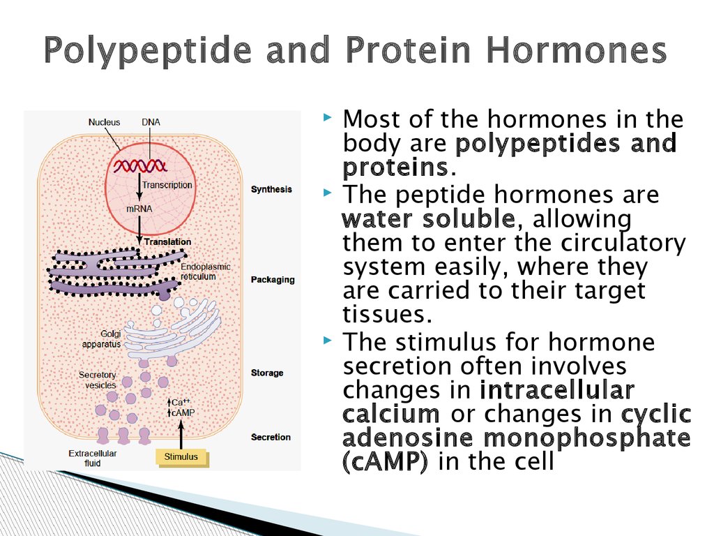
Polypeptide and Protein HormonesMost of the hormones in the
body are polypeptides and
proteins.
The peptide hormones are
water soluble, allowing
them to enter the circulatory
system easily, where they
are carried to their target
tissues.
The stimulus for hormone
secretion often involves
changes in intracellular
calcium or changes in cyclic
adenosine monophosphate
(cAMP) in the cell
9.
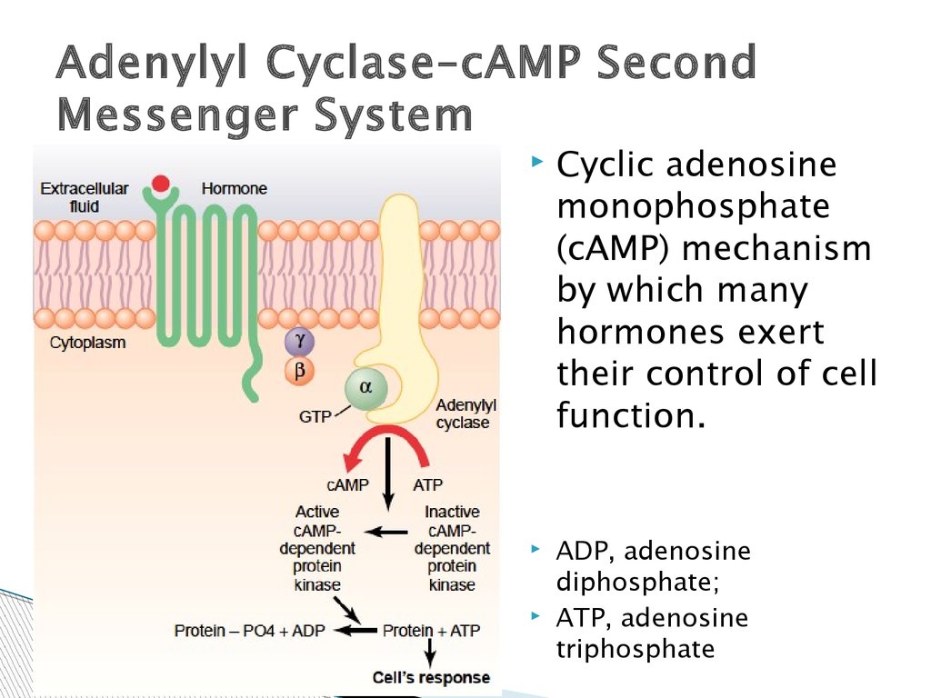
Adenylyl Cyclase–cAMP SecondMessenger System
Cyclic adenosine
monophosphate
(cAMP) mechanism
by which many
hormones exert
their control of cell
function.
ADP, adenosine
diphosphate;
ATP, adenosine
triphosphate
10.
Many hormones activate receptors (Protein–Linked Hormone Receptors) that indirectly
regulate the activity of target proteins (e.g.,
enzymes or ion channels) by coupling with groups
of cell membrane proteins called heterotrimeric
GTP-binding proteins (G proteins).
When the ligand (hormone) binds to the
extracellular part of the receptor, a
conformational change occurs in the receptor that
activates the G proteins and induces
intracellular signals that either
(1) open or close cell membrane ion channels or
(2) change the activity of an enzyme in the
cytoplasm of the cell.
11.
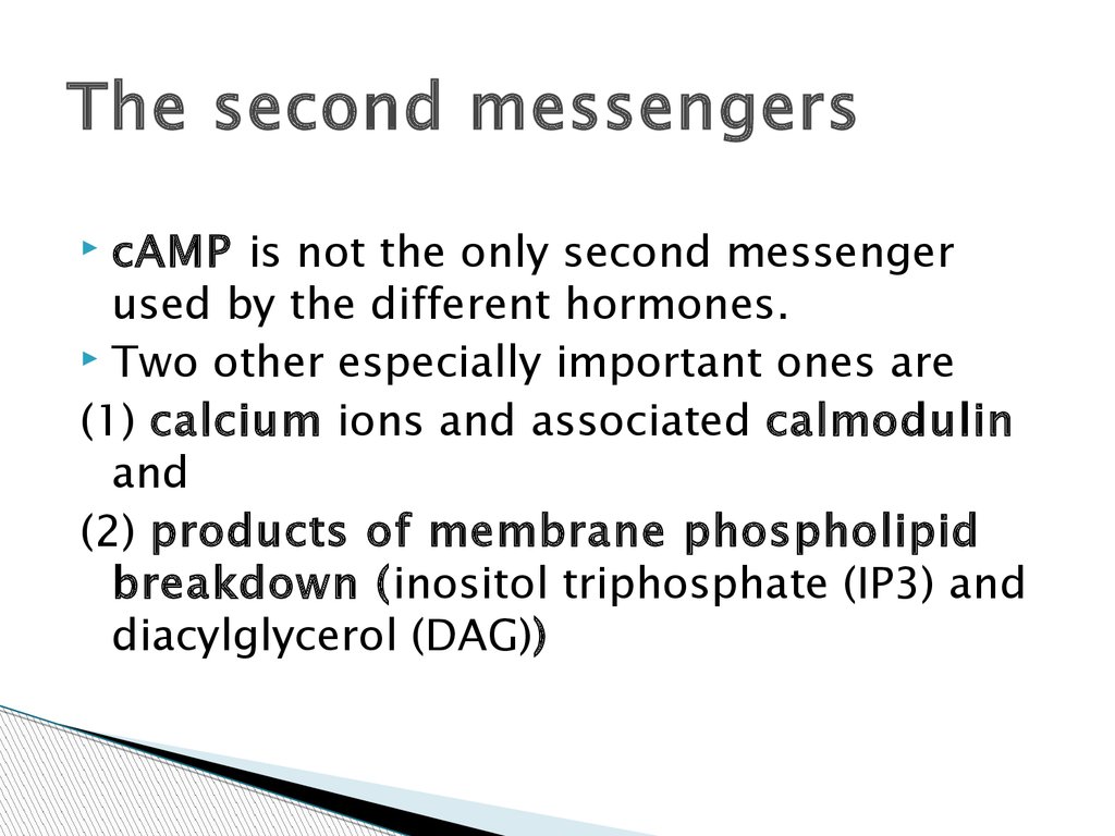
The second messengerscAMP is not the only second messenger
used by the different hormones.
Two other especially important ones are
(1) calcium ions and associated calmodulin
and
(2) products of membrane phospholipid
breakdown (inositol triphosphate (IP3) and
diacylglycerol (DAG))
12.
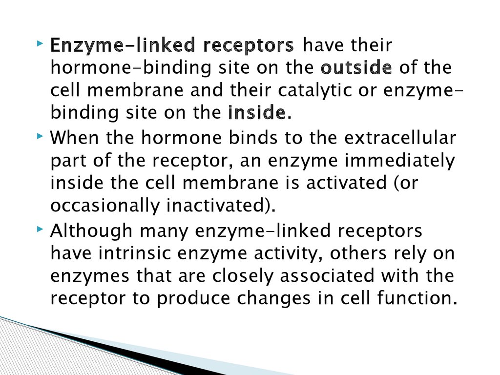
Enzyme-linked receptors have theirhormone-binding site on the outside of the
cell membrane and their catalytic or enzymebinding site on the inside.
When the hormone binds to the extracellular
part of the receptor, an enzyme immediately
inside the cell membrane is activated (or
occasionally inactivated).
Although many enzyme-linked receptors
have intrinsic enzyme activity, others rely on
enzymes that are closely associated with the
receptor to produce changes in cell function.
13.
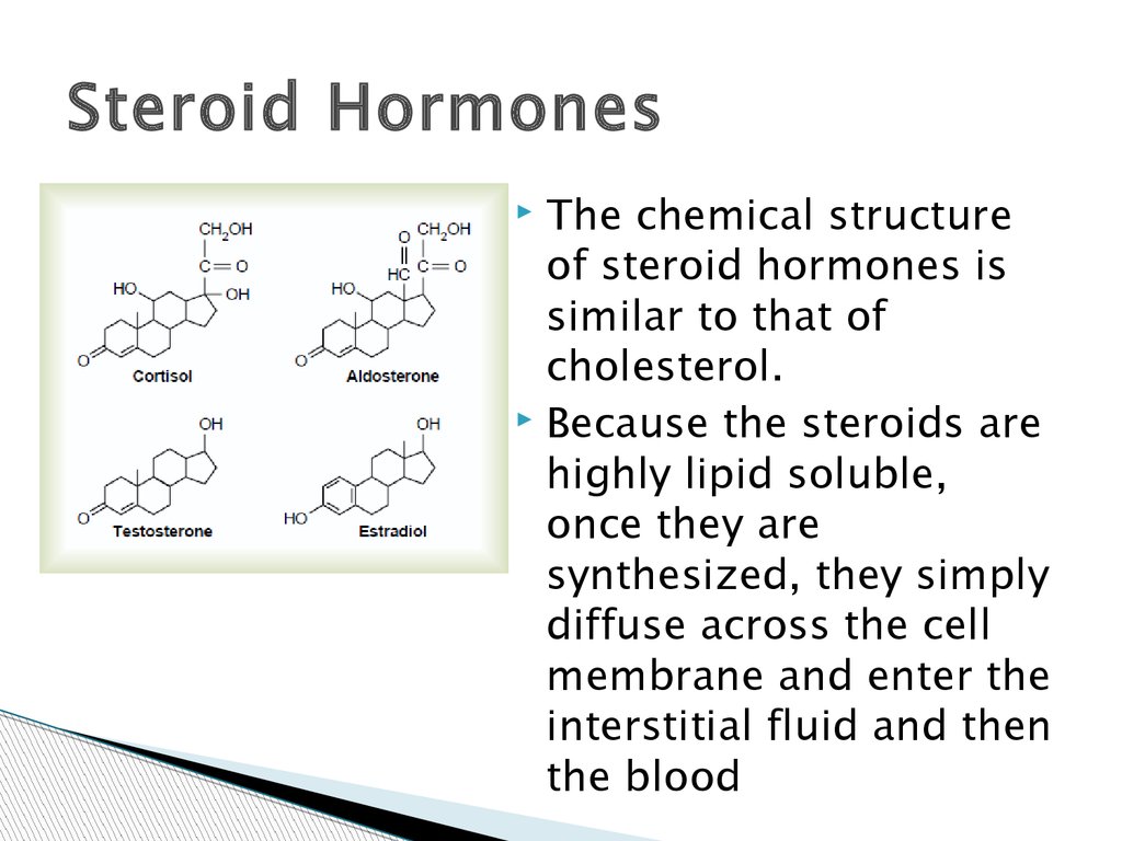
Steroid HormonesThe chemical structure
of steroid hormones is
similar to that of
cholesterol.
Because the steroids are
highly lipid soluble,
once they are
synthesized, they simply
diffuse across the cell
membrane and enter the
interstitial fluid and then
the blood
14.
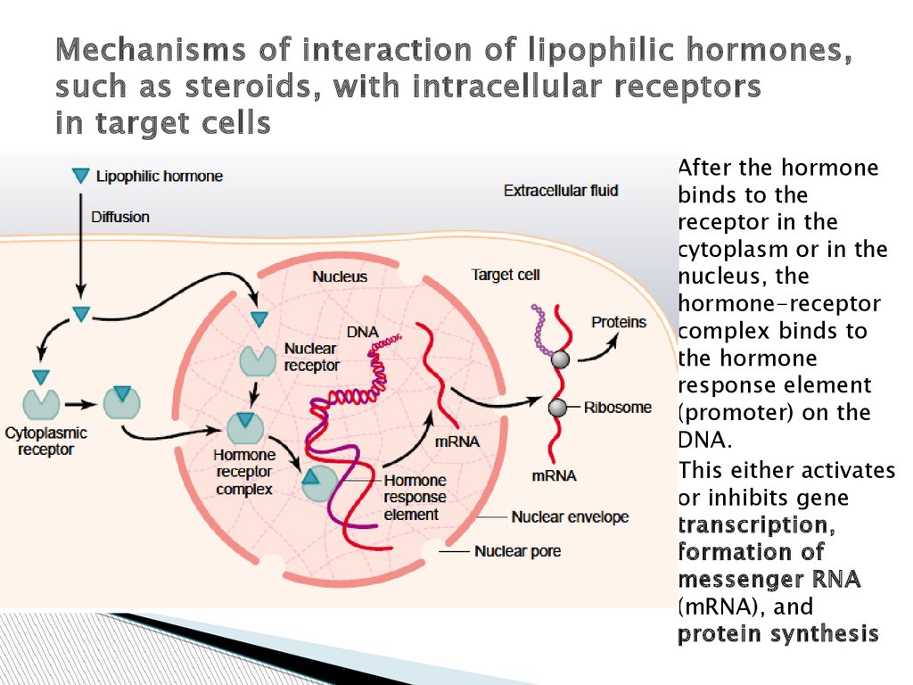
Mechanisms of interaction of lipophilic hormones,such as steroids, with intracellular receptors
in target cells
After the hormone
binds to the
receptor in the
cytoplasm or in the
nucleus, the
hormone-receptor
complex binds to
the hormone
response element
(promoter) on the
DNA.
This either activates
or inhibits gene
transcription,
formation of
messenger RNA
(mRNA), and
protein synthesis
15.
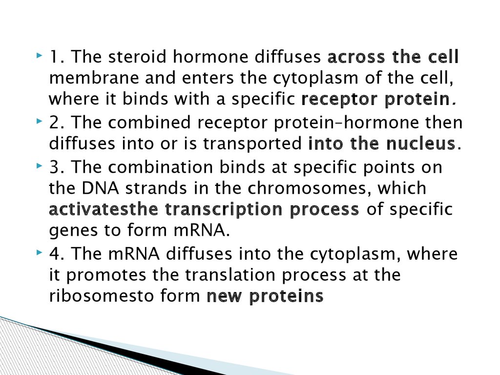
1. The steroid hormone diffuses across the cellmembrane and enters the cytoplasm of the cell,
where it binds with a specific receptor protein .
2. The combined receptor protein–hormone then
diffuses into or is transported into the nucleus.
3. The combination binds at specific points on
the DNA strands in the chromosomes, which
activatesthe transcription process of specific
genes to form mRNA.
4. The mRNA diffuses into the cytoplasm, where
it promotes the translation process at the
ribosomesto form new proteins
16.
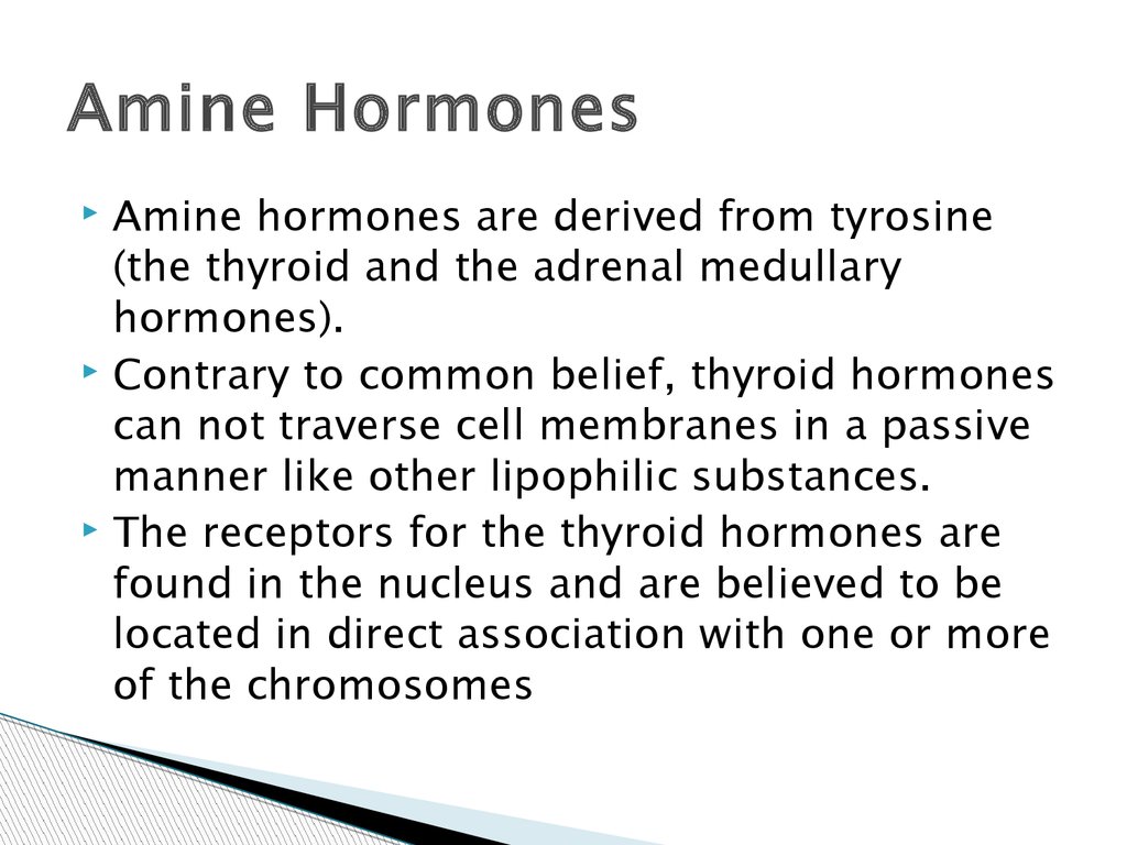
Amine HormonesAmine hormones are derived from tyrosine
(the thyroid and the adrenal medullary
hormones).
Contrary to common belief, thyroid hormones
can not traverse cell membranes in a passive
manner like other lipophilic substances.
The receptors for the thyroid hormones are
found in the nucleus and are believed to be
located in direct association with one or more
of the chromosomes
17.
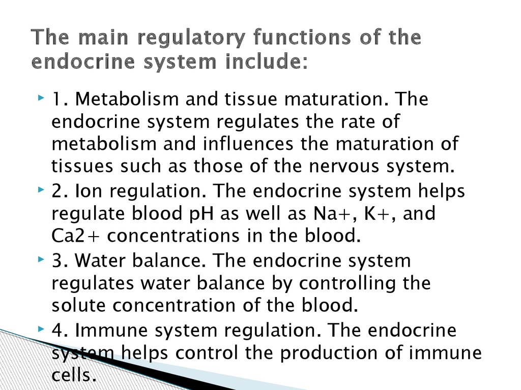
The main regulatory functions of theendocrine system include:
1. Metabolism and tissue maturation. The
endocrine system regulates the rate of
metabolism and influences the maturation of
tissues such as those of the nervous system.
2. Ion regulation. The endocrine system helps
regulate blood pH as well as Na+, K+, and
Ca2+ concentrations in the blood.
3. Water balance. The endocrine system
regulates water balance by controlling the
solute concentration of the blood.
4. Immune system regulation. The endocrine
system helps control the production of immune
cells.
18.
5. Heart rate and blood pressure regulation. Theendocrine system helps regulate the heart rate
and blood pressure and helps prepare the body
for physical activity.
6. Control of blood glucose and other nutrients.
The endocrine system regulates blood glucose
levels and other nutrient levels in the blood.
7. Control of reproductive functions. The
endocrine system controls the development and
functions of the reproductive systems in males
and females.
8. Uterine contractions and milk release. The
endocrine system regulates uterine contractions
during delivery and stimulates milk release from
the breasts in lactating females.
19.
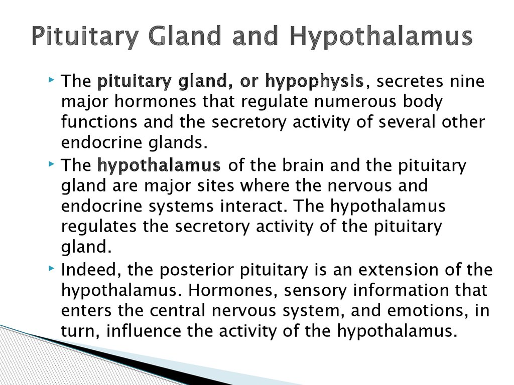
Pituitary Gland and HypothalamusThe pituitary gland, or hypophysis, secretes nine
major hormones that regulate numerous body
functions and the secretory activity of several other
endocrine glands.
The hypothalamus of the brain and the pituitary
gland are major sites where the nervous and
endocrine systems interact. The hypothalamus
regulates the secretory activity of the pituitary
gland.
Indeed, the posterior pituitary is an extension of the
hypothalamus. Hormones, sensory information that
enters the central nervous system, and emotions, in
turn, influence the activity of the hypothalamus.
20.
Relationship Among the Hypothalamus,Posterior Pituitary, and Target Tissues
21.
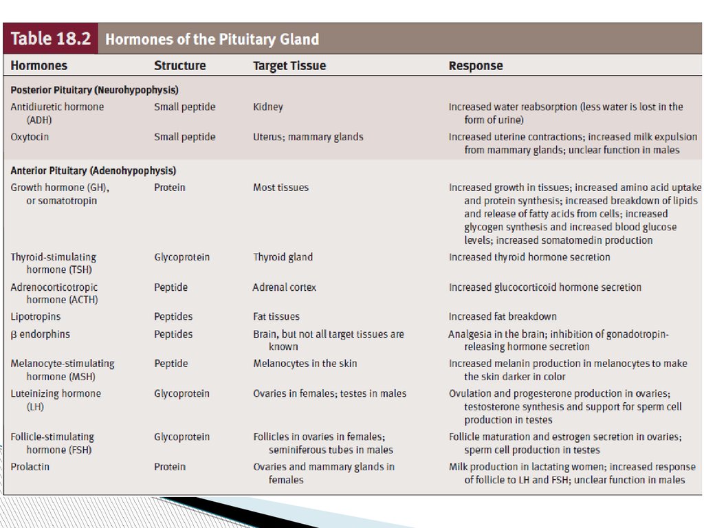
Posterior Pituitary HormonesAntidiuretic Hormone is so named because it
prevents the output of large amounts of urine
(diuresis). ADH is sometimes called vasopressin)
because it constricts blood vessels and raises blood
pressure when large amounts are released.
ADH is synthesized by neuron cell bodies in the
supraoptic nuclei of the hypothalamus and
transported within the axons of the
hypothalamohypophysial tract to the posterior
pituitary, where it is stored in axon terminals.
ADH is released from these axon terminals into the
blood and carried to its primary target tissue, the
kidneys, where it promotes the retention of water and
reduces urine volume
22.
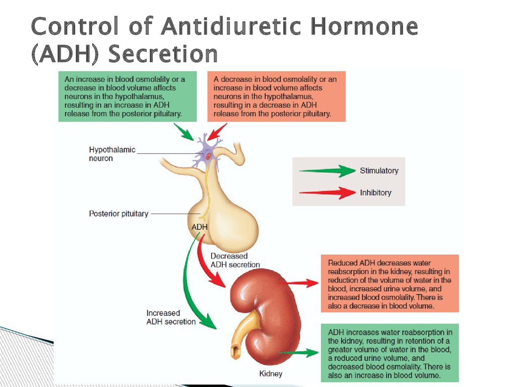
Control of Antidiuretic Hormone(ADH) Secretion
23.
When blood osmolality increases, the frequencyof action potentials in the osmoreceptors
increases, resulting in a greater frequency of
action potentials in the neurosecretory cells. As
a consequence, ADH secretion increases.
Alternatively, an increase in blood osmolality
can directly stimulate the ADH neurosecretory
cells.
Because ADH stimulates the kidneys to retain
water, it functions to reduce blood osmolality
and resists any further increase in the
osmolality of body fluids.
As the osmolality of the blood decreases, the
action potential frequency in the osmoreceptors
and the neurosecretory cells decreases.
24.
Diabetes InsipidusA lack of ADH secretion is one cause of
diabetes insipidus and leads to the production
of a large amount of dilute urine, which can
approach 20 L/day.
The loss of many liters of water in the form of
urine causes an increase in the osmolality of
the body fluids, and a decrease in extracellular
fluid volume, but negative-feedback
mechanisms fail to stimulate ADH release.
25.
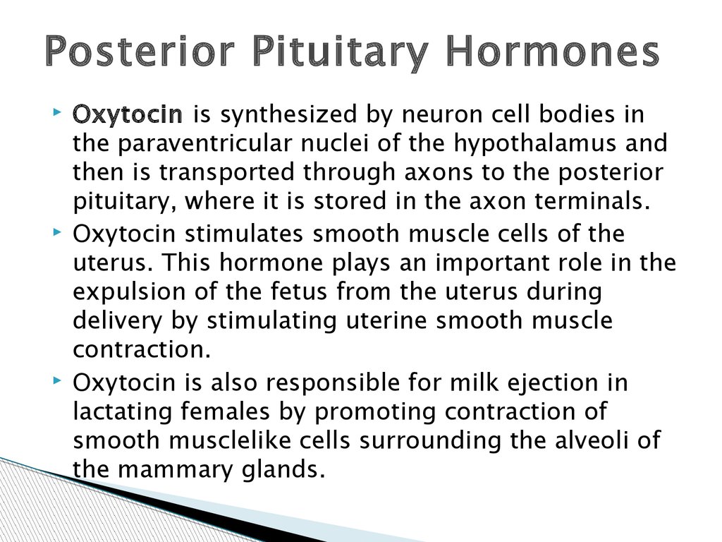
Posterior Pituitary HormonesOxytocin is synthesized by neuron cell bodies in
the paraventricular nuclei of the hypothalamus and
then is transported through axons to the posterior
pituitary, where it is stored in the axon terminals.
Oxytocin stimulates smooth muscle cells of the
uterus. This hormone plays an important role in the
expulsion of the fetus from the uterus during
delivery by stimulating uterine smooth muscle
contraction.
Oxytocin is also responsible for milk ejection in
lactating females by promoting contraction of
smooth musclelike cells surrounding the alveoli of
the mammary glands.
26.
Action potentials are carried by sensoryneurons from the uterus and from the nipples
to the spinal cord.
Action potentials are then carried up the
spinal cord to the hypothalamus, where they
increase action potentials in the oxytocinsecreting neurons.
Action potentials in the oxytocin-secreting
neurons pass along the axons in the
hypothalamohypophysial tract to the
posterior pituitary, where they cause the axon
terminals to release oxytocin.
27.
Relationship of the Pituitary to theBrain
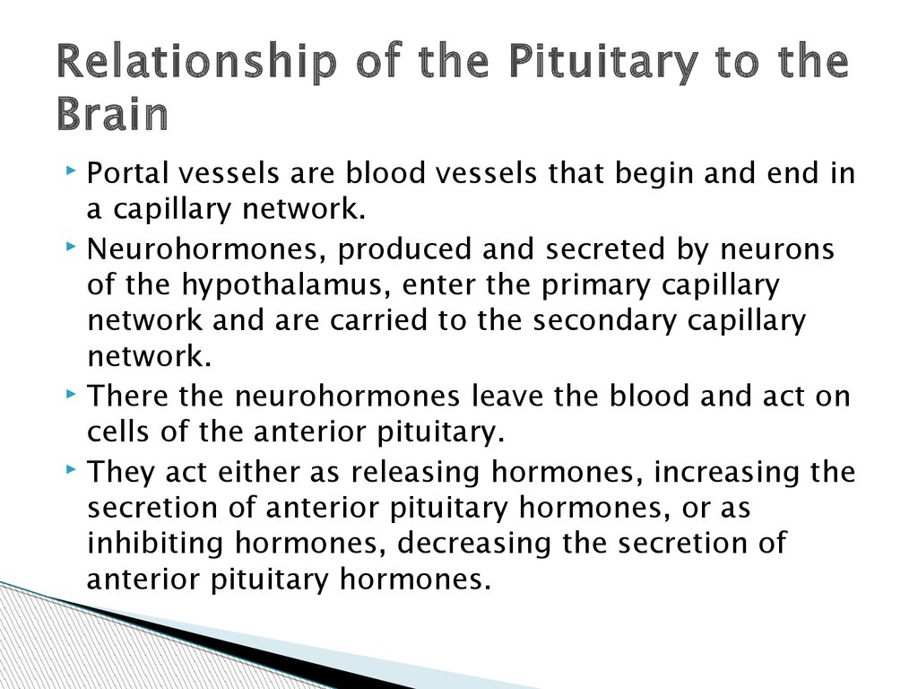
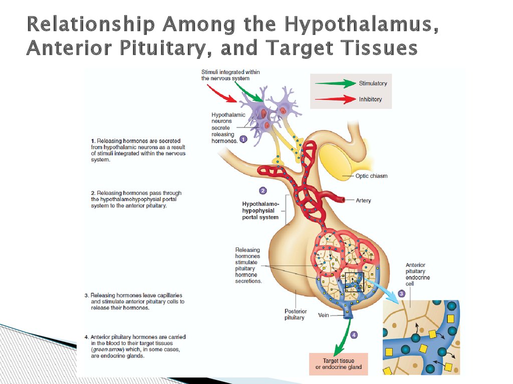
Portal vessels are blood vessels that begin and end in
a capillary network.
Neurohormones, produced and secreted by neurons
of the hypothalamus, enter the primary capillary
network and are carried to the secondary capillary
network.
There the neurohormones leave the blood and act on
cells of the anterior pituitary.
They act either as releasing hormones, increasing the
secretion of anterior pituitary hormones, or as
inhibiting hormones, decreasing the secretion of
anterior pituitary hormones.
28.
Relationship Among the Hypothalamus,Anterior Pituitary, and Target Tissues
29.
30.
31.
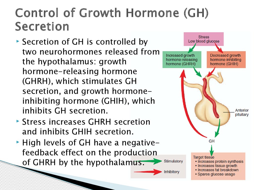
Control of Growth Hormone (GH)Secretion
Secretion of GH is controlled by
two neurohormones released from
the hypothalamus: growth
hormone-releasing hormone
(GHRH), which stimulates GH
secretion, and growth hormoneinhibiting hormone (GHIH), which
inhibits GH secretion.
Stress increases GHRH secretion
and inhibits GHIH secretion.
High levels of GH have a negativefeedback effect on the production
of GHRH by the hypothalamus.
32.
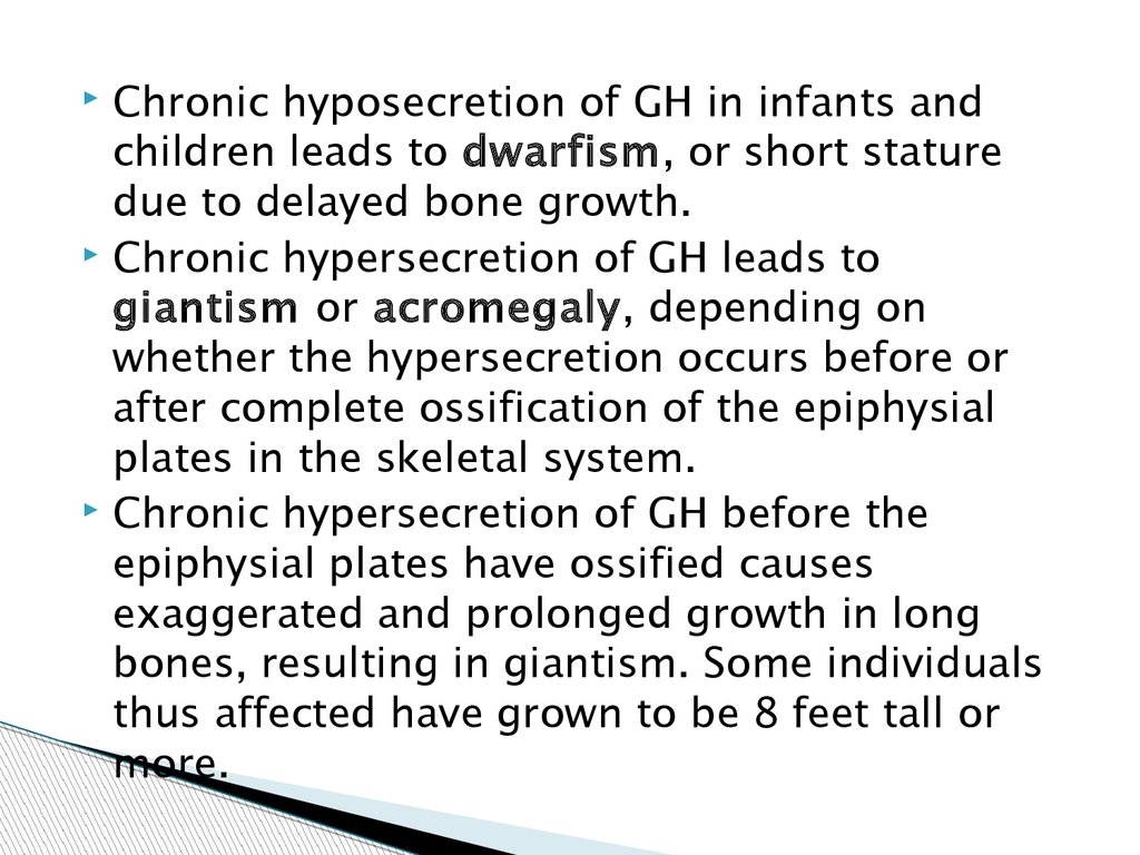
Chronic hyposecretion of GH in infants andchildren leads to dwarfism, or short stature
due to delayed bone growth.
Chronic hypersecretion of GH leads to
giantism or acromegaly, depending on
whether the hypersecretion occurs before or
after complete ossification of the epiphysial
plates in the skeletal system.
Chronic hypersecretion of GH before the
epiphysial plates have ossified causes
exaggerated and prolonged growth in long
bones, resulting in giantism. Some individuals
thus affected have grown to be 8 feet tall or
more.
33.
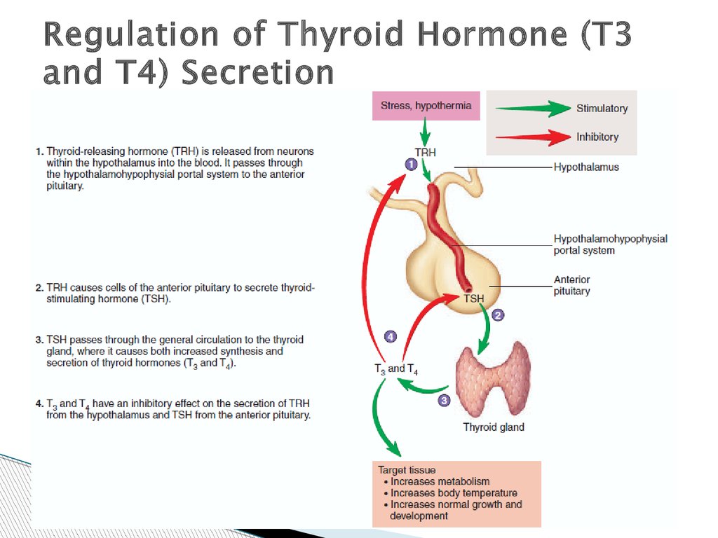
Regulation of Thyroid Hormone (T3and T4) Secretion
34.
GoiterAn abnormal enlargement of the thyroid gland is called a
goiter. Goiters can result from conditions that cause
hypothyroidism as well as conditions that cause
hyperthyroidism.
An iodine deficiency goiter results when dietary iodine
intake is very low and there is too little iodine to synthesize
T3 and T4. As a result, blood levels of T3 and T4 decrease
and the person may exhibit symptoms of hypothyroidism.
The reduced negative feedback of T3 and T4 on the
anterior pituitary and hypothalamus result in elevated TSH
secretion. TSH causes hypertrophy and hyperplasia of the
thyroid gland and increased thyroglobulin synthesis even
though there is not enough iodine to synthesize T3 and T4.
Consequently, the thyroid gland enlarges.
Toxic goiter secretes excess T3 and T4, and it can result
from elevated TSH secretion or elevated TSH-like immune
globulin molecules (Graves’ Disease).
35.
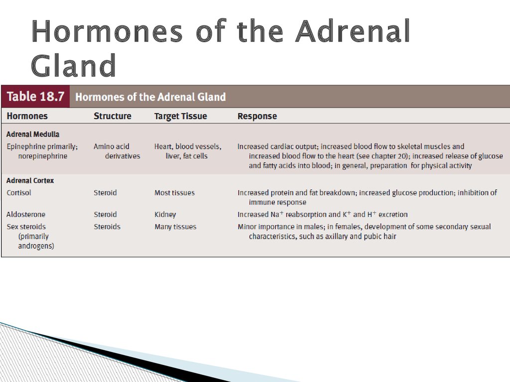
Hormones of the AdrenalGland
36.
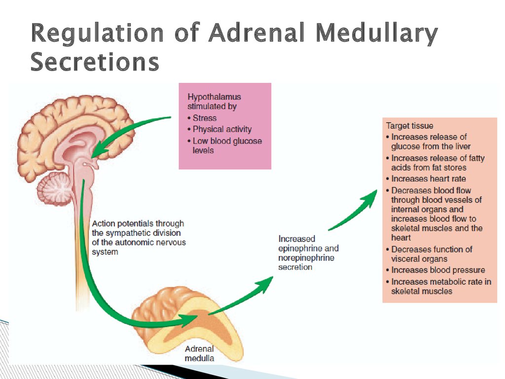
Regulation of Adrenal MedullarySecretions
37.
Hormones of the Adrenal CortexThe adrenal cortex secretes three hormone types:
mineralocorticoids, glucocorticoids, and
androgens.
All are similar in structure in that they are steroids,
highly specialized lipids that are derived from
cholesterol.
Because they are lipidsoluble, they are not stored in
the adrenal gland cells but diffuse from the cells as
they are synthesized.
Adrenal cortical hormones are transported in the
blood in combination with specific plasma proteins;
they are metabolized in the liver and excreted in the
bile and urine.
38.
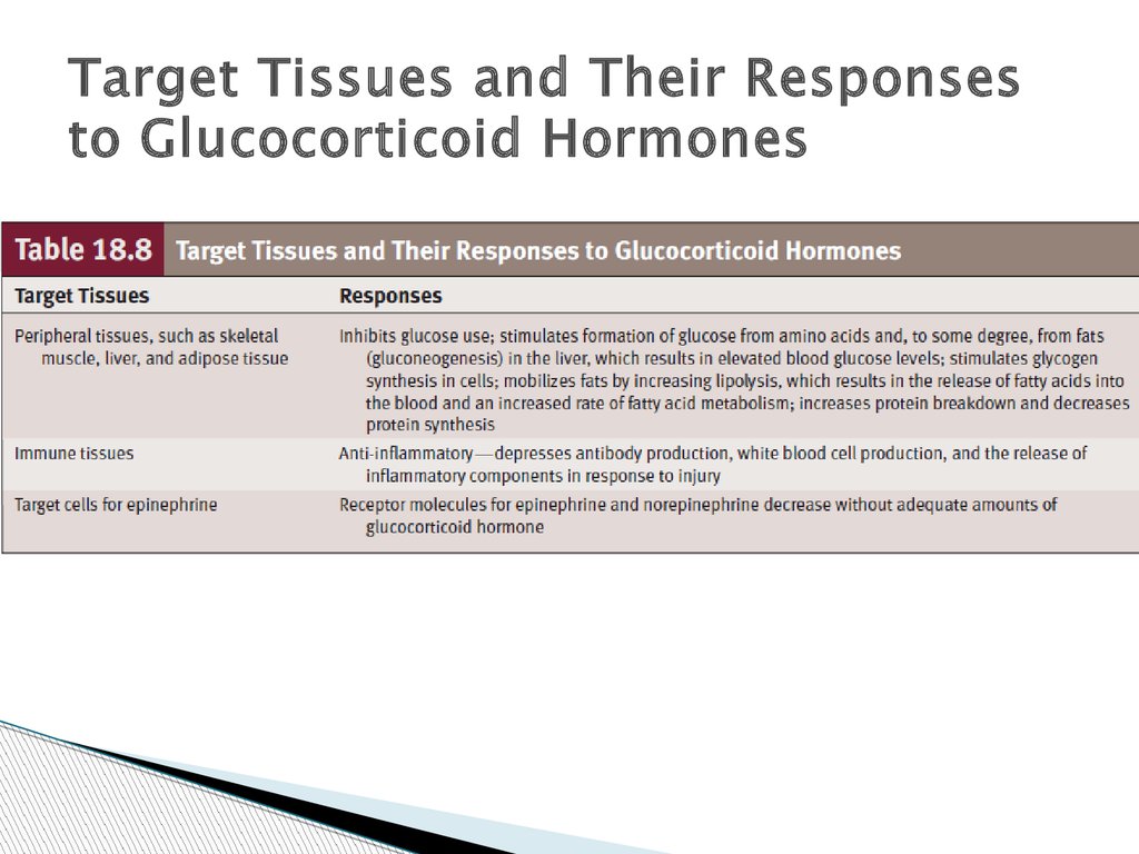
Target Tissues and Their Responsesto Glucocorticoid Hormones
39.
Regulation of Cortisol Secretion40.
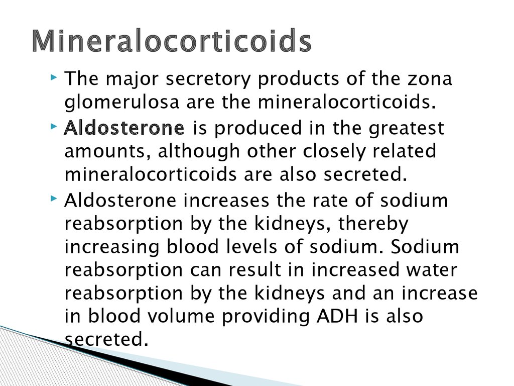
MineralocorticoidsThe major secretory products of the zona
glomerulosa are the mineralocorticoids.
Aldosterone is produced in the greatest
amounts, although other closely related
mineralocorticoids are also secreted.
Aldosterone increases the rate of sodium
reabsorption by the kidneys, thereby
increasing blood levels of sodium. Sodium
reabsorption can result in increased water
reabsorption by the kidneys and an increase
in blood volume providing ADH is also
secreted.
41.
Aldosterone increases K excretion into theurine by the kidneys, thereby decreasing
blood levels of K. It also increases the rate of
H excretion into the urine.
When aldosterone is secreted in high
concentrations, it can result in reduced blood
levels of K and alkalosis (elevated pH of body
fluids).
42.
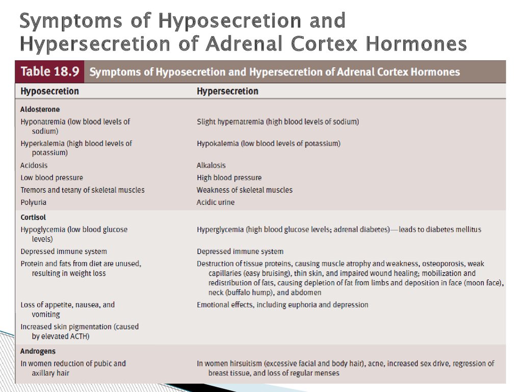
Symptoms of Hyposecretion andHypersecretion of Adrenal Cortex Hormones
43.
Adrenal AndrogensSome adrenal steroids, including
androstenedione are weak androgens.
They are secreted by the zona reticularis and
converted by peripheral tissues to the more
potent androgen, testosterone.
Adrenal androgens stimulate pubic and
axillary hair growth and sexual drive in
females. Their effects in males are negligible
in comparison to testosterone secreted by
the testes.