Похожие презентации:
General principles of anaesthesiology
1.
General principles ofanaesthesiology
Lecture for 5-year medical students
2. Lecture plan
◦◦
◦
◦
◦
◦
◦
◦
◦
Introduction
Preoperative History and Physical
IV’s and Premedication
Commonly Used Medications
Room Setup and Monitors
Induction and Intubation
Maintenance
Emergence
PACU Concerns
3.
Induction to anaesthesiologyDefinitions
Anesthesia - From the Greek
meaning lack of sensation;
particularly during surgical
intervention.
4.
Induction to anaesthesiologyOn October 16, 1846, in Boston,
William T.G. Morton - the first publicized
demonstration of general anesthesia
using ether.
The pre-existing word anesthesia was
suggested by Oliver Wendell Holmes, Sr.
in 1846 as a word to use to describe this
state.
5. History of anaesthesia
6. History of anaesthesia
7.
General anesthesia–a condition characterized by temporary
shutting down
consciousness
pain sensitivity
reflexes
relaxation of skeletal muscles
due to exposure to the anesthetics on the
Central nervous system
8.
9.
10.
AnaesthesiaReversible, drug-induced condition
◦ Amnesia & unconsciousness
◦ Analgesia
◦ Muscle relaxation
◦ Attenuation of autonomic
responses to noxious stimulation
◦ Homeostasis of Vital
◦ Functions
11.
Anaesthesiology is thescience of managing the life
functions of the patients
organism in time of surgery
or aggressive diagnostic
procedure.
12.
Induction to anaesthesiologyGeneral
Anesthesia
Preoperative evaluation
Intraoperative management
Postoperative management
13.
Preoperative History and PhysicalPhysical Examination
Physical exams of all systems.
Airway assessment to determine the
likelihood of difficult intubation
14. Preoperative History and Physical
Unlike the standard internal medicineH&P, ours is much more focused, with
specific attention being paid to the
airway and to organ systems at
potential risk for anesthetic
complications. The type of operation,
and the type of anesthetic will also
help to focus the evaluation.
15. Classification of operation
• Elective: operation at a time to suitboth patient and surgeon; for example
hip replacement, varicose veins.
• Scheduled: an early operation but
not immediately life saving; operation
usually within 3 weeks; for example
surgery for malignancy.
.
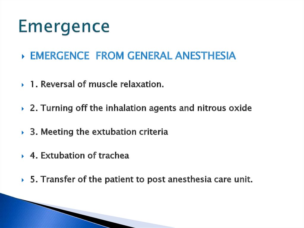
16. Classification of operation
• Urgent: operation as soon as possibleafter resuscitation and within 24 h; for
example intestinal obstruction, major
fractures.
• Emergency: immediate life-saving
operation, resuscitation simultaneous with
surgical treatment; operation usually within
1h; for example major trauma with
uncontrolled haemorrhage, extradural
haematoma
17. Preoperative History and Physical
Of particular interest in the history portion of theevaluation are:
◦
◦
◦
◦
◦
◦
◦
◦
◦
◦
◦
◦
◦
Coronary Artery Disease
Hypertension
Asthma
Kidney or Liver disease
Reflux Disease
Smoking
Alcohol Consumption or Drug Abuse?
Diabetes
Medications
Allergies
Family History
Anesthesia history
Last Meal
18. Preoperative History and Physical
Coronary Artery Disease◦ What is the patient’s exercise tolerance? How well
will his or her heart sustain the stress of the
operation and anesthetic.
◦ Asking a patient how he feels (ie. SOB, CP) after
climbing two or three flights of stairs can be very
useful as a “poor man’s stress test”.
19.
Preoperative History and PhysicalCoronary Artery Disease
◦ What is the patient’s exercise tolerance? How well
will his or her heart sustain the stress of the
operation and anesthetic.
◦ Asking a patient how he feels (ie. SOB, CP) after
climbing two or three flights of stairs can be very
useful as a “poor man’s stress test”.
20.
Preoperative History and PhysicalHypertension
◦ How well controlled is it?
Intraoperative blood pressure
management is affected by
preoperative blood pressure
control
21. Preoperative History and Physical
Asthma◦ How well controlled is it? What triggers it? Many of
the stressors of surgery as well as intubation and
ventilation can stimulate bronchospasm.
◦ Is there any history of being hospitalized,
intubated, or prescribed steroids for asthma? This
can help assess the severity of disease
22.
Preoperative History and PhysicalKidney or Liver disease
◦ Different anesthetic drugs have
different modes of clearance and
organ function can affect our choice
of drugs.
23. Preoperative History and Physical
Reflux Disease◦ Present or not? Anesthetized and relaxed patients
are prone to regurgitation and aspiration,
particularly if a history of reflux is present
24. Preoperative History and Physical
Smoking◦ Currently smoking? Airway and secretion
management can become more difficult in smokers.
25. Preoperative History and Physical
Alcohol Consumption or Drug Abuse?◦ Drinkers have an increased tolerance to many
sedative drugs (conversely they have a decreased
requirement if drunk), and are at an increased risk
of hepatic disease, which can impact the choice of
anesthetic agents.
26. Preoperative History and Physical
Diabetes◦ Well controlled? The stress response to surgery and
anesthesia can markedly increase blood glucose
concentrations, especially in diabetics
27. Preoperative History and Physical
Medications◦ Many medications interact with anesthetic agents,
and some should be taken on the morning of
surgery (blood pressure medications) while others
should probably not (diuretics, diabetes
medications).
28. Preoperative History and Physical
Allergies◦ We routinely give narcotics and antibiotics
perioperatively, and it is important to know the
types of reactions that a patient has had to
medications in the past.
29. Preoperative History and Physical
Family History◦ There is a rare, but serious disorder known as
malignant hyperthermia that affects susceptible
patients under anesthesia, and is heritable
30. Preoperative History and Physical
Anesthesia history◦ Has the patient ever had anesthesia and surgery
before? Did anything go wrong?
31. Preoperative History and Physical
Last Meal◦ Whether the patient has an empty stomach or not
impacts the choice of induction technique
32. Preoperative History and Physical
All patients must have an assessment madeof their airway, the aim being to try and
predict those patients who may be difficult to
intubate.
33. Preoperative History and Physical
Finding any of these suggests that intubationmay be more difficult.
• limitation of mouth opening;
• a receding mandible;
• position, number and health of teeth;
• size of the tongue;
• soft tissue swelling at the front of the neck;
• deviation of the larynx or trachea;
• limitations in flexion and extension of the
cervical spine.
34. Preoperative History and Physical
Also, any loose or missing teeth should benoted, as should cervical range of motion,
mouth opening, and thyromental distance,
all of which will impact the actual intubation
prior to surgery.
During the physical examination, particular
attention is paid to the airway by asking the
patient to “open your mouth as wide as you
can and stick out your tongue” The
classification scale of Mallampati is
commonly used.
35. Preoperative History and Physical
Mallampati ClassificationClass I: Entire uvula and tonsillar pillars
visible
Class II: Tip of uvula and pillars hidden by
tongue
Class III: Only soft palate visible
Class IV: Only hard palate visible
36. Mallampati Classification
37. Preoperative History and Physical
Finally, a physical status classification isassigned, based on the criteria of the American
Society of Anesthesiologists (ASA1-5), with
ASA-1 being assigned to a healthy person
without medical problems other than the
current surgical concern, and ASA-5 being a
moribund patient, not expected to survive for
more then twenty-four hours without surgical
intervention. An “E” is added if the case is
emergent. The full details of the classification
scale can be found below.
38. Preoperative History and Physical
ASA Physical Status ClassificationASA-I:
Healthy patient with no systemic disease
ASA-II:
ASA-III:
ASA-IV:
ASA-V:
ASA-VI:
Mild systemic disease , no functional
limitations
Moderate to severe systemic disease, some
functional limitations
Severe systemic disease, incapacitating, and a
constant threat to life
Moribund patient, not expected to survive >
24 hours without surgery
Brain-dead patient undergoing organ harvest
E: Added when the case is emergent
39. Classification of anaesthesia
general anesthesiaSimple (one-component) anaesthesia
Inhalation
mask
endotracheal
Noninhalation
intravenous
Combined (multi-component anaesthesia
Inhalation + Inhalation
Noninhalation + Noninhalation
Noninhalation + Inhalation
Combined with miorelaxanthams
40. Clasification of anaesthesia
local anesthesiaTerminal anesthesia
Infiltration anesthesia
Nerve block anesthesia
trunk
plexus
regional anesthesia
Spinal anesthesia
Epidural anesthesia
41.
42. IV’s and Premedication
Every patient (with the exception of somechildren that can have their IV’s inserted
following inhalation induction) will require IV
access prior to being brought to the
operating room.
Normal saline, Lactated Ringer’s solution, or
other balanced electrolyte solutions
(Plasmalyte, Isolyte) are all commonly used
solutions intraoperatively.
43.
PremedicationPremedication refers to the administration of any
drugs in the period before induction of
anaesthesia.
a wide variety of drugs are used with a variety of
aims
The 6 As of premedication
• Anxiolysis
• Amnesia
• Antiemetic
• Antacid
• Antiautonomic
• Analgesia
44. IV’s and Premedication
Many patients are understandably nervouspreoperatively, and we often premedicate
them, usually with a rapid acting
benzodiazepine such as intravenous
midazolam (which is also fabulously effective
in children orally or rectally).
Metoclopramide and an H2 blocker are also
often used if there is a concern that the
patient has a full stomach,
and anticholinergics such as glycopyrrolate or
atropin can be used to decrease secretions.
45. Room Setup and Monitors
Before bringing the patient to the room, theanesthesia machine, ventilator, monitors,
and cart must be checked and set up.
The anesthesia machine must be tested to
ensure that the gauges and monitors are
functioning properly, that there are no leaks
in the gas delivery system, and that the
backup systems and fail-safes are
functioning properly.
46.
47. Room Setup and Monitors
The monitors that we use on most patientsinclude the pulse oximeter, blood pressure
monitor, and electrocardiogram, all of which
are ASA requirements for patient safety.
Each are checked and prepared to allow for
easy placement when the patient enters the
room.
You may see some more complicated cases
that require more invasive monitoring such as
arterial or central lines
48. Room Setup and Monitors
The anesthesia cart is set up to allow easyaccess to intubation equipment including
endotracheal tubes, laryngoscopes, stylets,
oral/nasal airways and the myriad of drugs
that we use daily.
A properly functioning suction system is also
vital during any type of anesthetic
49. Room Setup and Monitors
Other preparations that can be done beforethe case focus on patient positioning and
comfort, since anesthesiologists ultimately
are responsible for intraoperative positioning
and resultant neurologic or skin injuries.
Heel and ulnar protectors should be available,
as should axillary rolls and other pads
depending on the position of the patient.
50. General Anesthesia
Four PhasesInduction
Maintenance
Emergence
Recovery
51.
General AnesthesiaFour Phases
Induction
Maintenance
Emergence
Recovery
52. Induction and Intubation
You now have your sedated patient in theroom with his IV and he’s comfortably lying
on the operating table with all of the
aforementioned monitors in place and
functioning. It is now time to start induction
of anesthesia.
Induction is the process that produces a state
of surgical anaesthesia in a patient.
53. Guedel’s stages of anaesthesia
StageStage
Stage
Stage
I – Amnesia
II – Excitement
III – Surgical Intervention (4 planes)
IV – Overdose
54. Guedel’s stages of anaesthesia
Stage I (stage of analgesia ordisorientation): from beginning of induction
of general anesthesia to loss of
consciousness.
Stage II (stage of excitement or delirium):
from loss of consciousness to onset of
automatic breathing. Eyelash reflex
disappear but other reflexes remain intact
and coughing, vomiting and struggling may
occur; respiration can be irregular with
breath-holding.
55. Guedel’s stages of anaesthesia
Stage III (stage of surgical anesthesia): fromonset of automatic respiration to
respiratory paralysis.
It is divided into four planes:
Plane I - from onset of automatic
respiration to cessation of eyeball
movements. Eyelid reflex is lost, swallowing
reflex disappears, marked eyeball
movement may occur but conjunctival
reflex is lost at the bottom of the plane
56. Guedel’s stages of anaesthesia
PlaneII
- from cessation of eyeball
movements to beginning of paralysis of
intercostal muscles. Laryngeal reflex is lost
although inflammation of the upper
respiratory tract increases reflex irritability,
corneal reflex disappears, secretion of tears
increases (a useful sign of light anesthesia),
respiration is automatic and regular,
movement and deep breathing as a
response to skin stimulation disappears.
57. Guedel’s stages of anaesthesia
Plane III - from beginning to completion ofintercostal muscle paralysis. Diaphragmatic
respiration persists but there is progressive
intercostal paralysis, pupils dilated and light
reflex is abolished. The laryngeal reflex lost
in plane II can still be initiated by painful
stimuli arising from the dilatation of anus or
cervix. This was the desired plane for surgery
when muscle relaxants were not used.
58. Guedel’s stages of anaesthesia
Plane IV - from complete intercostal paralysis to diaphragmatic paralysis (apnea).
Stage IV: from stoppage of respiration till
death. Anesthetic overdose cause medullary
paralysis with respiratory arrest and
vasomotor collapse.
59. Induction and Intubation
The first part of induction of anesthesiashould be preoxygenation with 100%
oxygen delivered via a facemask.
Again, using the example of a normal
smooth induction in a healthy patient with
an empty stomach, the next step is to
administer an IV anesthetic until the patient
is unconscious. A useful guide to
anesthetic induction is the loss of the lash
reflex, which can be elicited by gently
brushing the eyelashes and looking for
eyelid motion.
60. Induction and Intubation
Patients frequently become apneicafter induction and you may have to
assist ventilation.
The most common choices used for
IV induction are Propofol, Thiopental,
and Ketamine.
61.
62. IV Anesthetics
• PropofolTypical adult induction
dose 1.5–2.5 mg/kg
Popular and widely used drug associated with
rapid and ‘clear-headed' recovery. Rapid
metabolism and lack
Pro: Prevents nausea/vomiting, Quick recovery if
used as solo anesthetic agent
Con: Pain on injection, Expensive, Supports
bacterial growth, Myocardial depression),
Vasodilation
63.
64. IV Anesthetics
Thiopental Sodium
Typical adult induction dose 3–5 mg/kg (2.5%
solution)
The ‘gold-standard’ against which all other drugs
are judged. Smooth induction in one arm–brain
circulation time
Pro: Excellent brain protection, Stops convulsion ,
Cheap
Con: Myocardial depression, Vasodilation,
Histamine release.
65.
66. IV Anesthetics
KetamineTypical adult induction dose 0.5–2 mg/kg
Useful for sedation with profound analgesia.
Increases pulse rate and blood pressure and useful
for the induction of patients suffering from acute
trauma
Pro: Works IV, PO, PR, IM – good choice in
uncooperative patient without IV, Stimulation of
SNS, often preserves airway reflexes
Con: Dissociative anesthesia with postop.
dysphoria and hallucinations, bad for patients
with compromised cardiac function, increases
airway secretions
67.
68. Ketamine
69.
70.
71.
72. Induction and Intubation
Assuming that you are now able tomask ventilate the patient, the next
step is usually to administer a
neuromuscular blocking agent such as
succinylcholine (a depolarizing
relaxer).
Once the patient is adequately
anesthetized and relaxed, it’s time to
intubate, assuming you have all
necessary supplies at the ready.
73. Induction and Intubation
. Hold the laryngoscope in your left hand (whether you’re right orleft handed) then open the patient’s mouth with your right hand,
either with a head tilt, using your fingers in a scissors motion, or
both. Insert the laryngoscope carefully and advance it until you can
see the epiglottis, sweeping the tongue to the left. Advance the
laryngoscope further into the vallecula (assuming you’re using a
curved Macintosh blade), then using your upper arm and NOT your
wrist, lift the laryngoscope toward the juncture of the opposite wall
and ceiling. There should be no rotational movement with your
wrist, as this can cause dental damage. When properly done, the
blade should never contact the upper teeth. Once you see the vocal
cords, insert the endotracheal tube until the balloon is no longer
visible, then remove the laryngoscope, hold the tube tightly, remove
the stylet, inflate the cuff balloon, attach the tube to your circuit and
listen for bilateral breath. If you have chest rise with ventilation,
misting of the endotracheal tube, bilateral breath sounds and end
tidal CO2, you’re in the right place and all is well! Tape the tube
securely in place, place the patient on the ventilator, and set your
gas flows appropriately.
74. Intubation
75. Maintenance
Careful and continues vigilance of vital sings anddepth of anesthesia is the integral part of the
maintenance phase.
Pulse oximetry, End-tidal carbon dioxide tension,
patient's temperature, ECG and blood pressure are
continuously monitored during the maintenance
phase. End-tidal concentration of nitrous oxide
and inhalation agents (isoflurane, halothane etc) is
continuously monitored for the proper depth of
anesthesia (analgesia, amnesia, sedation and
muscle relaxation).
76. Maintenance
It is important to keep track of theblood loss during the case and should
be replaced hourly with crystalloid.
Fluid therapy should be guided by
monitoring hourly urine output (0.5
cc/Kg/Hr).
77. Maintenance
It is also vital to pay attention to thecase itself, since blood loss can occur
very rapidly, and certain parts of the
procedure can threaten the patient’s
airway, especially during oral surgery
or ENT cases. It is also important to
keep track of the progress of the case.
Vigilance is key to a good anesthesia.
78. Maintenance
One can also prepare for potentialpost-operative problems during the
case, by treating the patient
intraoperatively with long-acting antiemetics and pain medications.
79.
80.
81. Commonly Used Medications
Volatile AnestheticsHalothane
◦ Pro: Cheap, Nonirritating so can be
used for inhalation induction
◦ Con: Long time to onset/offset,
Significant Myocardial Depression,
Sensitizes myocardium to
catecholamines, Association with
Hepatitis
82.
83. Commonly Used Medications
Volatile AnestheticsSevoflurane
◦ Pro: Nonirritating so can be used for
inhalation induction, Extremely rapid
onset/offset
◦ Con: Expensive, Due to risk of
“Compound A” exposure must be
used at flows >2 liters/minute,
Theoretical potential for renal
toxicity from inorganic fluoride
metabolites
84.
85. Commonly Used Medications
Volatile AnestheticsIsoflurane
◦ Pro: Cheap, Excellent renal, hepatic,
coronary, and cerebral blood flow
preservation
◦ Con: Long time to onset/offset,
Irritating so cannot be used for
inhalation induction
86. Commonly Used Medications
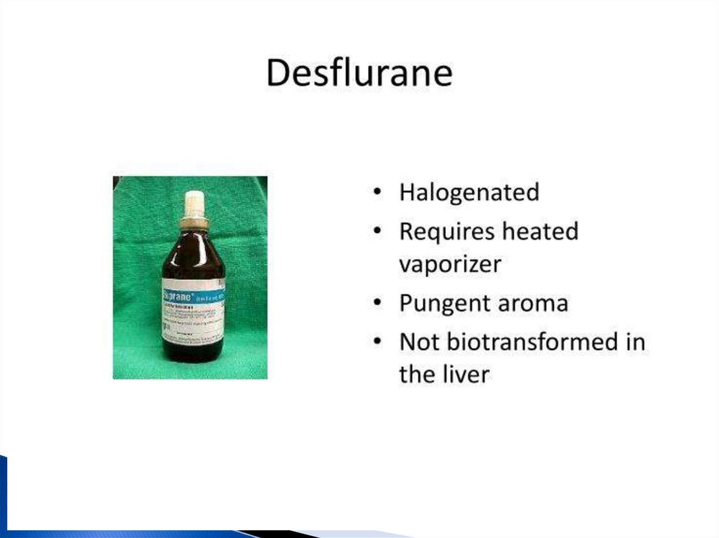
Volatile AnestheticsDesflurane
◦ Pro: Extremely rapid onset/offset
◦ Con: Expensive, Stimulates
catecholamine release, Possibly
increases postoperative nausea and
vomiting, Requires special activetemperature controlled vaporizer due
to high vapor pressure, Irritating so
cannot be used for inhalation induction
87.
88. Commonly Used Medications
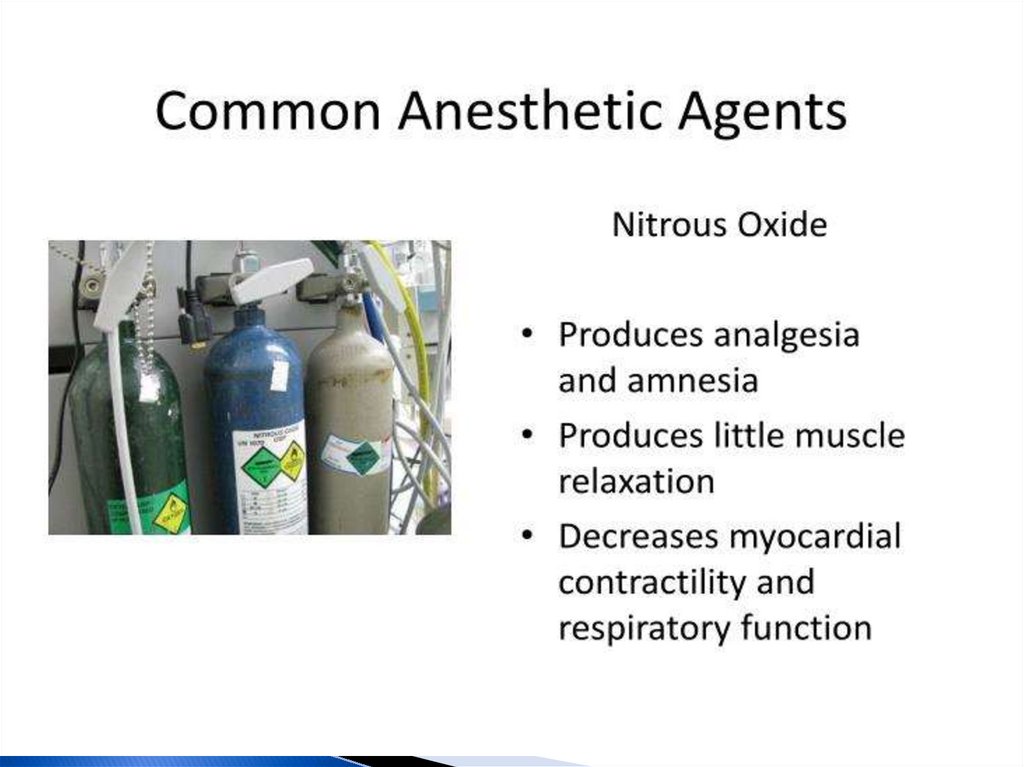
Nitrous OxidePro: Decreases volatile anesthetic
requirement, Dirt cheap, Less
myocardial depression than volatile
agents
Con: Diffuses freely into gas filled
spaces (bowel, pneumothorax, middle
ear, gas bubbles used during retinal
surgery), Decreases FiO2, Increases
pulmonary vascular resistance
89.
90.
91. Commonly Used Medications
Muscle RelaxantsDepolarizing
Succinylcholine inhibits the postjunctional receptor
and passively diffuses off the membrane, while
circulating drug is metabolized by plasma
esterases. Associated with increased ICP/IOP,
muscle fasciculations and postop muscle aches,
triggers MH, increases serum potassium
especially in patients with burns, crush injury,
spinal cord injury, muscular dystrophy or disuse
syndromes.
Rapid and short acting.
92.
NondepolarizingMany different kinds, all ending in “onium” or
“urium”. Each has different site of metabolism,
onset, and duration making choice depend on
specific patient and case. Some examples:
Pancuronium - Slow onset, long duration,
tachycardia due to vagolytic effect. Cisatracurium
- Slow onset, intermediate duration, Hoffman
(nonenzymatic) elimination so attractive choice in
liver/renal disease. Rocuronium - Fastest onset
of nondepolarizers making it useful for rapid
sequence induction, intermediate duration.
93. Emergence
EMERGENCE FROM GENERAL ANESTHESIA1. Reversal of muscle relaxation.
2. Turning off the inhalation agents and nitrous oxide
3. Meeting the extubation criteria
4. Extubation of trachea
5. Transfer of the patient to post anesthesia care unit.
94. Emergence
First, the patient’s neuromuscular blockade mustbe re-assessed, and if necessary reversed and then
rechecked with a twitch monitor.
Next, the patient has to be able to breathe on his
own, and ideally follow commands, demonstrating
purposeful movement and the ability to protect his
airway following extubation. Suction must always
be close at hand, since many patients can become
nauseous after extubation, or simply have copious
oropharyngeal secretions
95. Emergence
. Once the patient is reversed, awake,suctioned, and extubated, care must be
taken in transferring him to the gurney and
oxygen must be readily available for
transportation to the recovery room/PostAnesthesia Care Unit (PACU). Finally,
remember that whenever extubating a
patient, you must be fully prepared to
reintubate if necessary, which means having
drugs and equipment handy.
96. PACU Concerns
The anesthesiologist’s job isn’t over once thepatient leaves the operating room. Concerns
that are directly the responsibility of the
anesthesiologist in the immediate
postoperative period include
nausea/vomiting, hemodynamic stability, and
pain.
97. PACU Concerns
Other concerns include continuingawareness of the patient’s airway and level
of consciousness, as well as follow-up of
intraoperative procedures such as central
line placement and postoperative X-rays to
rule out pneumothorax. A resident and
staff member are usually assigned to the
PACU specifically to follow up on these
concerns, since we frequently have to return
to the OR for subsequent cases, and may
not be available if problems should arise.
98. Commonly Used Medications
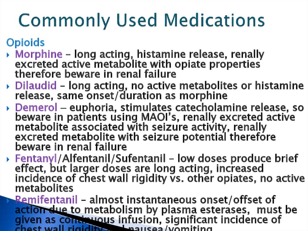
OpioidsMorphine – long acting, histamine release, renally
excreted active metabolite with opiate properties
therefore beware in renal failure
Dilaudid – long acting, no active metabolites or histamine
release, same onset/duration as morphine
Demerol euphoria, stimulates catecholamine release, so
beware in patients using MAOI’s, renally excreted active
metabolite associated with seizure activity, renally
excreted metabolite with seizure potential therefore
beware in renal failure
Fentanyl/Alfentanil/Sufentanil – low doses produce brief
effect, but larger doses are long acting, increased
incidence of chest wall rigidity vs. other opiates, no active
metabolites
Remifentanil – almost instantaneous onset/offset of
action due to metabolism by plasma esterases, must be
given as continuous infusion, significant incidence of
99. Commonly Used Medications
Reversal Agents/AnticholinergicsReversal Agents: all are acetylcholinesterase inhibitors, thereby
allowing more acetylcholine to be available to overcome the
neuromuscular blocker effect at the nicotinic receptor, but also
causing muscarinic stimulation
Neostigmine – shares duration of action with glycopyrrolate (see
below)
Edrophonium – shares duration of action with atropine (see below)
Physostigmine – crosses the BBB, therefore useful for atropine
overdose
.
Anticholinergics: given with reversal agents to block the muscarinic
effects of cholinergic stimulation, also excellent for treating
bradycardia and excess secretions
Atropine – used in conjunction with edrophonium, crosses the BBB
causing drowsiness, so maybe bad at end of surgery for reversal,
some use as premed for all children since they tend to become
bradycardic with intubation and produce copious drool
Glycopyrrolate – used in conjunction with neostigmine, does not
cross the BBB
Questions?
Thank you for listening






 Медицина


