


")


 Медицина
Похожие презентации:


 õppeainesse")

")

Clinical anatomy of the head
1. Clinical anatomy of the head
*2. Topographic anatomy & operative surgery
** TOPOGRAPHIC ANATOMY is a science which
studies relations between organs and tissues
in various regions of the human body
* Operative surgery is a subject about
techniques, ways of surgical operations
3. Cranial bones
*The cranial bones enclose and protect thebrain and associated sensory organs.
*They consist of one frontal, two parietals,
two temporals, one occipital, one sphenoid
and one ethmoid bone.
*
4. SKULL
* The human skull, consisting of 8 cranial and 14facial bones, contains several cavities that
house the brain and sensory organs.
* Each bone of the skull articulates with the
adjacent bones and has diagnostic and
functional processes, surface features and
foramina.
*
5.
* The eight bones of the cranium articulate firmly with oneanother to enclose and protect the brain and sensory organs.
* The 14 fascial bones form the framework for the facial region
and support the teeth.
* Variation in size, shape and density of the facial bones is a
major contributor to the individuality of each human face.
* The facial bones, with the exeption of the mandible, are also
firmly interlocked with one another and the cranial bones.
6. A lateral view of the skull
*7. An anterior view of the skull
*8. Regio fronto-parieto-occipitalis
** 1 - кожа;
* 2 - подкожная клетчатка;
* 3 - сухожильный шлем;
* 4 - диплоическая вена;
* 5 - подапоневротическая клетчатка;
* 6 - надкостница;
* 7 - поднадкостничная клетчатка:
* 8 - пахионовы грануляции;
* 11 - твердая мозговая оболочка:
* 12 - паутинная оболочка;
* 13 - спинномозговая жидкость
*
*
*
подпаутинного пространства;
14 - мягкая мозговая оболочка;
15 - кора полушарий большого мозга;
16 - серповидный отросток твердой
мозговой оболочки; 19 - верхняя
сагиттальная пазуха твердой мозговой
оболочки;
9. Sinuses of dura mater encephali
** 1 - confluens sinuum;
* 2 - sinus rectus;
* 3 - incisura tentorii;
* 4 - v. cerebri magna;
* 6 - sinus petrosus superior sinister;
* 7 - sinus petrosus inferior;
* 8 - falx cerebri;
* 9 - sinus sagittalis superior;
* 10 - sinus sagittalis inferior;
* 15 - sinus intercavernosus anterior;
* 16 - sinus sphenoparietalis;
* 19 - sinus intercavernosus posterior;
* 21 - sinus cavernosus;
* 23 - bulbus v. jugularis internae superior;
* 24 - sinus sigmoideus;
* 25 - tentorium cerebelli; 27 - sinus
transversus.
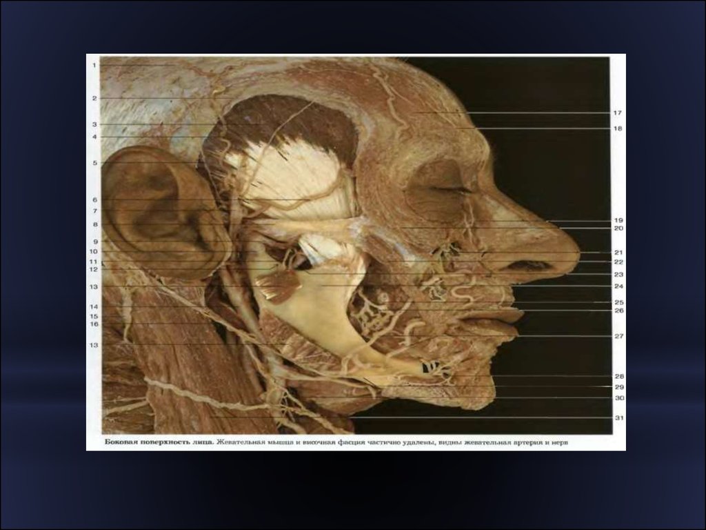
10. Regio temporalis
*11. Regio temporalis
** 1 - r. parietalis a. temporalis
*
*
*
*
*
*
*
*
*
*
*
*
*
*
*
*
superficialis;
2 - r. frontalis a. temporalis
superficialis;
3 - n. supraorbitalis;
4 - a. angularis;
5 - v. facialis;
6 - rr. zygomatici n. facialis; 7 - a.
facialis;
8 - rr. buccales n. facialis;
9 - r. marginalis mandibulae;
10 - r. superior n. transversi colli
(plexus cervicalis);
11 - n. auricularis magnus;
12 - m. masseter;
13 - gl. parotis;
14 - ductus parotideus;
15 - a. transversa faciei;
16 - a. et v. temporales superficiales;
17 - n. auriculotemporalis;
18 - rr. temporofrontales n. facialis.
12. )
** 1 — a. meningea media; 2
— r. frontalis a meningea
media; 3 — r. parietalis a.
meningea media
* Ствол a. meningea media
*
проецируется на точку
пересечения передней
вертикали с нижней
горизонталью, то есть у
середины верхнего края
скуловой дуги.
Лобная ветвь a.
meningea media
проецируется на точку
пересечения передней
вертикали с верхней
горизонталью, а
теменная ветвь — на
точку пересечения этой
горизонтали с задней
вертикалью.
13. Bony-plastic trpanation
** I — выкраивание кожно-апоневротического лоскута; II — в кости после
отслоения надкостницы распатором фрезами сделано три отверстия; III —
промежутки между отверстиями пропиливаются пилкой Джильи с
использованием проводника Поленова; IV - надкостнично-костный лоскут
отвернут, разрезана твердая мозговая оболочка; V — отвернут лоскут
твердой мозговой оболочки, обнажено вещество мозга; VI — после выполнения
оперативного приёма накладывается непрерывный шов на твердую мозговую
оболочку.
14. Regio mastoidea
**
1 - linea temporalis;
* 2 - cellulae mastoideae
(проекция);
* 3 - spina suprameatica;
* 4 - проекция лицевого нерва;
* 5 - crista mastoidea;
* 6 - foramen mastoideum;
* 7 - проекция sinus sigmoideus
15. An inferior view of the skull
*16. The floor of the cranial cavity
*17. Fontanels
*18. The fetal skull: superior view
*19. The fetal skull: sagittal view
*20. Regio lateralis
*21.
22. Parapharyngeal space
**
1 — mandibula; 2 — m. masseter; 3 — ductus
parotideus; 4 — fascia masseterica; 5 — n.
facialis; 6, 10 — nodi lymphatici parotidei
superficiales; 7 — a. facialis, v, retromandibularis
и nodus lymphaticus parotideus profundus; 8 — v.
jugularis externa; 9 — gl. parotis; 11 — m.
digastricus; 12 — m. sternocleidomastoideus; 13
— задний отдел окологлоточного
пространства; 14 — верхняя группа глубоких
шейных лимфатических узлов; 15 — v. jugularis
interna и n. glossopharyngeus; 16 — верхний
шейный узел симпатического ствола, n. vagus
и n. accessorius; 17 — предпозвоночные мышцы
и покрывающая их предпозвоночная фасция;
18 — nodi lymphatici retropharyngeales и
spatium retropharyngeum; 19 — a. carotis interna
и n. hypoglossus; 20 — глоточно-позвоночный
апоневроз (перегородка Шарли); 21 —
шилоглоточный апоневроз; 22 — processus
styloideus с начинающимися от него мышцами;
23 — глоточный отросток околоушной железы;
24 — глоточно-основной апоневроз; 25 —
передний отдел окологлоточного
пространства; 26 — tonsilla palatina; 27 — m.
constrictor pharyngis superior; 28 — m.
pterygoideus medialis.
23.
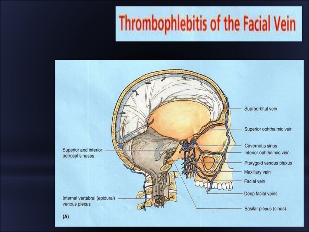
24. Danger triangle of the face
*25.
26.
27.
28.
29. Bones of the orbit
*30. Bones forming the orbit
*31. The lateral wall of the nasal cavity
*32. Bones that enclose the nasal cavity
*33. Radiographs of the skull
*34. Cavities of the scull
*The s k u l lhas several cavities. The cranial cavity is the
largest, with an approximate capacity of 1,300 to 1,350
cc.
*The n a s a l c a v i t y
is formed by both cranial and
facial bones and is partitioned into two chambers by a
nasal septum of bone and cartilage.
*Four sets of p a ra n a s a l s i n u s e s
(sinus maxillaris,
sinus frontalis, sinus sphenoidalis, labyrhintus
ethmoidalis), located within the bones surrounding the
nasal area, communicate via ducts into the nasal cavity.
*
35. Cavities of the scull
*M i d d l ea n d i n n e r - e a r c a v i t i e s are
positioned inferior to the cranial cavity and
house the organs of hearing and balance.
*The two o r b i t s
for the eyeballs are formed
by facial and cranial bones.
*The o ra l , or buccal cavity (mouth), which is
only partially formed by bone, is completely
within the facial region
*