


 Медицина
Похожие презентации:


Osta of lumbar & retroperitoneal space
1. OSTA
TOPIC:- OSTA OF LUMBAR & RETROPERITONEALSPACE
2. TOPGRAPHY & OPERATIVE SURGERY OF LUMBAR & RETROPERITONEUM
TOPGRAPHY & OPERATIVE SURGERYOF LUMBAR & RETROPERITONEUM
CONTENTS:1. TOPOGRAPHY OF LUMBAR REGION AND
RETROPERITONEAL SPACE
2. LAYERED TOPOGRAPHY, PROJECTION VESSELS
AND NERVES
3. CUTS IN PHLEGMON
3. TOPOGRAPHY OF LUMBAR REGION
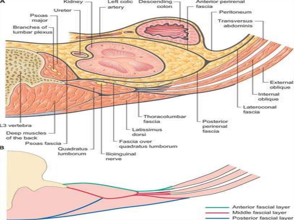
1. BORDERS:• SUPERIOR-12TH Rib• Inferior- iliac crest
Lateral –lesgaft’s line (vertical line passing
through the 11th rib from mid axillary line)
• According to the erector spinae muscle, the
lumbar region is divided into the medial and
lateral departments.
4.
2. LAYERS:• The skin is thick• Subcutaneous tissue: It contains the superficial fascia which divides the fatty
tissue into 2-3 layers
The fat tissue continued in the gluteal region is called
massa adiposa lumboglutealis.
• Properfascia: It forms a sheath for the erector spinae muscle and is called
the fascia thoracolumbalis.\
It is divided into superficial and deep layers
Deep layer is fixed to spinous process
• Muscles: They are divided into the superficial and deep groups
Superficial:- latissmus dorsi
Deep muscles are divided into thr medial and lateral groups
according to the margin of erector spinae muscle.
5.
6.
3. WEAK PLACES:• Petit’s lumbar triangle:i. Borderso Medial:- margin of latissmus dorsi muscle
o Lateral:- nmargin of external oblique muscle
o Inferior:- iliac crest
o Floor:- internal oblique muscle
ii. Clinical importance:o Herniation
o This place contains fat, where abscess and
phlegmones tend to occur.
7.
4.5.
ARTERIAL SUPPLY
Lumbar arteries (branches of abdominal aorta)
VENOUS DRAINAGE:Lumbar veins, then drained into the inferior vena
cava
6. NERVE SUPPLY:• Subcostal nerve
• Posterior branches of the spinal nerve
8.
9. RETROPERITONEAL SPACE
• The retroperitoneal space (retroperitoneum) isthe anatomical space (sometimes a potential space) in
the abdominal cavity behind (retro) the peritoneum. It has
no specific delineating anatomical structures. Organs are
retroperitoneal if they have peritoneum on their anterior
side only. Structures that are not suspended
by mesentery in the abdominal cavity and that lie
between the parietal peritoneum and abdominal wall are
classified as retroperitoneal.
• The retroperitoneum can be further subdivided into the
following:_
• Perirenal space
• Anterior pararenal space
• Posterior pararenal space
10. RETROPERITONEAL STRUCTURES
• Structures that lie behind the peritoneum are termed "retroperitoneal". Organsthat were once suspended within the abdominal cavity by mesentery but
migrated posterior to the peritoneum during the course of embryogenesis to
become retroperitoneal are considered to be secondarily retroperitoneal
organs.
• Primarily retroperitoneal, meaning the structures were retroperitoneal during
the entirety of development:
– urinary
• adrenal glands
• kidneys
• ureter
– circulatory
• aorta
• inferior vena cava
• Secondarily retroperitoneal, meaning the structures initially were suspended
in mesentery and later migrated behind the peritoneum during development
– the duodenum, except for the proximal first segment, which is
intraperitoneal
– ascending and descending portions of the colon (but not the transverse
colon, sigmoid or the cecum)
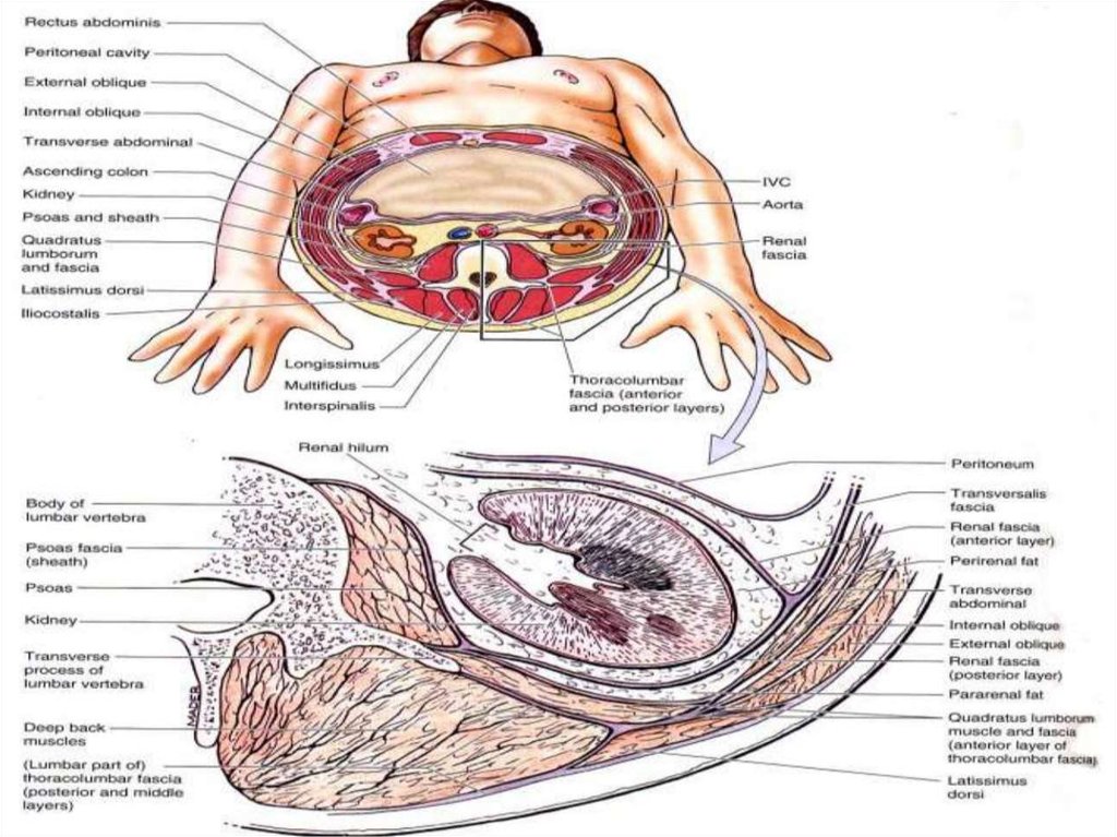
11. FASCIA AND FATTY LAYERS OF RETROPERITONEAL SPACE
12.
13.
14.
VESSELS OF RETROPERITONIUM
ARTERIES
Abdominal Aorta
The abdominal aorta is the principal artery of the abdomen, pelvis and lower limb.
Course
The abdominal aorta is the continuation of the descending thoracic aorta. It begins at the level of T 12, slightly to
the left of midline, posterior to the diaphragm and anterior to the T12 vertebral body. It passes inferiorly,
remaining anterior to the lumbar vertebrae, before dividing into the paired common iliac arteries.
Relations
The relations of the abdominal aorta are:
Left, with the left kidney
Right, with the azygos vein (superiorly), inferior vena cava and right coeliac plexus
Posteriorly, with the vertebral bodies of T12 to L4.
Anteriorly, with the right lobe of the liver, stomach, pancreas and small bowel. The left renal vein passes
anterior to the aorta
Branches
The principal branches of the abdominal aorta are:
The coeliac trunk or axis, a short vessel that contributes to supply of the liver, stomach, pancreas and spleen
The superior mesenteric artery which supplies the small bowel, proximal large bowel, and the proximal
pancreas
The paired renal arteries which supply the kidneys. The right renal artery passes posterior to the inferior vena
cava
The paired gonadal arteries, the course of which varies between men and women.
The inferior mesenteric artery which supplies the descending and sigmoid colon, and the rectum.
Smaller branches include lumbar arteries which supply the vertebrae and spinal canal, and inferior phrenic
branches which supply the diaphragm.
Coeliac Axis / Trunk
The coeliac axis is a 2 cm stub that arises from the anterior aspect of the abdominal aorta, at about T12. It
rapidly divides into numerous branches.
15. VESSELS OF RETROPERITONIUM
• Common Hepatic Artery and BranchesThe common hepatic artery is the larger branch of the coeliac axis, and passes laterally to the porta
hepatis within the lesser omentum. It gives off the gastroduodenal artery as it passes superior to
the pylorus, followed by the right gastric artery which passes back along the lesser omentum to
supply the lesser curvature of the stomach. It continues as the hepatic artery into the porta hepatis,
giving off the cystic artery before dividing into right and left hepatic arteries.
Splenic Artery and Branches
The splenic artery passes to the left in the retroperitoneum. It gives off numerous branches to the
pancreas, which lies inferiorly. It also gives off the left gastroepiploic artery and short gastric
arteries to the lateral greater curvature of the stomach.
Left Gastric Artery
The smallest branch of the coeliac axis, the left gastric passes to the gastro-oesophageal junction,
where it gives of an oesophageal branch. It then passes along the lesser curvature of the stomach
to anastamose with the right gastric
• Superior Mesenteric Artery
The superior mesenteric is the second anterior artery to arise from the abdominal aorta, about
1 cm below the coeliac axis and posterior to the pancreas. It passes inferiorly, laterally and slightly
anteriorly, in front of the uncinate process of the pancreas. The left renal vein passes between this
artery and the aorta, as does the third part of the duodenum. The superior mesenteric gives off
numerous branches to the small bowel and proximal large bowel
16.
• VEINS:• Inferior Vena Cava• The inferior vena cava is the major vessel for the return of blood to the
heart from the abdomen and pelvis. Many abdominal viscera drain via the
portal system to the liver; but hepatic veins still empty into the inferior
vena cava just prior to its entry into the right atrium. The IVC is typically
considered in four parts:
• The long abdominal section which runs from L5 to L1
• The intrahepatic part that lies within the substance of the liver
• The short suprahepatic segment between the liver and the diaphragm
• The short thoracic part that empties into the right atrium
• Portal Vein
• The portal venous system drains blood from the spleen, pancreas and
gastrointestinal tract to the liver, separate to the systemic venous return.
It is not covered in this section
17.
TOPOGRAPHY OF KIDNEYS• MORPHOLOGY:
• kidney has two histologically parts
i.
Cortex &
ii. Medulla
It has 2 poles which are distinguished, namely superior and
inferior poles
It has 2 margins which are distinguished, namely medial and
lateral margins.
FUNCTIONS:
Filtartion
Reabsorption
Excretion
Production of erythropoietin for erythropoiesis
18. TOPOGRAPHY OF KIDNEYS
19.
• SYNTOPY:RIGHT KIDNEY:i. SUPERIOR:- rt. Adrenal gland and liverii. Inferior:- loops of small intestine and right colon
iii. Anterior:-transverse mesocolonnear hilus
iv. Posterior:- psoas major muscle, quadratus lumborum
muscle
v. Medial:- descending part of duodenum
LEFT KIDNEY
i. SUPERIOR:- left adrenal gland, stomach and spleen
ii. Inferior:- loops of sma;ll intestine
iii. Ant.:- transverse mesocolon, pancreas, left colic flexure and
loops of small intestine
iv. Posterior:- psoas major muscle, quadratus lumborum
muscle, transverse abdominis muscle
v. Lateral:- spleen and descending colon.
20.
• ARTERIAL SUPPLY:Renal arteryRenal vein, then drained into inf. Vena cava
Lymphatics:- para aortic and coeliac lymph nodes
Nerve supply:Renal plexus
Sympathetic fibers from T10th-L1
Parasympathetic fibers:- vagus nerve
21.
TOPOGRAPHY OF ADRENAL GLANDSIT HAS 2 PARTS CORTEX AND MEDULLA
FUNCTIONS:• Secretion of glucocorticoids, mineralocorticoids
and androgens in the cortex
• Secretion of adrenaline and noradrenaline in
medulla
HOLOTOPY:- they are projected rt. And lft.
Hypochondriac regions
SKELETOPY:- 11th and 12th thoracic vertebra
22. TOPOGRAPHY OF ADRENAL GLANDS
23.
• ARERIAL SUPPLY:• Superior suprarenal artery(br. Of inf. Phrenic A.)• Middle and suprarenal artery(branches of
abdominal aorta and renal artery)
• VENOUS DRAINAGE:• right and left suprarenal gland(drained into inf.
Vena cava and left renal vein then into inf. Vena
cava)
• NERVE SUPPLY:• Suprarenal nerve plexus
24.
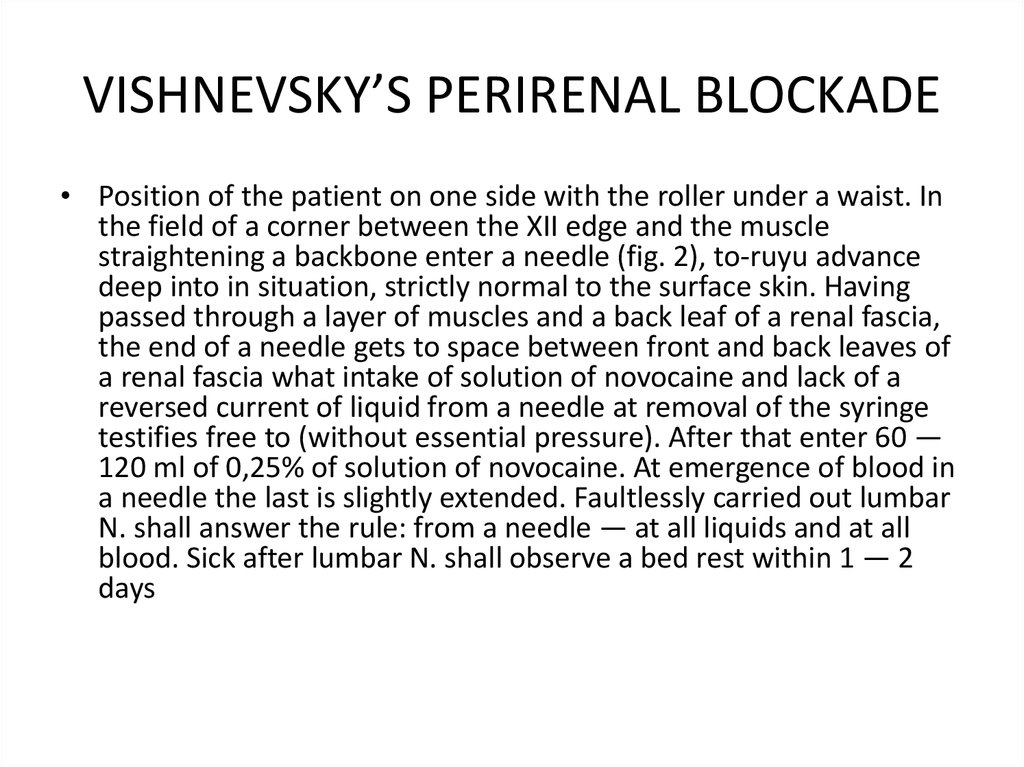
VISHNEVSKY’S PERIRENAL BLOCKADE• Position of the patient on one side with the roller under a waist. In
the field of a corner between the XII edge and the muscle
straightening a backbone enter a needle (fig. 2), to-ruyu advance
deep into in situation, strictly normal to the surface skin. Having
passed through a layer of muscles and a back leaf of a renal fascia,
the end of a needle gets to space between front and back leaves of
a renal fascia what intake of solution of novocaine and lack of a
reversed current of liquid from a needle at removal of the syringe
testifies free to (without essential pressure). After that enter 60 —
120 ml of 0,25% of solution of novocaine. At emergence of blood in
a needle the last is slightly extended. Faultlessly carried out lumbar
N. shall answer the rule: from a needle — at all liquids and at all
blood. Sick after lumbar N. shall observe a bed rest within 1 — 2
days
25. VISHNEVSKY’S PERIRENAL BLOCKADE
• INDICATIONS:• it has to be regarded only as one of to lay down. thefactors applied in a complex with others.
• The main indications to use. inflammatory processes,
disturbance of a tone of muscles of bodies, the
pathology which is followed by pain are (injuries,
wounds, an obliterating endarteritis, hepatic and renal
colic, etc.). By Vishnevsky, under the influence of
inflammatory process in a stage of serous treatment of
fabrics can be suspended, in a stage of abscessing — is
quicker delimited and allowed, in an infiltrative stage,
and also at subacute , forms positive trophic shifts are
observed, destructive processes quite often break and
replaced by recovery. At disturbance of a tone of
bodies (a gut, a uterus) . promotes permission of
spasms, on the one hand, and to increase of a tone at
an atony — with another.
26.
NEPHROPEXYNephroptosis (also called floating kidney or renal ptosis) is an abnormal condition
in which the kidney drops down into the pelvis when the patient stands up. It is
more common in women than in men.
Operation is carried out in nephroptosis
INDICATIONS:SIGNIFICANT DISPLACEMENT OF KIDNEY
Haemorrhage
Pyelonephritis
Renal hypertension
PROCEDURE:THE pelvis and ureter are examined to exclude any organic obstruction to the
urinary outflow.
The kidney is stiched against the quadratus lumborum muscle
The kidney is then placed to the lateral part of the muscle with 3 stiches
A sheet of polyvinyl alcohol sponge is placed between the kidney and muscle to
promote adhesion.
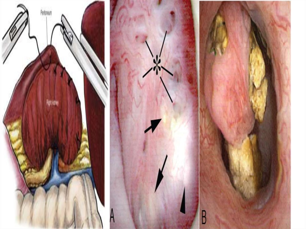
27. NEPHROPEXY
• Potential complications of nephropexy includethe following :
• Urinary tract infection.
• Uncorrected ptotic kidney.
• Retroperitoneal hematoma.
• Bowel injury or puncture during trocar
placement.
• Conversion to open nephropexy.
• Muscle paresthesia.
• Genitofemoral nerve injury or entrapment