


 Биология
Похожие презентации:

")

 and blood, muscle, nervous")


Urinary system
1. МОЧЕВЫВОДЯЩАЯ СИСТЕМА
Лектор: д.м.н., профессорСырцов В.К.
2.
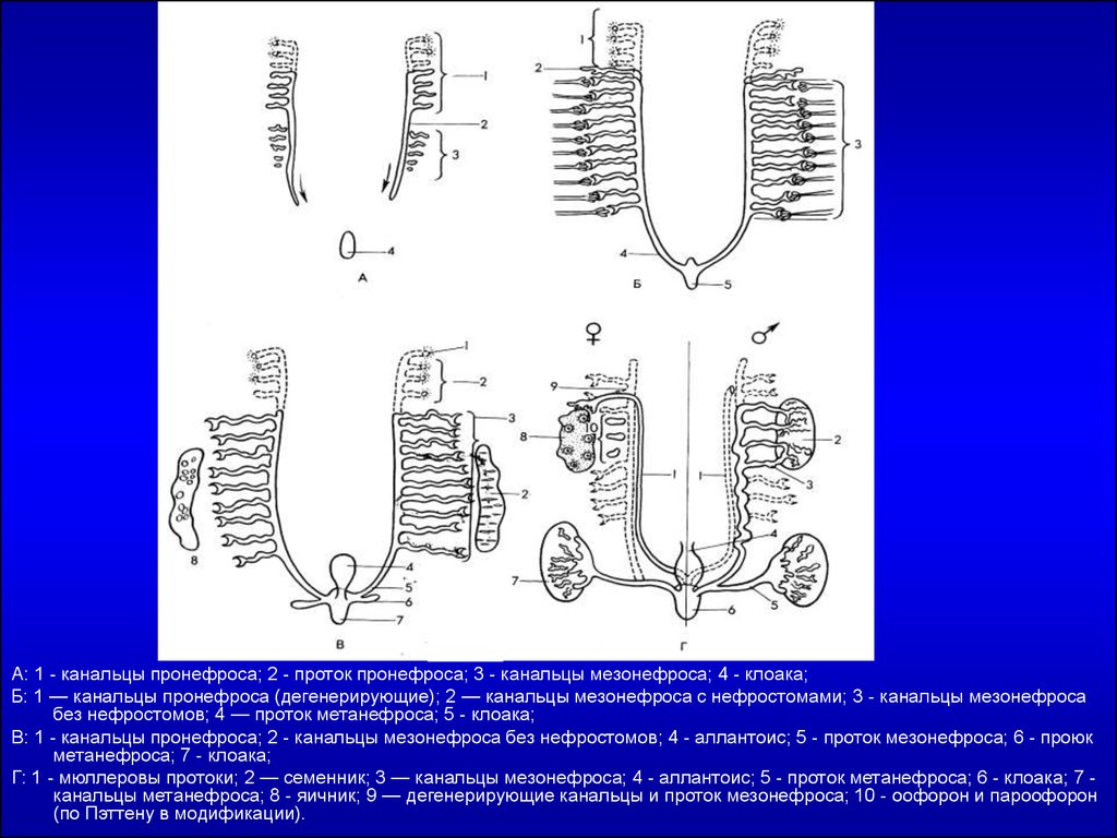
А: 1 - канальцы пронефроса; 2 - проток пронефроса; 3 - канальцы мезонефроса; 4 - клоака;Б: 1 — канальцы пронефроса (дегенерирующие); 2 — канальцы мезонефроса с нефростомами; 3 - канальцы мезонефроса
без нефростомов; 4 — проток метанефроса; 5 - клоака;
В: 1 - канальцы пронефроса; 2 - канальцы мезонефроса без нефростомов; 4 - аллантоис; 5 - проток мезонефроса; 6 - проюк
метанефроса; 7 - клоака;
Г: 1 - мюллеровы протоки; 2 — семенник; 3 — канальцы мезонефроса; 4 - аллантоис; 5 - проток метанефроса; 6 - клоака; 7 канальцы метанефроса; 8 - яичник; 9 — дегенерирующие канальцы и проток мезонефроса; 10 - оофорон и пароофорон
(по Пэттену в модификации).
3.
4.
5.
6. RENAL CORTEX
Glomerular capsuleGlomerulus
Distal convoluted tubule
Distal straight segment
Proximal convoluted tubule
7.
Photomicrograph of renal cortex.A macula densa is clearly seen (arrow) at the vascular pole of a renal
corpuscle. Picrosirius-hematoxylin (PSH) stain. Medium magnification.
8.
The renal corpuscle.The upper part of the
drawing shows the vascular
pole, with afferent and
efferent arterioles and the
macula densa. Note the
juxtaglomerular cells in the
wall of the afferent arteriole.
Podocyte processes cover
the outer surfaces of the
glomerular capillaries; the
part of the podocyte
containing the nucleus
protrudes into the urinary
space. Note the flattened
cells of the parietal layer of
Bowman’s capsule. The
lower part of the drawing
shows the urinary pole and
the proximal convoluted
tubule.
9.
Photomicrograph of an afferent arteriole entering a renal corpuscle.The wall of this arteriole shows the renin-producing juxtaglomerular
(JG) cells (broken line). At the upper right is a distal convoluted tubule
(DCT) with many elongated mitochondria. PT stain. High magnification.
10.
Schematic representation of a glomerular capillary with the visceral layer ofBowman’s capsule (formed of podocytes). In this capillary, endothelial cells are
fenestrated, but the basal lamina on which they rest is continuous. At left is a
podocyte shown in partial section. As viewed from the outside, the part of the
podocyte that contains the nucleus protrudes into the urinary space. Each
podocyte has many primary processes, from which arise an even greater number
of secondary processes that are in contact with the basal lamina.
11.
12.
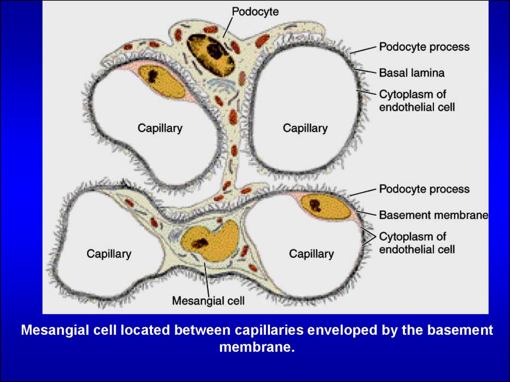
Mesangial cell located between capillaries enveloped by the basementmembrane.
13.
14.
Renal cortex section showing a proximal convoluted tubule (PCT) withits large cuboidal cells presenting a brush border formed by numerous
microvilli. Distal convoluted tubules (DCT) are also present.
15.
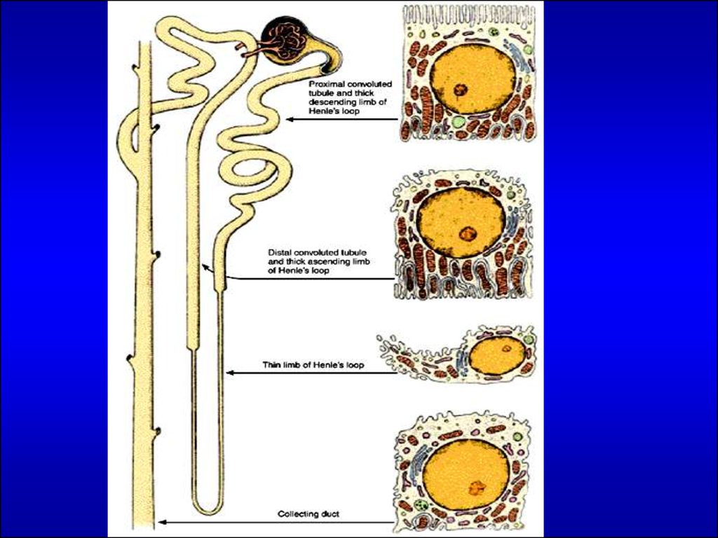
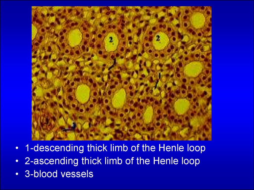
• 1-descending thick limb of the Henle loop• 2-ascending thick limb of the Henle loop
• 3-blood vessels
16. Collecting tubule
Basalmembrane
prysmatic
Nucle
us
Cytoplasma
Collecting tubule
17.
18. THE URINARY BLADDER
Transitional epitheliumSubmucosa layer
Muscle layer
THE URINARY BLADDER
19. Transitional epithelium
20.
Compare the structure of thetransitional epithelium when the
urinary bladder is empty (A) or
full (B). When the bladder is full,
the capacity of epithelial cells to
slide upon one another reduces
the thickness of the epithelium.
As a result, the interior surface
of the bladder increases. In B,
note the thin strands of collagen
fibers separating bundles of
smooth muscle cells. PSH stain.
Medium magnification.