

")


:")
:")


")


 Muscarinic cholinergic receptor")


agonist = mimic or block the NT receptor (Ant)agonist = mimic or block secretion, reuptake or degradation of NT")
agonists")


 Медицина
 Биология
Похожие презентации:


Efferent peripheral NS: the autonomic votor divisions
1. Efferent Peripheral NS: The Autonomic Motor Divisions
2.
Autonomic nervous system: A part ofthe nervous system that regulates key
involuntary functions of the body,
including the activity of the heart
muscle; the smooth muscles, including
the muscles of the intestinal tract; and
the glands.
3. Review (again)
4. Autonomic Nervous System
Responsible for control of involuntary orvisceral bodily functions
cardiovascular cardiovascular
respiratory respiratory
digestive digestive
urinary urinary
reproductive functions
Key role in the bodies response to
stress
5.
6. Overview: The Parts of a Reflex
7. Autonomic Targets
Smooth MuscleCardiac Muscle
Exocrine Glands
Some Endocrine glands
Lymphoid Tissue
Adipose
8. Divisions of ANS
SympatheticParasympathetic
Metasympathetic
9.
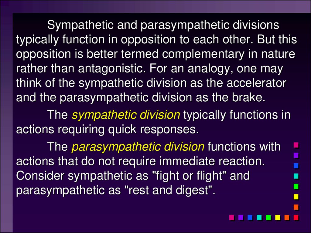
Sympathetic and parasympathetic divisionstypically function in opposition to each other. But this
opposition is better termed complementary in nature
rather than antagonistic. For an analogy, one may
think of the sympathetic division as the accelerator
and the parasympathetic division as the brake.
The sympathetic division typically functions in
actions requiring quick responses.
The parasympathetic division functions with
actions that do not require immediate reaction.
Consider sympathetic as "fight or flight" and
parasympathetic as "rest and digest".
10. ANS
2 divisions:Sympathetic
“Fight or flight”
“E” division
Exercise, excitement,
emergency, and
embarrassment
Parasympathetic
“Rest and digest”
“D” division
Digestion, defecation,
and diuresis
11.
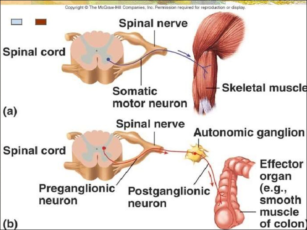
1. The autonomic nervous system (ANS) isan involuntary motor (efferent) system.
2. Autonomic nerves are typically composed
of a two-neuron chain. One neuron has its
cell body in the central nervous system while
the other is outside the CNS.
12. Autonomic pathway: Two Efferent Neurons in Series
Two EfferentNeurons in Series
Autonomic pathway:
Preganglionic neuron cell body in CNS
Synapse in autonomic ganglion outside CNS (often
divergence!)
Postganglionic neurons
target cells
N1
N2
13.
14.
3. Although “involuntary”, the autonomicnervous system is regulated by higher
centers. The best known of these centers
is the hypothalamus which has
descending projections to cell bodies of
the preganglionic neurons. Other areas
of the central nervous system affect the
activities of the hypothalamus.
15.
4. The autonomic nervous system consists oftwo divisions:
a) the sympathetic (or thoracolumbar)
division in which the preganglionic cells are
located in the thoracic and first two lumbar
segments of the spinal cord.
b) the parasympathetic (or craniosacral)
division in which the preganglionic neurons
are located in the brain stem and in sacral
(S2 - S4) segments of the spinal cord.
16. Sympathetic “Fight or flight” “E” division Exercise, excitement, emergency, and embarrassment
17.
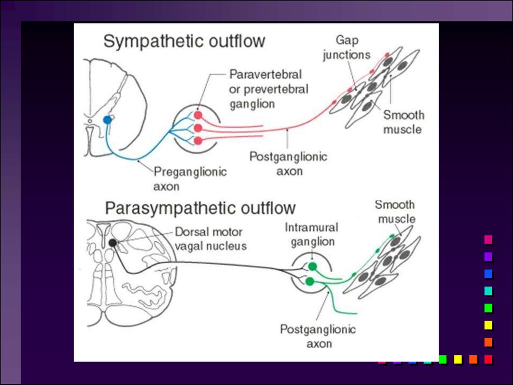
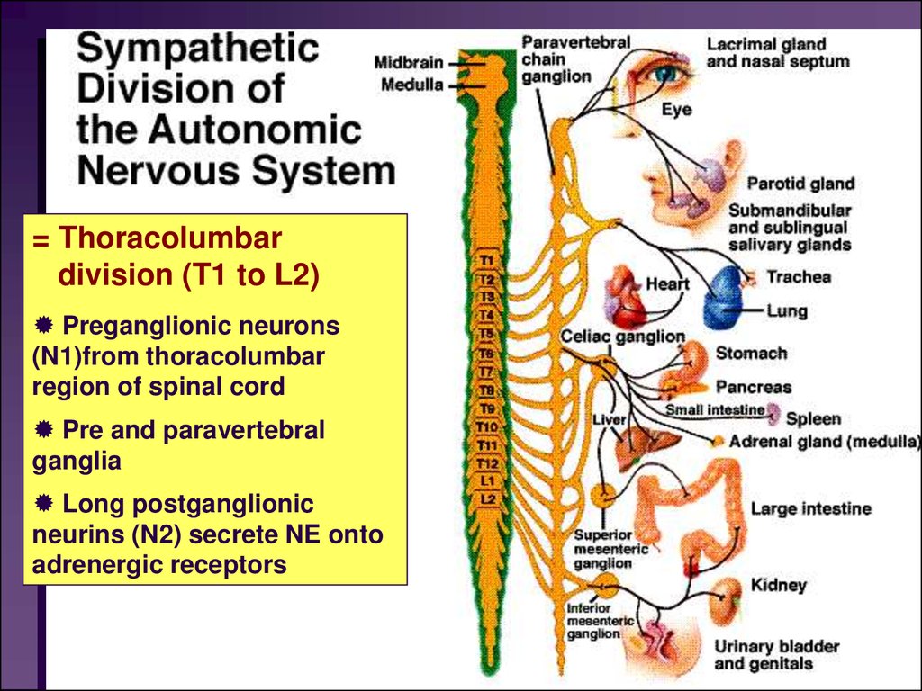
= Thoracolumbardivision (T1 to L2)
Preganglionic neurons
(N1)from thoracolumbar
region of spinal cord
Pre and paravertebral
ganglia
Long postganglionic
neurins (N2) secrete NE onto
adrenergic receptors
18. Sympathetic (preganglionic ):
1. The cell bodies giving rise to preganglionicneurons (N1) are located in the
intermediolateral column (lateral horn) of the
gray matter in spinal cord segments T1 through
L2.
2. Preganglionic fibers leave the spinal cord
with the ventral roots of spinal nerves arising
from cord segments T1 - L2.
19. Sympathetic (postganglionic ):
1. The cell bodies giving rise to postganglionicneurons (N2) are located in the paravertebral
ganglia (sympathetic trunk (vertebral chain)).
2. Prevertebral (collateral) ganglia: celiac,
superior mesenteric, inferior mesenteric,
aorticorenal and renal.
.
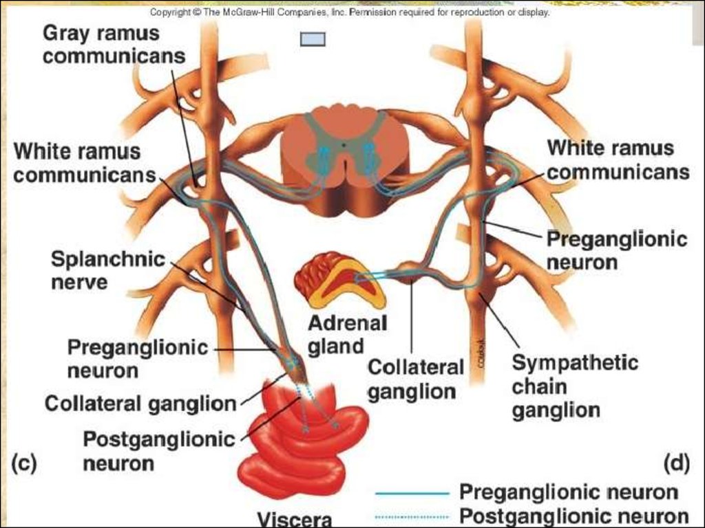
20. Sympathetic ganglia
Sympathetic chain ganglia(paravertebral ganglia) – preganglionic
fibers of the sympathetic NS that carry
motor impulses to the body wall or
thoracic cavity synapses in chain
ganglia
Collateral ganglia (prevertebral ganglia)
– group of second order neurons that
innervate organs in the abdominopelvic
region
21. Sympathetic Trunk Ganglia
Located on both sides of the vertebralcolumn
Linked by short nerves into sympathetic
trunks
Joined to ventral rami by white and gray
rami communicantes
Right and left sympathetic trunks extend from
the base of the skull to the region of the coccyx;
at their distal ends, the right and left trunks are
fused.
22. Prevertebral Ganglia
Unpaired, not segmentally arrangedOccur only in abdomen and pelvis
Lie anterior to the vertebral column
Main ganglia
Celiac, superior mesenteric, inferior
mesenteric, inferior hypogastric ganglia
23. The Organization of the Sympathetic Division of the ANS
24. Sympathetic Pathways to Periphery
CopyrightFigure 15.9
25. Postganglionic fibers
Rejoin spinal nerves and reach theirdestination by way of the dorsal and ventral
rami
Those targeting structures in the thoracic
cavity form sympathetic nerves
Go directly to their destination
26. Sympathetic Pathways to Thoracic Organs
Copyright27. Abdominopelvic viscera
Sympathetic innervation via preganglionicfibers that synapse within collateral ganglia
Splanchic nerves – carry fibers that synapse in
collatheral ganglia
28. Abdominopelvic viscera
Celiac ganglionSuperior mesenteric ganglion
Innervates stomach, liver, gall bladder,
pancreas, spleen
Innervates small intestine and initial portion of
large intestine
Inferior mesenteric ganglion
Innervates kidney, urinary bladder, sex organs,
and final portion of large intestine
29. Sympathetic Pathways to the Abdominal Organs
Copyright30. Sympathetic Pathways to the Pelvic Organs
Copyright31. Other important considerations:
ganglion cells are usually located at somedistance from the effectors. Accordingly,
postganglionic sympathetic fibers are usually
long fibers.
Acetylcholine (Ach) - pre-ganglionic ganglionic Neurotransmitter
Norepinephrine (NE) - post-ganglionic ganglionic Neurotransmitter
32. Sympathetic Division
A single sympathetic preganglionic fiberhas many axon collaterals and may
synapse with 20 or more postganglionic
neurons.
The postganglionic axons typically
terminate in several visceral effectors
and therefore the effects of sympathetic
stimulation are more widespread than
the effects of parasympathetic
stimulation.
33. Sympathetic Variosities
34. Effects of Sympathetic Division
cardiac output increasesSA node: heart rate (chronotropic) β1, : ↑cardiac
muscle: contractility (inotropic) β1 ↑conduction at AV
node β1 : increases
vascular smooth muscle: α = contracts; β2 = relaxes
smooth muscles of bronchioles β2: relaxes;
pupil of eye α1: relaxes
ciliary muscle β2 : relaxes
smooth muscles of GI tractα, β2: relaxes
sphincters of GI tract α1: contracts
glands of GI tract inhibits
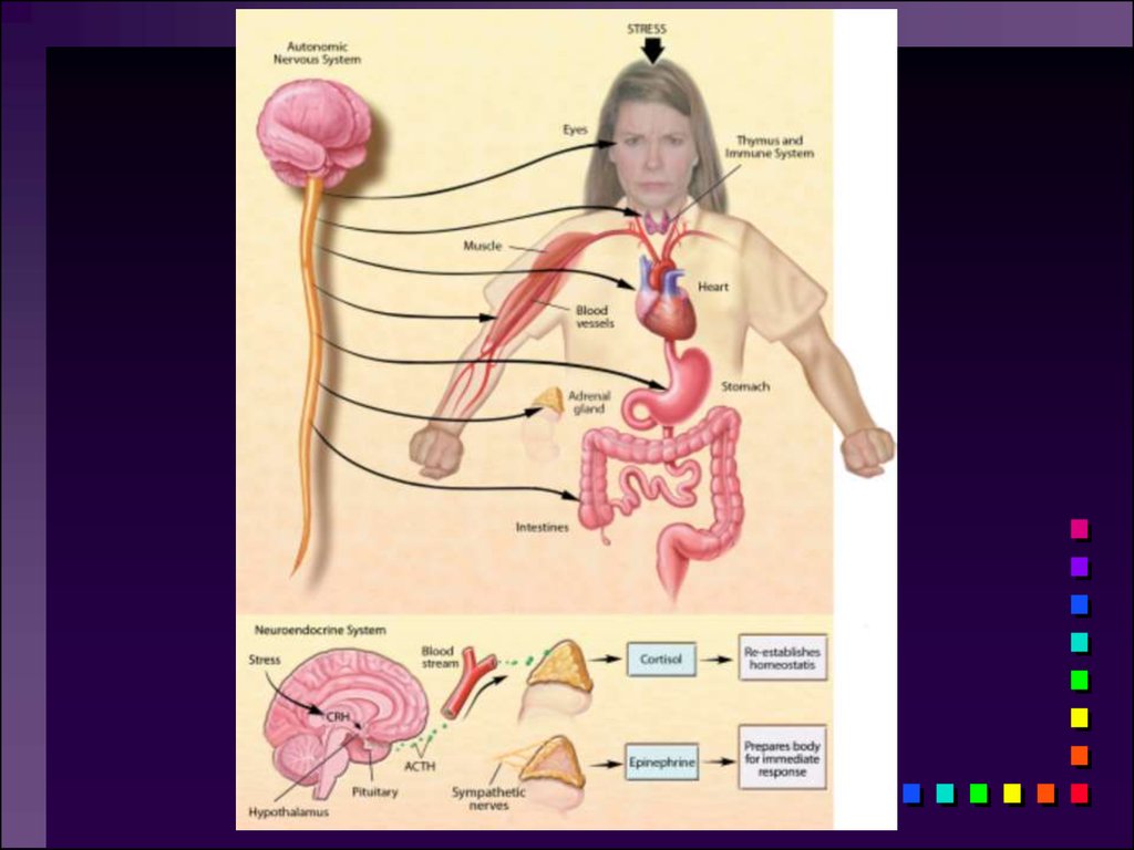
35. THE STRESS REACTION
A stressful situation activates three majorcommunication systems in the brain that regulate
bodily functions.
The first of these systems is the voluntary nervous
system, which sends messages to muscles so that
we may respond to sensory information.
The second communication system is the
autonomic nervous system.
The brain’s third major communication process is
the neuroendocrine system, which also maintains
the body’s internal functioning.
36. THE STRESS REACTION
When stress occurs, the sympathetic nervoussystem is triggered. Norepinephrine is released by
nerves; epinephrine and norepinephrine is secreted
by the adrenal glands. By activating receptors in
blood vessels and other structures, these substances
ready the heart and working muscles for action.
Acetylcholine is released in the
parasympathetic nervous system, producing calming
effects. The digestive tract is stimulated to digest a
meal, the heart rate slows, and the pupils of the eyes
become smaller. The neuroendocrine system also
maintains the body’s normal internal functioning.
37.
38.
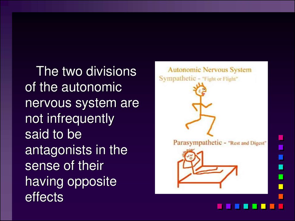
The two divisionsof the autonomic
nervous system are
not infrequently
said to be
antagonists in the
sense of their
having opposite
effects
39. Homeostasis and the Autonomic Division
BP, HR, Resp., H2O balance, Temp. . .Mostly dual reciprocal innervation
i.e., agonist/antagonist or excitatory/inhibitory
Sympathetic:
AKA Thoracolumbar
flight-or-fight
Parasympathetic:
AKA Craniosacral
rest and digest
40. Other important considerations:
ganglion cells are usually located at somedistance from the effectors. Accordingly,
postganglionic sympathetic fibers are usually
long fibers.
41.
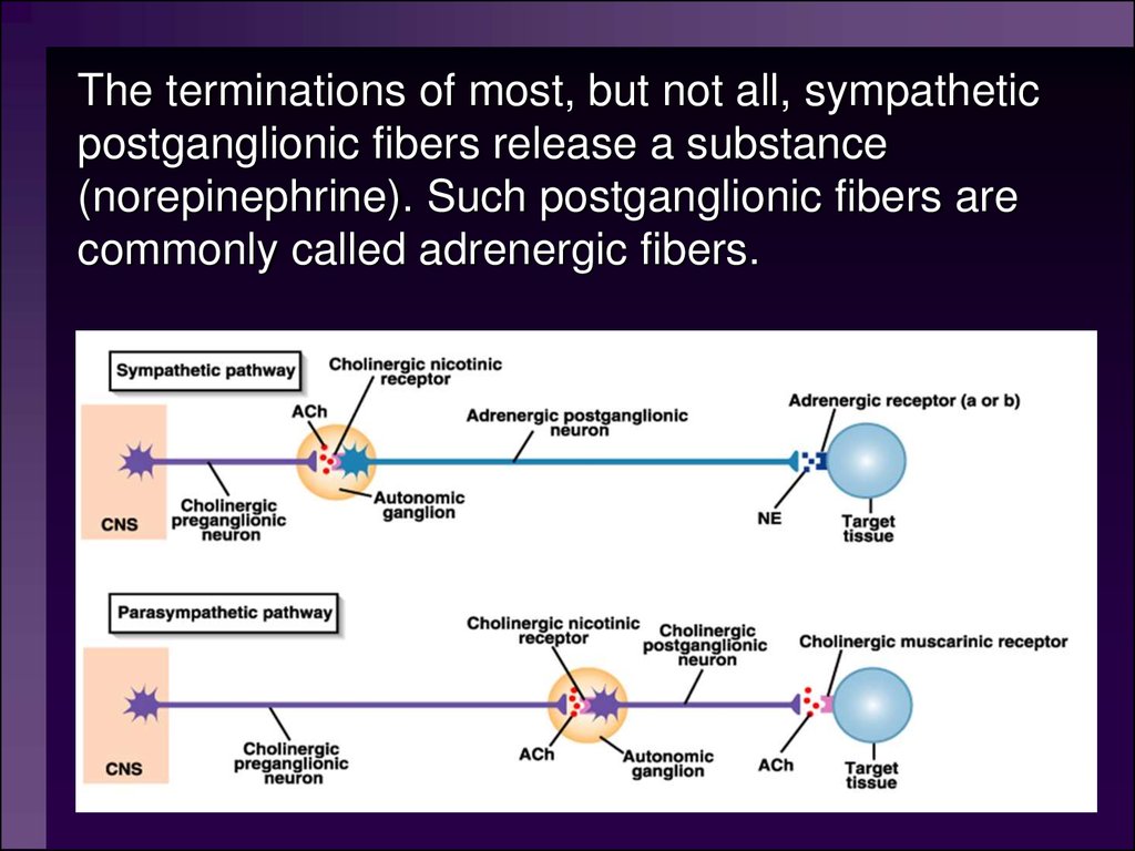
The terminations of most, but not all, sympatheticpostganglionic fibers release a substance
(norepinephrine). Such postganglionic fibers are
commonly called adrenergic fibers.
42.
43.
The effects elicited by theaction of the sympathetic
division of the ANS are
typically effects useful in
“fight or flight”. These
include dilation of the
pupil, increase in heart
rate, elevation of blood
pressure, diversion of
blood from the alimentary
tract to skeletal muscles,
etc.
44. Parasympathetic “Rest and digest” “D” division Digestion, defecation, and diuresis
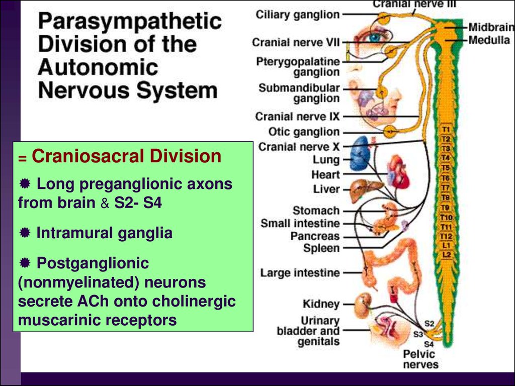
45. Parasympathetic: Craniosacral or rest and digest Center of parasympathetic division the ANS
Has preganglionic cell bodies (N2) in themidbrain and brainstem and in sacral segments
2, 3 and 4 of the spinal cord.
The fibers of cells in the midbrain and brainstem
are in the oculomotor (III), facial (VII),
glossopharyngeal (IX), and vagus (X) nerves.
They innervate smooth muscles of the eye (III),
lacrimal and salivary glands (VII and IX), and
smooth muscles of the thoracic and abdominal
viscera (X).
46. The Organization of the Parasympathetic Division of the ANS
47. The Distribution of Parasympathetic Innervation
48.
49.
= Craniosacral DivisionLong preganglionic axons
from brain & S2- S4
Intramural ganglia
Postganglionic
(nonmyelinated) neurons
secrete ACh onto cholinergic
muscarinic receptors
50. Parasympathetic: Craniosacral or rest and digest Center of parasympathetic division the ANS
The cell bodies giving rise to postganglionicneurons (N2) are located in the Intramural
ganglia.
51.
The ganglion cells of the parasympatheticsystem are located in or on the wall of the
organs supplied or in specific ganglia located
near the organs supplied. Hence the
postganglionic fibers are short.
Except for the vagus nerves, the area of
distribution of parasympathetic nerves is
somewhat limited. The number of synaptic
connections is smaller than in the sympathetic
division. Accordingly, the effects of the
parasympathetic division tend to be local rather
than widespread.
52.
53.
Most postganglionic parasympatheticfibers release acetylcholine at their
terminations. These fibers are, hence,
often called cholinergic fibers. They
may also release a variety of peptides
that influence smooth muscle activity.
54. Summary: Pre- & Postganglionic Parasympathetic Neurons Release ACh
Summary: Pre- & PostganglionicParasympathetic Neurons Release ACh
N1
N2
nicotinic
muscarinic
Receptors
55. Neurotransmitters and parasympathetic functions
All parasympathetic fibers release AChShort-lived response as ACH is broken down
by AChE and tissue cholinesterase
56. Parasympathetic (muscarinic)
cardiac output M2: decreasesSA node: heart rate (chronotropic) M2: decreases
cardiac muscle: contractility (inotropic) M2: decreases
(atria only)
conduction at AV node M2: decreases
smooth muscles of bronchioles M3: contracts
pupil of eye M3: contracts
ciliary muscle M3: contracts
salivary glands: secretions stimulates watery secretions
GI tract motility M1, M3: increases
smooth muscles of GI tract M3: contracts
sphincters of GI tract M3: relaxes
glands of GI tract M3: secretes
57. Parasympathetic activation
Effects produced by the parasympatheticdivision
relaxation
food processing
energy absorption
58.
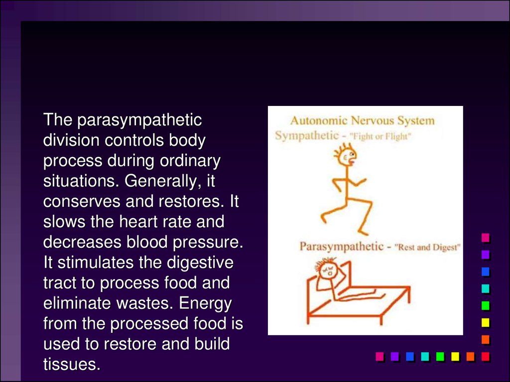
The parasympatheticdivision controls body
process during ordinary
situations. Generally, it
conserves and restores. It
slows the heart rate and
decreases blood pressure.
It stimulates the digestive
tract to process food and
eliminate wastes. Energy
from the processed food is
used to restore and build
tissues.
59. Most Common Autonomic NTs:
Acetylcholine (ACh)ACh neurons & ACh receptors are called
cholinergic (nicotinic or muscarinic). Located
at autonomic preganglionic & parasympathetic postganglionic synapses
Norepinephrine (NE)
NE neurons & receptors are called (nor) adrenergic
( and ). Located at sympathetic
postganglionic synapses
Fig 11-7
60. NTs of Autonomic NS
Compare to Fig 11-7N1
N2
N1
N2
and β
61. Neuroeffector Junction
= Synapse betweenpostganglionic cell and target
Most are different from model
synapse (compare to Fig 8-20,
p. 270)
ANS synapse: axon has
varicosities containing
neurotransmitter
May supply many cells,
resulting in less specific
communication
Synthesis of NT is in the
varicosity
Fig 11-8
62. Summary: Pre- & Postganglionic Parasympathetic Neurons Release ACh
Summary: Pre- & PostganglionicParasympathetic Neurons Release ACh
N1
N2
nicotinic
muscarinic
Receptors
63. Two Types of Cholinergic Receptors: Nicotinic and Muscarinic
1) Nicotiniccholinergic
receptor
2.
Nicotine = agonist
In autonomic ganglia & somatic NS
3.
Directly opens a Na+ & K+ channel: ?
4.
Curare = antagonist
1.
64.
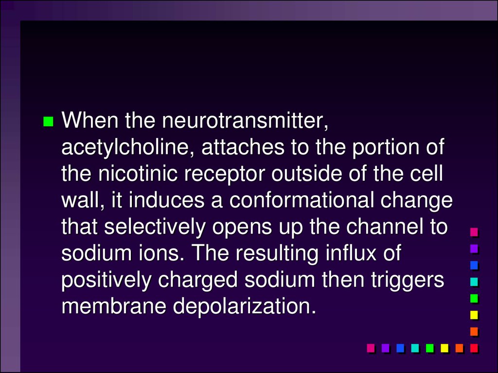
When the neurotransmitter,acetylcholine, attaches to the portion of
the nicotinic receptor outside of the cell
wall, it induces a conformational change
that selectively opens up the channel to
sodium ions. The resulting influx of
positively charged sodium then triggers
membrane depolarization.
65. 2) Muscarinic cholinergic receptor
Muscarine = agonistAmanita muscarina
Found in neuro-effector junctions of
parasympathetic branch
G-protein coupled mechanisms
Atropine = antagonist
N1
N2
66. Muscarinic ACh are G-protein Mediated Receptor Mechanism of Sweat Glands:
Also some 2nd messenger mechanisms67. Note on G-Proteins:
Many functions of the nervous system (e.g.,memory) require prolonged changes in
neurons after the initial neurotransmitter is
gone. Ligand-gated channels (such as those
found in nicotinic receptors) are not suitable
for this because the channels close in
milliseconds. Prolonged changes can be
achieved, however by activating G-proteins
inside the post-synaptic neuron. It is then the
G-proteins that trigger the prolonged effects.
68. Adrenergic Receptors
Found in neuroeffector junctions ofsympathetic branch
G protein linked, with various 2nd mess. Mech
NT is NE
α- and β- Receptors
69. NE Action
Sympathetic Receptorsα Receptors:
•NT is NE
•(most common) Excitation [Ca2+] In
muscle contraction or secretion by exocytosis.
• Inhibition of GI tract and pancreas
70. Sympathetic Receptors
- Receptors Clinically more important1 Excitation heart ([E] = [NE])
“ - blockers” = Antagonists (e.g.: Propranolol)
2 usually inhibitory: smooth muscle relaxation of some
blood vessels and bronchioles ([E] > [NE])
3 Adipose; [NE]>[E]
“ -blockers” = Antagonists (e.g.: Propranolol)
71.
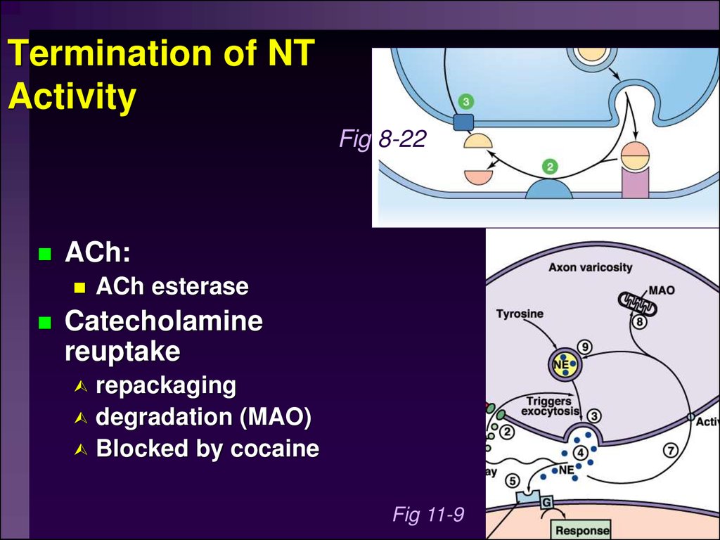
Termination of NTActivity
Fig 8-22
ACh:
ACh esterase
Catecholamine
reuptake
repackaging
degradation (MAO)
Blocked by cocaine
Fig 11-9
72. Termination of NT Activity
Somatic Motor DivisionPathway consists of single
neuron from CNS to target
Neuromuscular junction:
nicotinic cholinergic receptors
Similar to synapse; post –
synaptic membrant called Motor
End Plate
Recall Motor Unit
Always excitatory muscle
contracts
All Ach mediated
Degraded by Ach esterase
Fig 11-13
73. Somatic Motor Division
Myasthenia gravisMG: Antibodies block, alter, or destroy the
receptors for acetylcholine at the neuromuscular
junction
74.
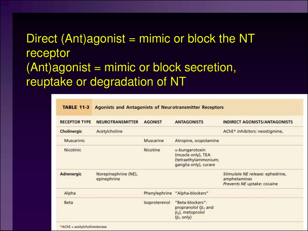
Direct (Ant)agonist = mimic or block the NTreceptor
(Ant)agonist = mimic or block secretion,
reuptake or degradation of NT
75. Direct (Ant)agonist = mimic or block the NT receptor (Ant)agonist = mimic or block secretion, reuptake or degradation of NT
Direct AntagonistsAtropine muscarinic
Curare nicotinic
Propranolol 1 and 2
Metoprolol 1
Strychnos Toxifera
(Curare) from
Koehler's MedicinalPlants 1887
76.
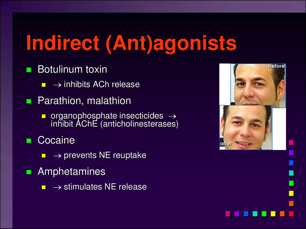
Indirect (Ant)agonistsBotulinum toxin
Parathion, malathion
organophosphate insecticides
inhibit AChE (anticholinesterases)
Cocaine
inhibits ACh release
prevents NE reuptake
Amphetamines
stimulates NE release
77. Indirect (Ant)agonists
Comparison of the twodivisions
Important physiological and functional
differences exist
78. Comparison of the two divisions
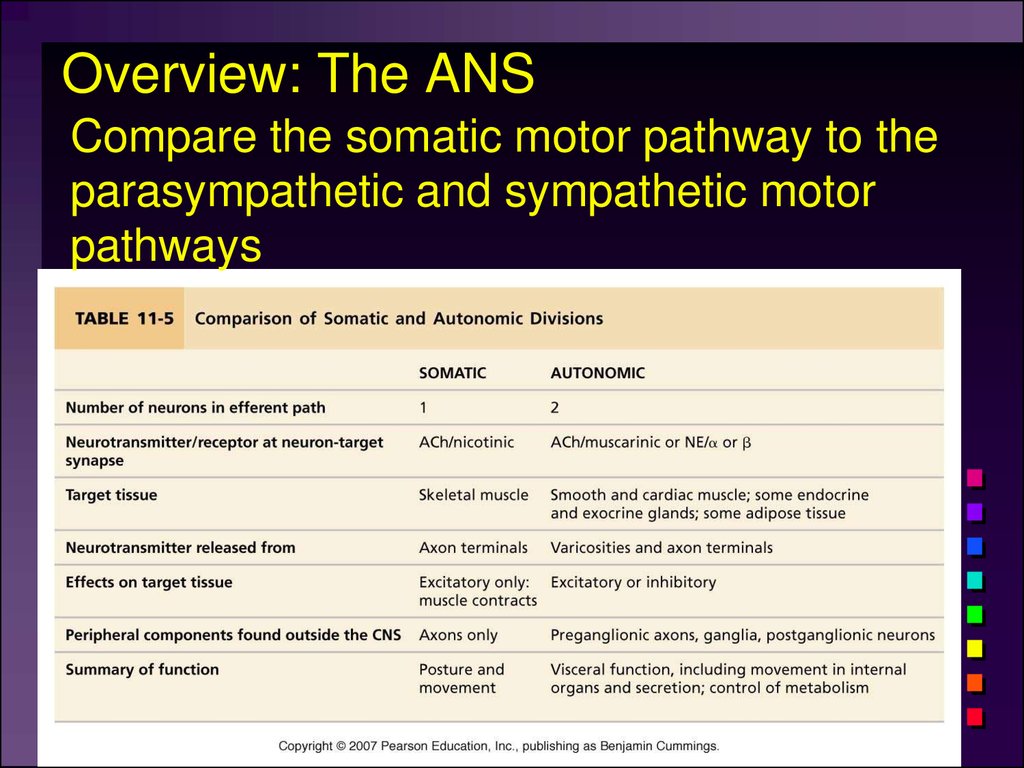
Overview: The ANSTable 11-4
79.
Overview: The ANSCompare the somatic motor pathway to the
parasympathetic and sympathetic motor
pathways
80.
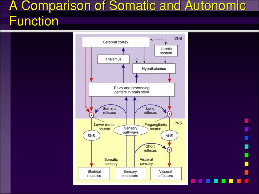
A Comparison of Somatic and AutonomicFunction
81. A Comparison of Somatic and Autonomic Function
Summary of Efferent NS82. Summary of Efferent NS
83.
Higher levels of autonomiccontrol
Activity in the ANS is controlled by centers in
the brainstem that deal with visceral
functioning
84. Higher levels of autonomic control
Levels of Autonomic ControlExample of higherlevel of autonomic
function would be
increased heart rate
when you see a
person that you
dislike.
85. Levels of Autonomic Control
ccLimbic system
anterior
nucleus
Craniosacral
ГИПОТАЛАМУС
posterior
nucleus
Thoracolumbar
86.
87.
Levels of Autonomic ControlExample of higherlevel of autonomic
function would be
increased heart rate
when you see a
person that you
dislike.
88. Levels of Autonomic Control
Visceral Afferents andReferred Pain