


")


 Биология
Похожие презентации:


Central nervous system
1. Kazan State Medical University Department of Anesthesiology and Emergency Medicine
KAZAN STATEMEDICAL UNIVERSITY
DEPARTMENT OF ANESTHESIOLOGY AND
EMERGENCY MEDICINE
CENTRAL NERVOUS SYSTEM
Mohammad Meher Alam
2. The Human Brain
THE HUMAN BRAIN• Complex
• 1.4 kg in weight
• Pre frontal cortex
• 2% of body weight
• 20% of oxygen
• 15% of our cardiac input
• 10% of all energy
3. The Blood Brain barrier
THE BLOOD BRAIN BARRIER• Brain protection system
• The BBB is both;
• A physical barrier that restricts the
entrance of potentially harmful
substances
• A system of cellular transport
mechanisms that controls the entrance
of essential nutrients
4. Divisions of CNS
DIVISIONS OF CNS• CNS - central nervous system:
• consists of brain and spinal cord
• • Nerves and associated structures within the brain and spinal
cord Brain • Cerebrum • Brain stem Spinal cord • Gray matter
White matter • Meninges; dura mater, arachnoid, pia mater
Epidural space • Subarachnoid space(intrathecal space)
• CSF : Formed at choroid plexuses in the ventricles • Cushioning
effect • Normal: 10 mmHg in pressure, 1.002 – 1.009 in SG, 7.32 in
pH • Increased production, decreased absorption, and/or
obstruction of flow of CSF all contribute to hydrocephalus
symptom
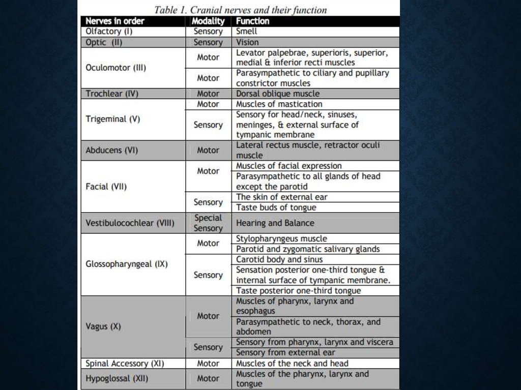
5. A. Cranial Nerves
A. CRANIAL NERVES• 12 pairs & their branches
• Some responsible for special senses: sight, hearing, taste, smell
• Others receive sensations: touch, pressure, pain, temperature
6.
7. C. Cerebrum
C. CEREBRUM• Largest section of the brain
• Responsible for:
• reasoning, thought, memory, speaking, sensstion, sight, hearing,
voluntary body movement
8. D. Cerebellum
D. CEREBELLUM• Responsible for:
• coordination of muscles, balance, posture, & muscle tone
9. E. Midbrain
E. MIDBRAIN• Responsible for:
• conducting impulses between brain parts
• certain eye reflexes
10. Pons
PONS• Responsible for:
• conducting messages to other parts of the brain
• Reflex actions such as chewing, production of saliva
11. G. Medulla Oblongata
G. MEDULLA OBLONGATA• Lowest part of brain stem
• Connects to the spinal cord
• Responsible for:
• regulating heart beat, respirations, swallowing, coughing, b/p
12. 2. Spinal Cord
2. SPINAL CORD• Goes down back of body from Medulla Oblongata
• Surrounded and protected by vertebrae
• Responsible for reflex actions
• Carries sensory and motor messages
13. 3. Meninges
3. MENINGES• Consists of 3 membranes
• Covers and protects the brain and spinal cord
14. Three Membranes
THREE MEMBRANES• C. Dura mater
• thick, tough outer layer
• D. Arachnoid membrane
• middle delicate weblike layer
• E. Pia mater
• inner most layer with blood vessels to nourish the nerves
15. 4. Ventricles
4. VENTRICLES• Four hallow spaces located in the middle of the brain.
• Connected to each other
• Filled with fluid called cerebrospinal fluid
16. Cerebrospinal Fluid
CEREBROSPINAL FLUID• Circulates continuously
• Serves as shock absorber to protect brain and spinal cord
• Carries nurients to parts of brain and spinal cord
• helps remove metabolic products & wastes
• after circulation, absorbed into the blood vessels of the dura
mater.
17. B. Spinal Nerves
B. SPINAL NERVES• 31 pairs & their branches
• carries messages to & from the spinal cord
• Both sensory and motor nerves
• 31 spinal nerves:
8 cervical
12 thoracic
5 lumbar
5 sacral
1 coccygeal
18. 3. Autonomic Nervous System
3. AUTONOMIC NERVOUSSYSTEM
• Autonomic nervous system • It is further subdivided into sympathetic
and parasympathetic divisions (see figure 3). • Because the autonomic
nervous system regulates involuntary or automatic functions, it is called
the involuntary nervous system. The Parasympathetic Nervous System
(craniosacral) • Acetylcholine is transmitter both at pre and
postganlionic (muscarinic) neurons • long preganglionic neurons, short
postganglionic neurons; ganglia are diffusely spread; allows for
discrete, localized innervation and control • Vagus nerve innervates
heart, lungs, esophagus, stomach, small intestine, proximal colon, liver,
gallbladder, pancreas, kidneys, upper ureters • Distribution of
innervation to the heart is to the AV node, SA node, and atria (essentially
none to the ventricles) • Sacral outflow from 2nd, 3rd, and 4th sacral
segments of the cord; form the pelvic nerves, and innervate the bladder,
distal colon, rectum, and sexual organs
19. Neurotransmission
NEUROTRANSMISSIONNeurotransmission • A nerve impulse is an electric current that passes along an axon to
the presynaptic membrane. Upon reaching the presynaptic membrane, it causes the
release of neurotransmitters into the synaptic cleft. • The neurotransmitter then
interacts with receptors on effector cells to induce a response in the effector cell.
Neuroregulators: Neurotransmitters are released into the synaptic cleft in response to
action potentials - release is voltage dependent and requires calcium influx
Neuropeptide modulators are released in smaller quantities than neurotransmitters in
response to action potentials - they serve to amplify or dampen neural activity.
Cholinergic transmission
Acetylcholine is the neurotransmitter • Primary means of terminating action is break
down of acetylcholine into acetate and
20. Neurotransmission
NEUROTRANSMISSION• and choline by acetylcholine esterase (AchE), found principally in
neurons and neuromuscular junctions .
• • Cholinergic receptors are present in the parasympathetic nervous
system, brain, ganglia of the sympathetic nervous system, and skeletal
muscle • Two main types of receptors present • Muscarinic (principally
autonomic nervous system) • Nicotinic (principally skeletal muscle)
• Adrenergic transmission • Catecholamines (dopamine,norepinephrine,
epinephrine) are the neurotransmitters • Primary means of terminating
action is by neural membrane reuptake of the transmitter, although
metabolism by catechol-O-methyltransferase (COMT) and monoamine
oxidase (MAO) is important in some tissues.
21. Neurotransmission
NEUROTRANSMISSIONAdrenergic receptors : Alpha receptors are mainly subdivided
into alpha-1 and alpha 2 receptors • Alpha-1 o principally found
in peripheral vascular smooth muscle • Alpha-2 o occur both
presynaptically and postsynaptically o those occurring
presynaptically on sympathetic nerve terminals reduce the
release of norepinephrine, thus producing a negative feedback
loop o also may modulate cholinergic, serotonergic, GABA-ergic
neurons o central alpha-2adrenergic receptor stimulation results
in sedation, analgesia, decreased sympathetic outflow,
tranquilization o indirectly affects cardiac function by decreased
sympathetic tone o act pre- and postjuntionally to decrease
motility and secretions in the GI tract o produces diuresis by
inhibiting ADH release, blocking ADH’s effect in the renal tubule,
increasing GFR, and inhibiting renin release o stimulate platelet
aggregation
22. Neurotransmission
NEUROTRANSMISSION• • Beta receptors, again, are mainly subdivided into beta-1 and beta 2
receptors • Beta-1 are located in the myocardium, SA node, ventricular
conduction system, and adipose tissue • Beta-2 are vascular smooth
muscle of the skin, muscles, mesentery and bronchial tree; stimulation
results in vasodilation and bronchodilation
• Dopaminergic receptors are dopamine: splanchnic and renal vasodilation
• NANC(nonadrenergic & noncholinergic) – NO • In the brain, spinal cord,
and peripheral nervous system. • L-Arginine and O2 produce L- Citrulline
and NO by NO synthases • It activates guanyl cyclase to increase cGMP
which leads to relaxation of smooth muscle. • NMDA glutamate receptor
activation releases NO and in turn results in excitatory neurotransmission
in the CNS. • NOS inhibitor causes dose-dependent MAC decrease
23. Neurotransmisison
NEUROTRANSMISISONNeuromuscular junction and neuromuscular blocker (NMB) • It consists of presynaptic
nerve terminal and postsynaptic muscular membrane. • Mainly cholinergic nicotinic
receptors, two at postsynaptic and one presynaptic • The neurotransmitter is the
quaternary ammonium ester, acetylcholine • Acetate and choline through choline
acetylase form Acetylcholine at motor nerve ending • Acetylcholinesterase at
cholinergic receptors is responsible for hydrolysing Ach into Acetic acid and choline
Choline can reenter nerve terminal to again participate in the synthesis of new
acetylcholine • Depolarizing neuromuscular blocker o Succinylcholine
(suxamethonium in Europe), mimics the action of Ach by occupying postsynaptic
nicotinic cholinergic receptor, thus depolarizing postsynaptic membrane. However,
hydrolysis of Sch is slower, so postjunctional membrane does not respond to
subsequently released Ach prolonging neuromuscular blockade (Phase I). o Side
effects include hyperkalemia, hypertension, myalgia, cardiac arrhythmia, and
increased intraocular pressure. Also known as a trigger for malignant hyperthermia in
susceptible patients. • Nondepolarising NMBs o Some examples of drugs falling into
this category are pancuronium, atracurium, doxacurium, vecuronium and mivacurium.
o These agents bind to the post synaptic nicotinic cholinergic receptors without
causing any activation of ion channel permeability, and yet impeding normal
postjunctional depolarization with less Ach availability at the receptor leading to the
neuromuscular blockade. o Occupation as many as 70 % does not produce
neuromuscular blockade, but 80-90 % occupation fails neuromuscular transmission,
indicating wide safety margin of the drug.
24. Neuromuscular junction and neuromuscular blocker (NMB)
NEUROMUSCULAR JUNCTION ANDNEUROMUSCULAR BLOCKER (NMB)
• • It consists of presynaptic nerve terminal and postsynaptic muscular membrane. • Mainly
cholinergic nicotinic receptors, two at postsynaptic and one presynaptic • The
neurotransmitter is the quaternary ammonium ester, acetylcholine • Acetate and choline
through choline acetylase form Acetylcholine at motor nerve ending
Acetylcholinesterase at cholinergic receptors is responsible for hydrolysing Ach into
Acetic acid and choline • Choline can reenter nerve terminal to again participate in the
synthesis of new acetylcholine • Depolarizing neuromuscular blocker o Succinylcholine
(suxamethonium in Europe), mimics the action of Ach by occupying postsynaptic nicotinic
cholinergic receptor, thus depolarizing postsynaptic membrane. However, hydrolysis of
Sch is slower, so postjunctional membrane does not respond to subsequently released Ach
prolonging neuromuscular blockade (Phase I). o Side effects include hyperkalemia,
hypertension, myalgia, cardiac arrhythmia, and increased intraocular pressure. Also
known as a trigger for malignant hyperthermia in susceptible patients. • Nondepolarising
NMBs o Some examples of drugs falling into this category are pancuronium, atracurium,
doxacurium, vecuronium and mivacurium. o These agents bind to the post synaptic
nicotinic cholinergic receptors without causing any activation of ion channel permeability,
and yet impeding normal postjunctional depolarization with less Ach availability at the
receptor leading to the neuromuscular blockade. o Occupation as many as 70 % does not
produce neuromuscular blockade, but 80-90 % occupation fails neuromuscular
transmission, indicating wide safety margin of the drug.
25. Theories of Anesthesia
THEORIES OF ANESTHESIA• Wide range of compounds produce anesthesia, without any unifying chemical structure or
activity • We don’t as yet understand how general anesthetics function • A key concept in
any theory regarding anesthetic mechanisms must be the ability of the anesthetic to
disrupt cellular and intercellular communication, particularly in the CNS. • Many
hypotheses have been proposed over the years; it appears that there is expansion and
fluidization of the cell membrane by anesthetic agents that result in depressed synaptic
transmission, and some anesthetic agents also hyperpolarize neurons by increasing
potassium permeability.
• • Meyer-Overton hypothesis asserts that, anesthesia results from the presence of a certain
concentration of the anesthetic at a hydrophobic site. Evidence for this has come from the
fact that potency is strongly correlated with the lipid solubility of the drug. • Critical
volume theory asserts that anesthetic’s direct action on proteins (ion channel proteins nicotinic Ach, GABA, glycine, NMDA; signal transduction pathways) will induce
conformation change on lipoprotein (expansion beyond the critical volume) and lead to
interruption of neurotransmission by obstructing ion flux with changes of electrical
conductivity in the neurons. • The reticular activating system, a multi-synaptic structure, is
believed to be the most important site within the central nervous system for anesthetic
action. • We do have an understanding of how certain classes of drugs work - those that
interact with specific receptor sites. o opioids (eg, morphine, butorphanol) o alpha-2
receptor agonists (eg, xylazine, medetomidine) o benzodiazepines (eg, diazepam,
midazolam)