


:")


 Медицина
Похожие презентации:


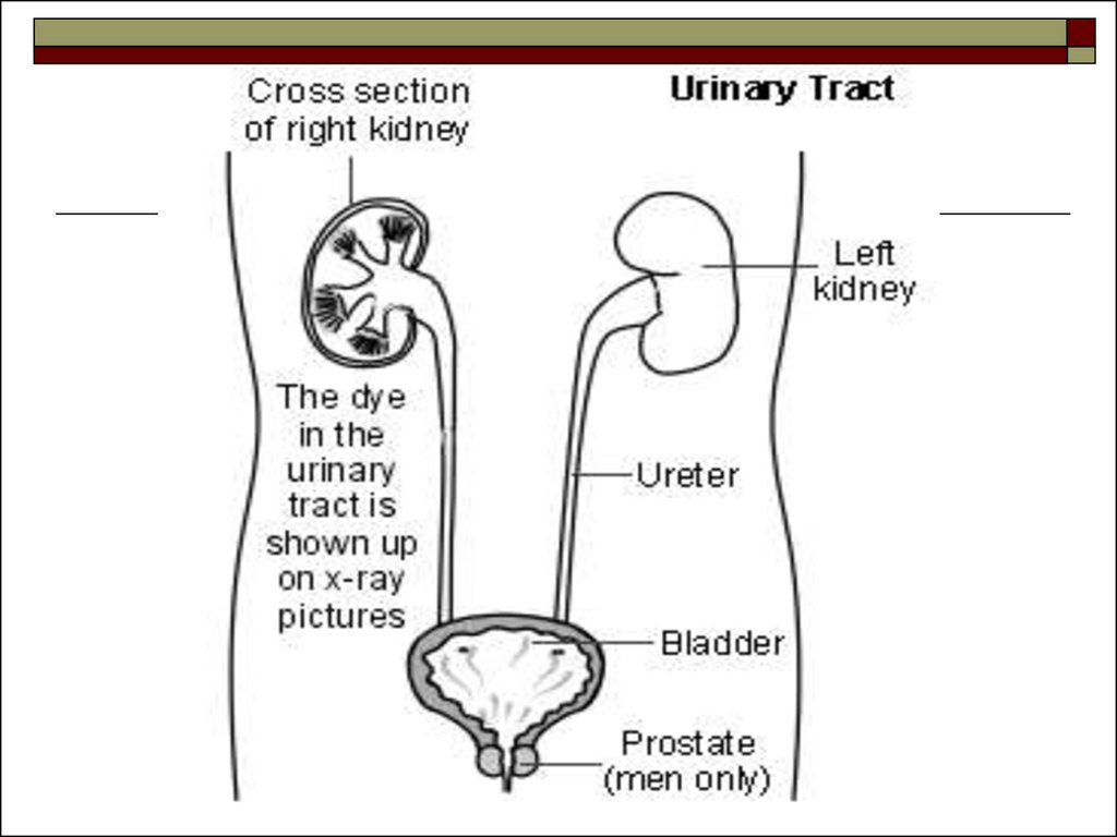
Investigation of the urinary system
1. Investigation of the urinary system
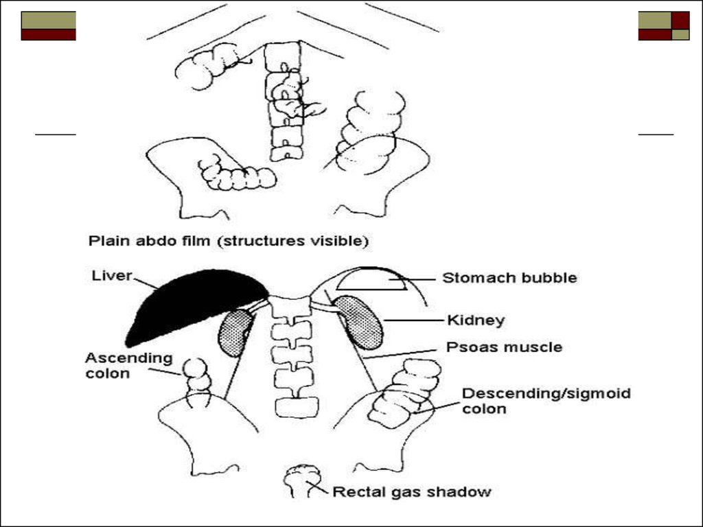
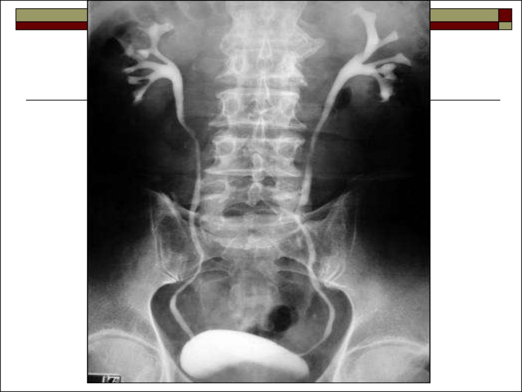
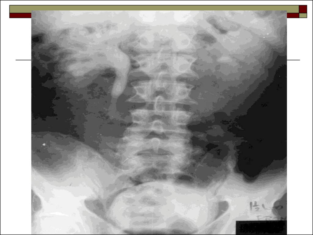
2. Plain Film:
Plain film is taken in supine position. Theradiograph should include the upper poles of
both the kidneys and lower border of
symphysis pubis (for prostatic urethra).
A plain abdominal film is essential prior to
urinary tract investigation.
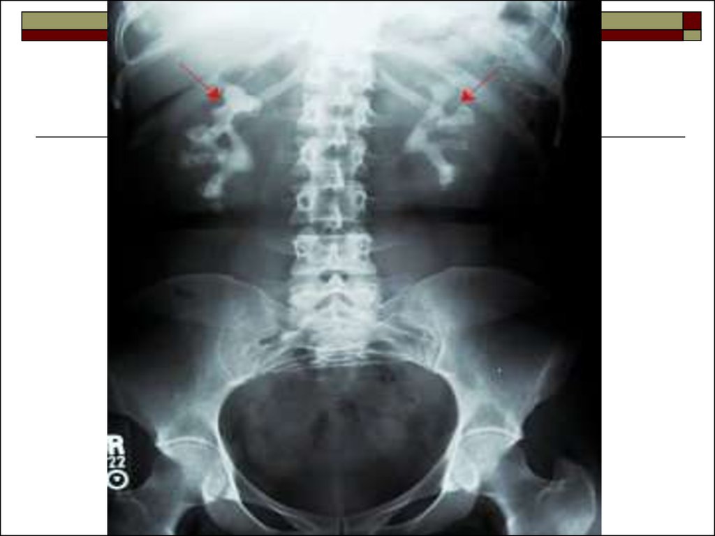
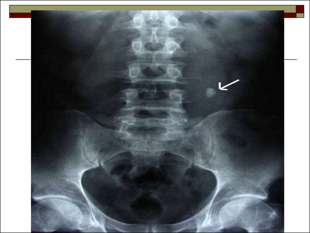
3. This may show
renal calculi in the pelvicalyceal systemrenal parenchymal calcification
ureteric calculi
bladder calcification and calculi
prostatic calcification or sclerotic bone
deposits
4.
Caution should be used in interpreting renaltract calcification as overlying calcifiedmesenteric glands and pelvic vein phlebolitis
are often mistaken for ureteric calculi.
Inspiration and expiration films change the
position of the kidneys and often confirm that
a calcified area in the upper abdomen is a
calculus.
5.
6.
7.
8. Intravenous Urography (IVU):
IVU is frequently performed in the evaluationof hematuria. Urography may also be
performed in the pre- or post theraupetic
evaluation of stone disease that has been
discovered with other imaging modalities.
9. Indications
obstructive calculihematuria or pyuria
diseases of renal collecting system and renal pelvis
abnormalities of the ureter
tuberculosis of the urinary tract
prior to endourological procedures and surgery of
the urinary tract
suspected renal injury
renal colic or flank pain
in children – polycystic kidney diseases, pelviureteric junction obstruction, anorectal anomalies
pelvic malignancies to see uretic involvement
10. Patient preparation
blood urea and serum creatinine level shouldbe within normal limits
if patient is asthmatic premedication in the
form of steroids is administered two days
prior
fasting after 10 pm (previous night) (as
contrast injection sometimes induces nausea
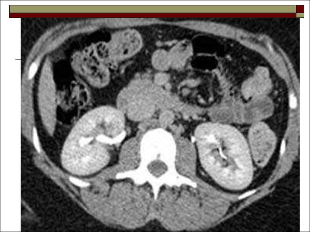
which might lead to vomiting and aspiration)
11.
patient should be well hydrated (dehydratedpatients are prone for renal damage)
bowel preparation is necessary, as gas and
faecal matter filled bowel loops will obscure
the kidney shadows
low residue diet with plenty of oral fluids, the
day previous to the IVU
12.
Bowel wash is given till bowel is clear offaecal matter on previous night.
Laxatives (ducolax, castor oil) are
recommended to eliminate faecal matter from
colon and gas absorbing agents (flatulex) are
given to reduce the amount of gas in the
bowel.
In young children no special preparation is
needed, only 4 hours fasting is sufficient.
13. Procedure
Patient is placed in supine position. Thepatient is asked to void the bladder before the
procedure.
A plain film is taken which includes the
kidneys, ureters, bladder and urethral regions
on a large size film, called as the scout film.
14.
Contrast media is injected intravenously into aprominent vein in the arm. Test injection of
1ml of contrast is given and patient observed
for 5 min for any contrast reactions. Then the
rest of the contrast is rapidly injected within
30-60 seconds.
The dose of contrast media is 2 ml/kg body
wt.
15. Contrast media
Contrast materials currently in use are excretedalmost exclusively by glomerular filtration, with
subsequent concentration in the renal tubules and
progressive opacification of the urinary tract.
They are two types:
ionic (urograffin, angiograffin)
non-ionic (omnipaque, ultravist)
Ionic contrast media have a higher incidence of
reaction but they are cheaper as compared to the
non-ionic contrast media.
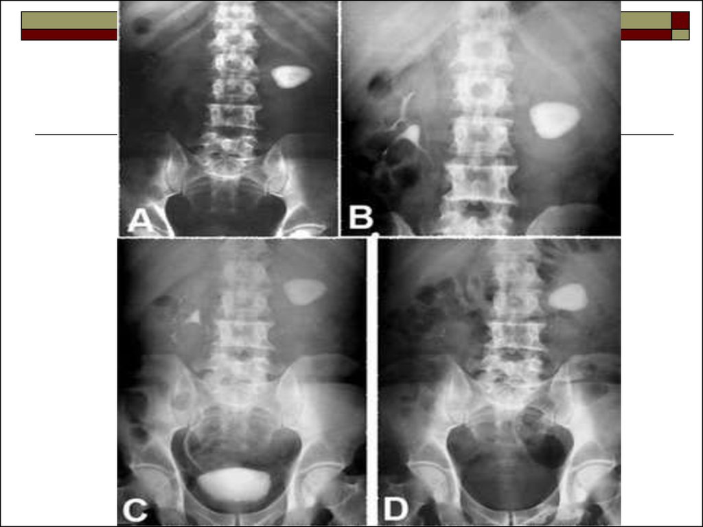
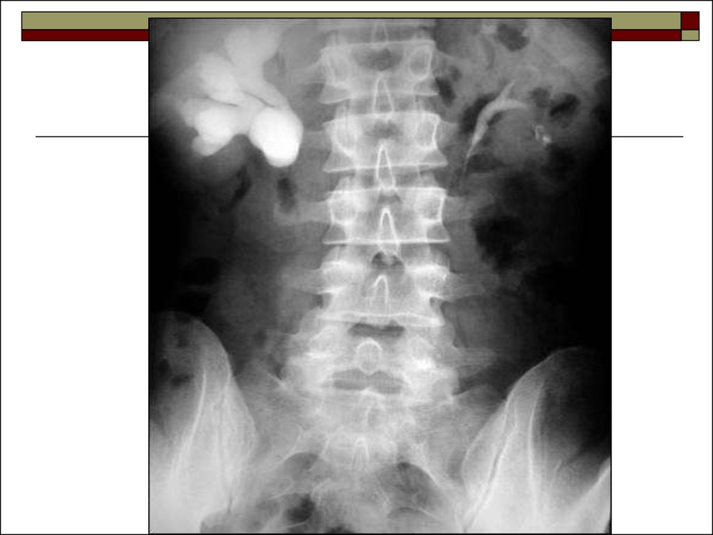
16. Filming technique and interpretation
Plain x-ray (scout film)It gives information about:
renal outlines
psoas muscles
bony structures such as vertebra and its appendages,
pelvis
any stones
abdominal mass
foreign body
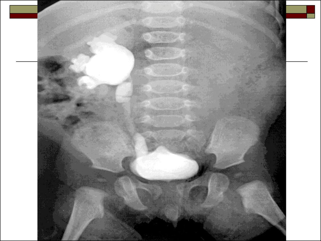
17.
5-10 min filmShows nephrogram, renal pelvis
15-20 min film
A complete visualization of the pelvicalyceal system
entire ureters is possible in this film, especially with
the patient in prone position as the ureters will be
antedependent in prone position.
18.
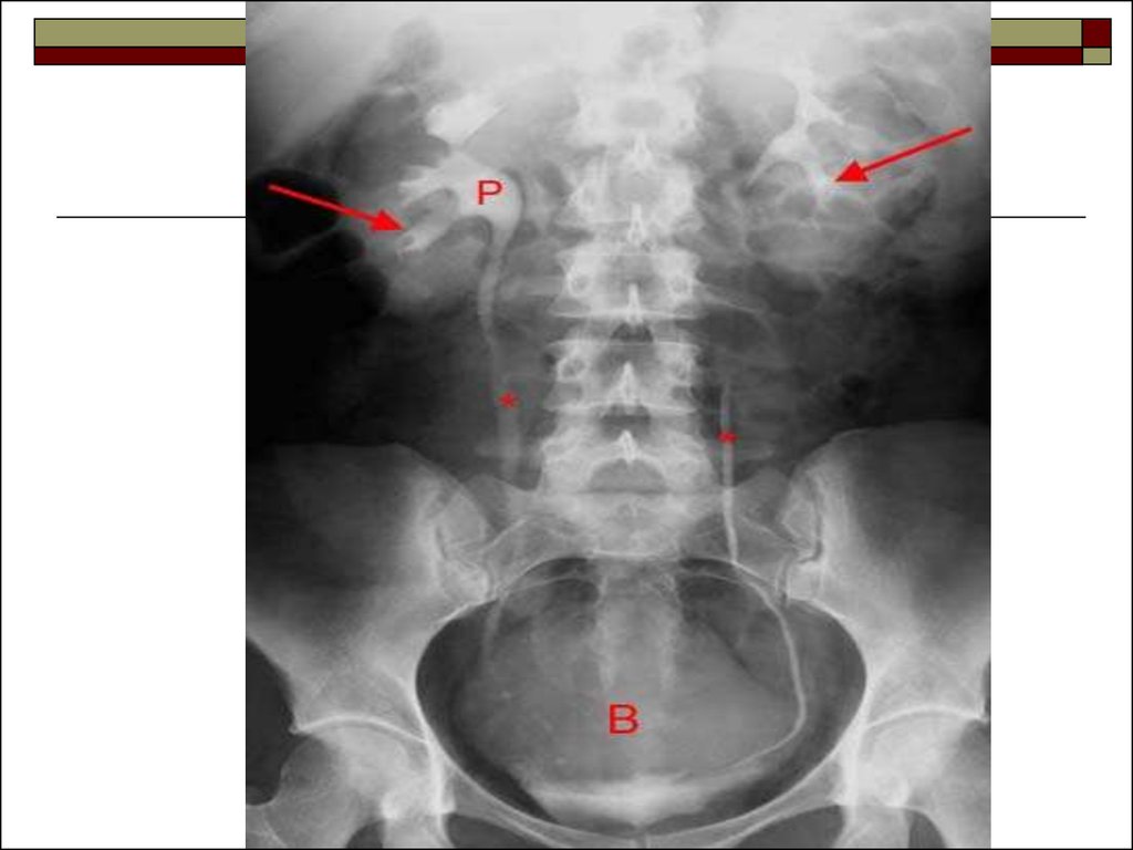
30-35 min filmA complete visualization of the urinary tract: kidney,
ureter, bladder can be done and bladder distension
can be evaluated in the later film.
The series is varied according to the individual
patient. Renal obstruction may require a delayed
study up to 24 hours to outline the pelvicalyceal
system.
19. Post void film
It taken immediately after voiding.To assess for:
residual urine
bladder mucosal lesions
diverticula
bladder tumors
outlet obstruction
20.
21.
22.
23.
24.
25.
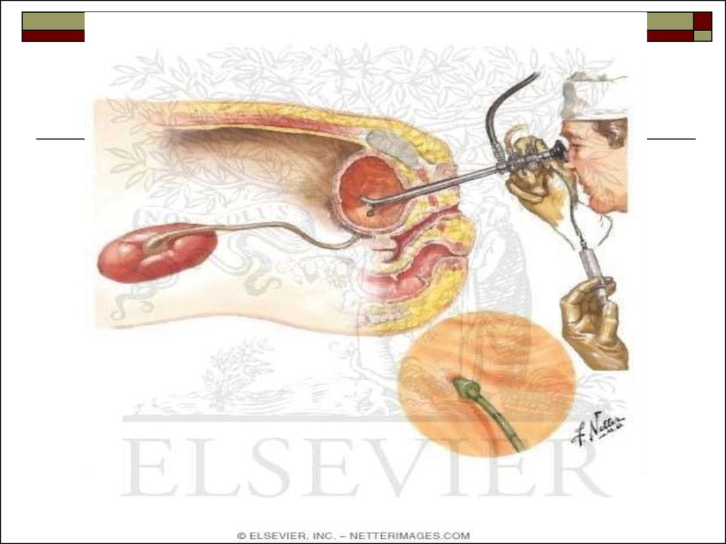
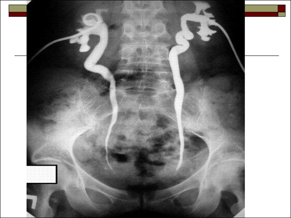
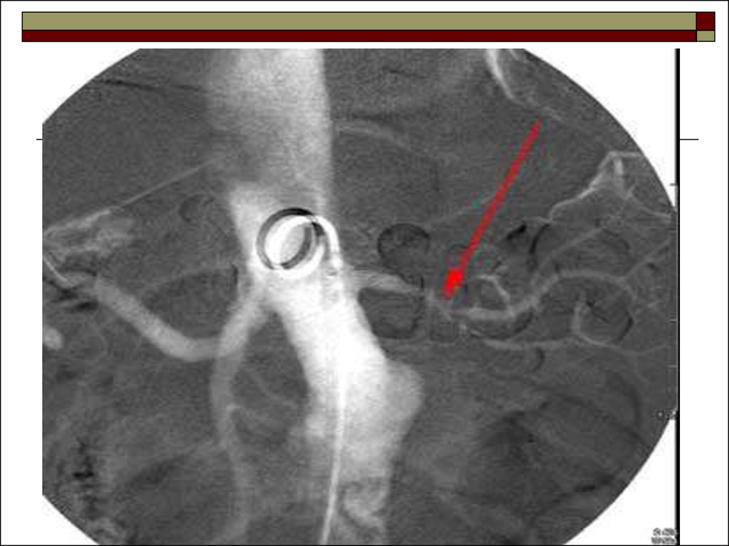
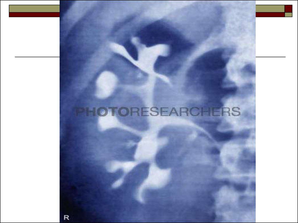
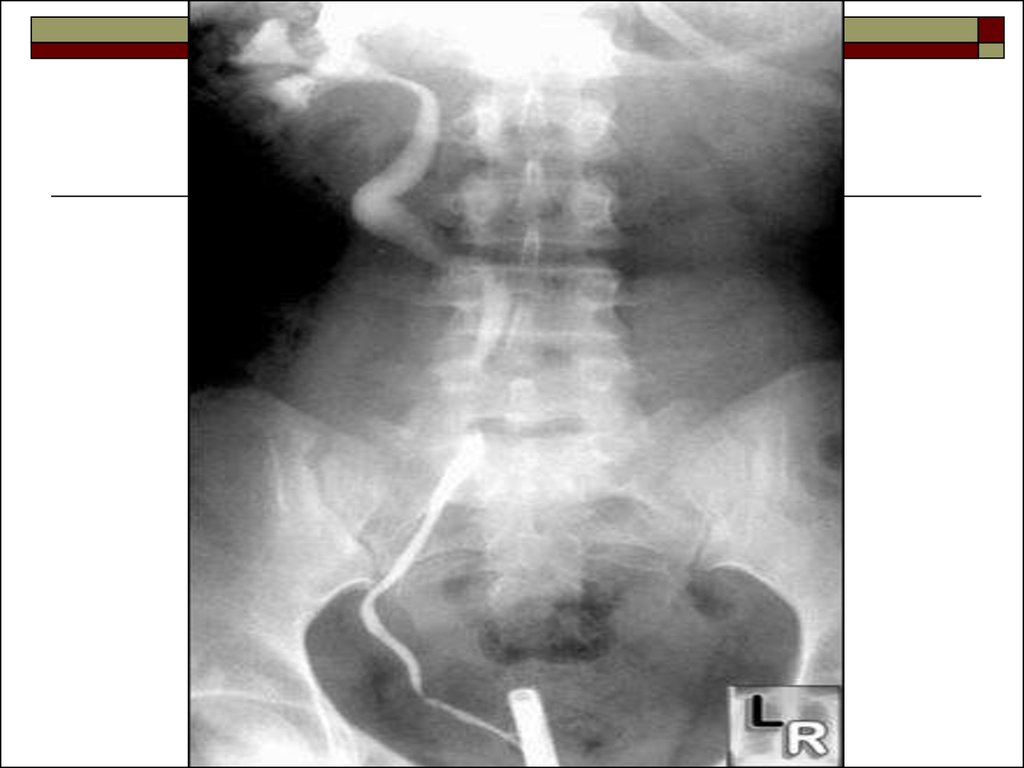
26. Retrograde pyelography
A retrograde pyelography is occasionally necessarywhen detail of the pelvicalyceal system and ureter is
not adequately delineated by intravenous contrast,
especially when there is suspect ion of an epithelial
tumor of the urinary tract.
A catheter is placed into the ureter after a
cystoscopy; contrast injected trough the catheter
outlines the pelvicalyceal system and ureter.
27.
28.
29.
30. Antegrade pyelography
A fine-gauge needle, under local anesthetic, can beinserted directly into the pelvicalyceal system and
contrast injected to visualize the calyces, pelvis and
ureter. The patient lies in a prone position and the
examination is carried out under either ultrasound or
fluoroscopic control. This procedure, not requiring a
general anesthetic, accurately localizes the site of an
obstructing lesion, such as a calculus or stricture.
31.
32. Micturating cystogram
A catheter is inserted in the bladder which isfilled to capacity with contrast. After catheter
removal, films are taken of the renal tract as
the patient is micturating, looking for vesicoureteric reflux. Careful examination of the
urethra in the oblique position is necessary in
suspected urethral valves, as they are usually
only demonstrated during micturition.
33. Indications
Children:vesico-ureteric reflux
post urinary tract infection
trauma
hematuria
posterior urethral valve
voiding difficulties like dysuria, thin stream,
frequency and urgency
in case of genitor-urinary anomalies
34.
Adults:trauma to urethra
urethral stricture
urethral diverticula
vesico-ureteric reflux
35.
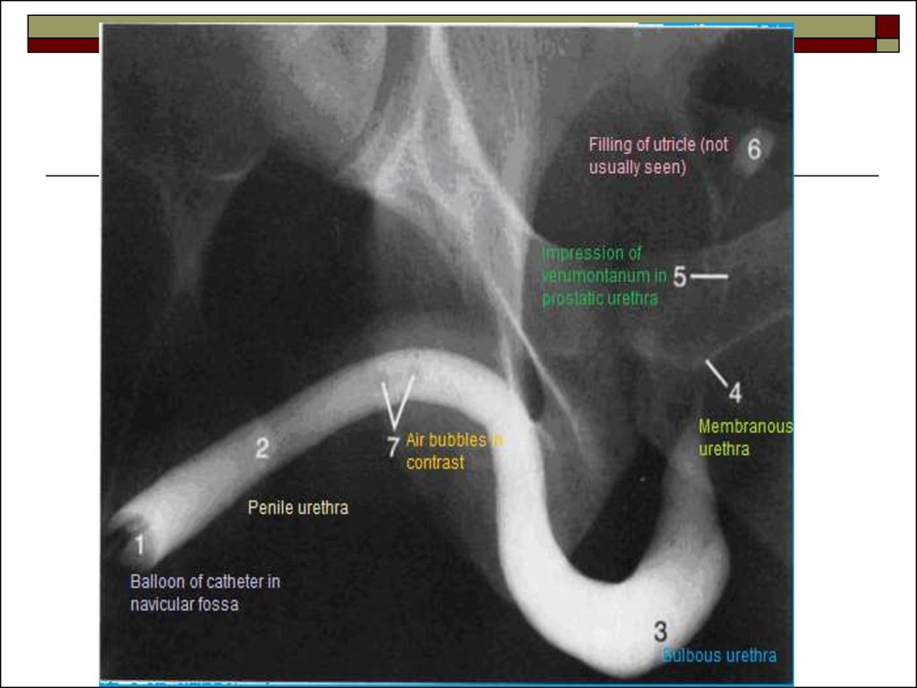
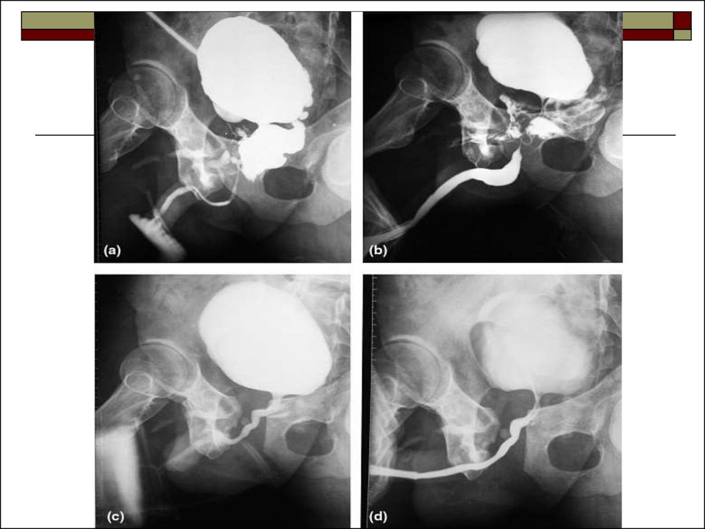
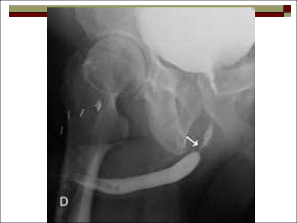
36. Urethrography
The adult male urethra can be visualized by:ascending urethrography: contrast is injected into the
meatus and films obtained of the urethra
descending urethrography: after filling the bladder
with contrast, the catheter is removed and films of
the urethra are taken during micturition
In both studies, the entire urethra must be studied.
37.
38.
39.
40. Ultrasound
Ultrasound is one of the most valuableinvestigations of the urinary tract and the
investigation of choice in children.
It is extremely effective in evaluating:
renal size
growth
masses
41.
renal obstructionurinary tract infection
hematuria
congenital abnormalities
renal failure
transplants
bladder residual volumes
prostatic size
it is non-invasive and can be repeated frequently.
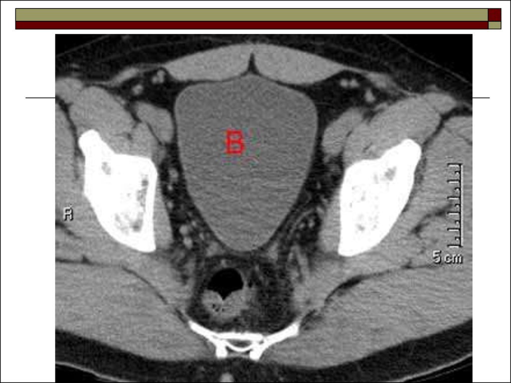
42. Urinary bladder
43. Isotope Scanning:
Static Scanning: Technetium-99m DMSA:Selective uptake by the renal cells with
stagnation in the proximal tubules produces
images of the renal parenchyma. The isotope
is used to assess function, position, size and
scarring of kidneys.
44.
Dynamic scanning: Technetium-99m DTPA:Isotope clearance by glomerular filtration
produces a dynamic scan, providing
information on renal blood flow and renal
function. The function of each individual
kidney can be assessed as well as total renal
function.
45.
46.
47. . Arteriography:
Evaluation of the renal arterial circulation may benecessary for:
further investigation of equivocal renal masses:
renal cell carcinoma are usually hypervascular with a
pathological circulation
arteriovenous malformation
renal artery stenosis
anatomical details prior to renal transplantation
suspected vascular occlusion after surgery
48.
49.
50. Computed tomography
This aids assessment of:renal masses – especially differentiation of solid and
cystic lesions
obstruction
retroperitoneal disease
staging of renal and bladder neoplasm
tumor invasion into the renal vein or inferior vena
cava
evaluation after trauma, surgery or chemotherapy
inflammation
trauma
51.
52.
53.
54. Congenital anomalies
Ectopic kidneyNormally the kidneys are located in the abdomen
adjacent to the upper three lumbar vertebrae. The
final position of kidney and associated length of
ureter is determined by extent of ureteral bud
elongation, which if ceases earlier than normal stage
will result in ectopic location of kidneys like:
pelvic
sacrum
lower lumbar levels
intrathoracic kidneys – commonly occurs on left side
of thorax
55.
56. Crossed fused ectopia
The two renal masses fuse with each otherhowever the ureters draining the two renal
masses are separate and insert into the bladder
trigone distally.
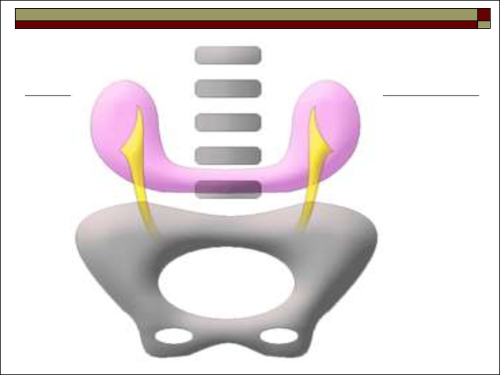
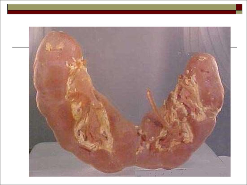
57. Horse shoe kidney
Is a fusion of lower poles of both the kidneysoccurs by either renal or fibrous tissue.
Plain radiograph: the axis of each kidney is
markedly altered, the upper pole being more
lateral and the lower pole being more medial.
58.
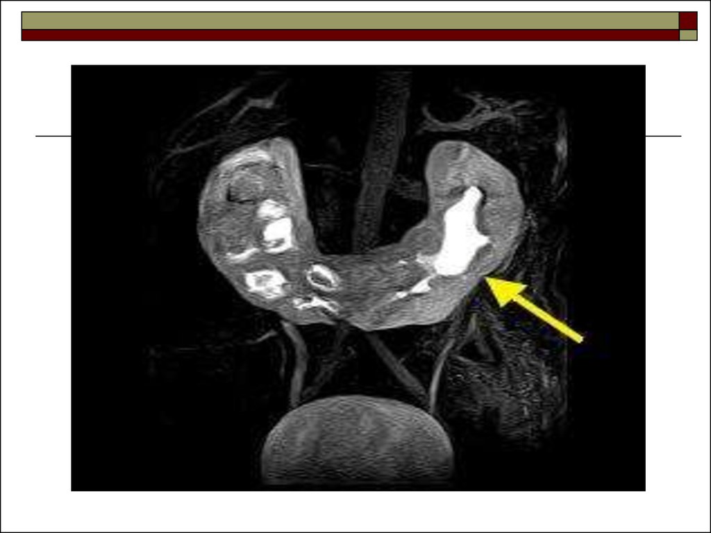
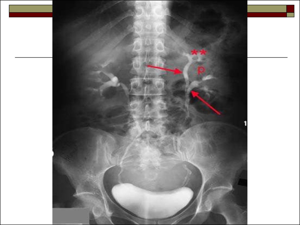
IVU: may demonstrate the isthmus which connectsthe two kidneys. There is some degree of malrotation
with renal pelvis lying anteriorly, and calyces lying
posteriorly, medially or laterally. The ureters are seen
to course anteriorly over the lower pole or over the
isthmus.
CT shows: the parenchyma of the horseshoe kidney
is well visualized. Isthmus can be very well depicted
in CT.
59.
60.
61.
62.
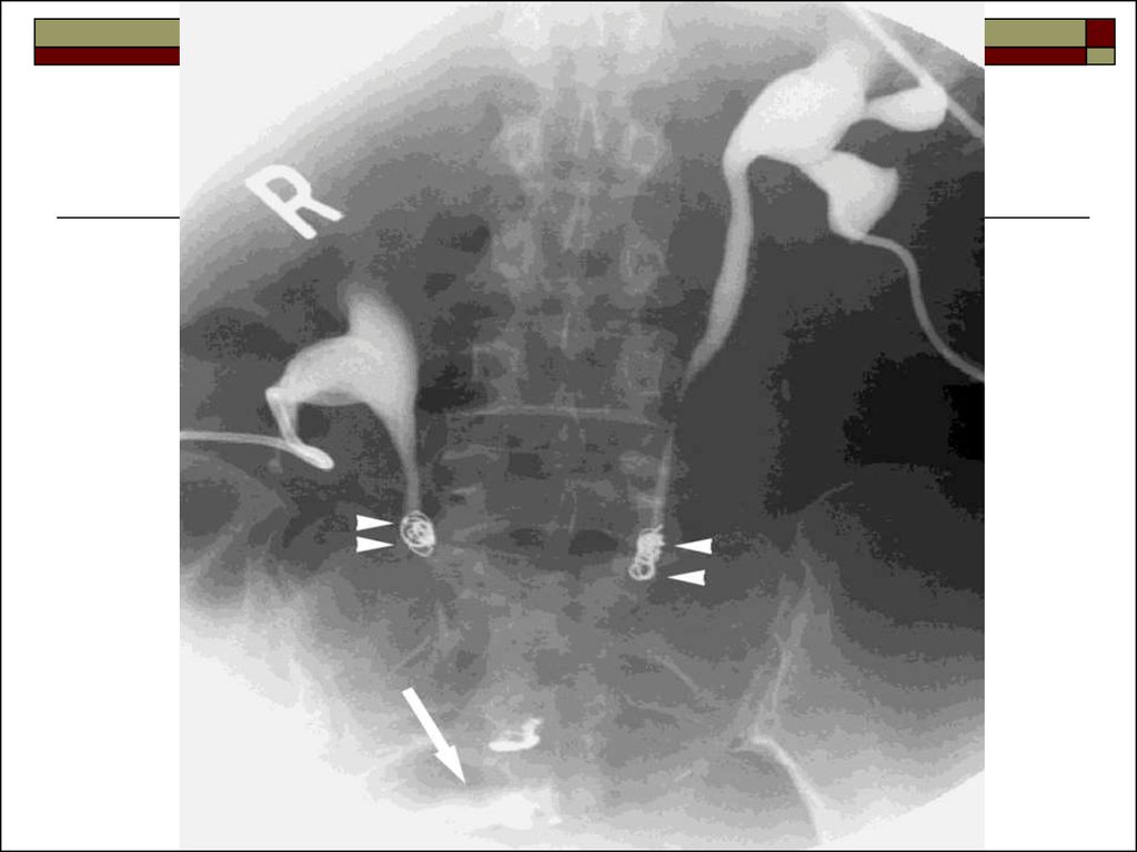
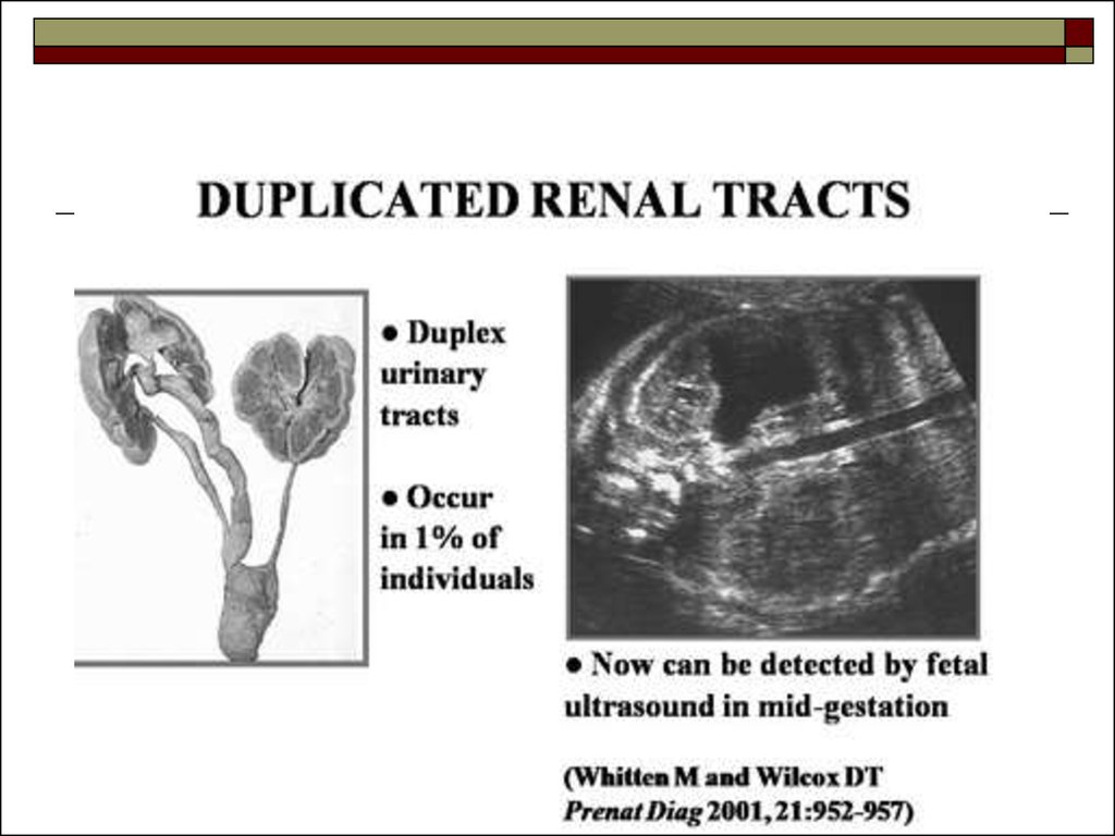
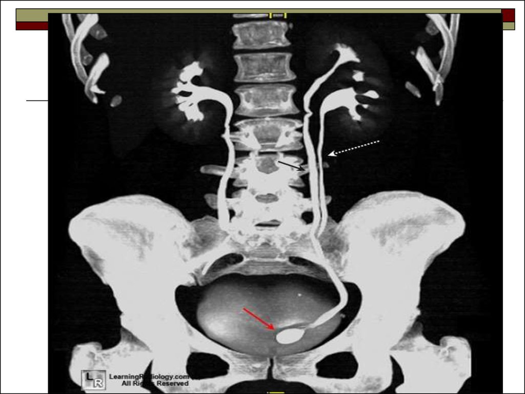
63. Duplex Kidney:
the commonest renal anomaly with a variabledegree of duplication ranging from minor
changes of the renal pelvis, to total
duplication of the renal pelvis and ureter
64.
65.
66.
67.
68. Agenesis
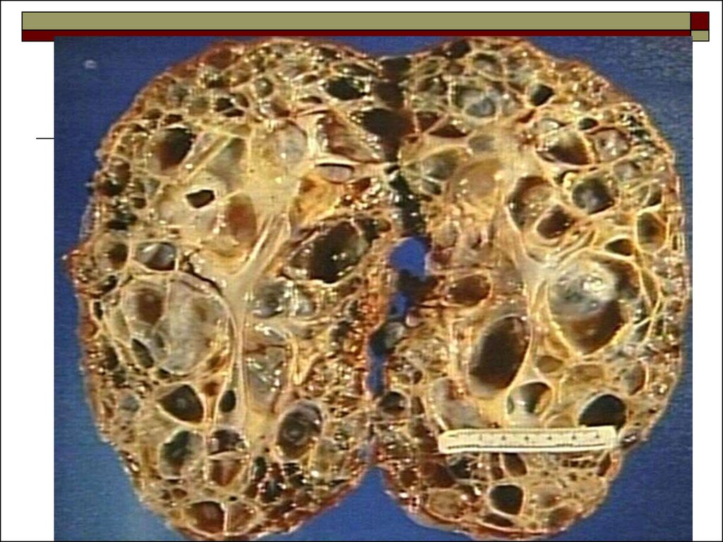
69. Polycystic kidney disease
Clinical featureshypertension
bilaterally enlargement kidneys as masses per
abdomen
loin pain rarely
Plain film
enlargement kidneys seen as soft tissue masses
bilaterally
occasionally dystrophic calcification in cyst seen
70.
IVUmajor calyces may be displaced, narrowed
and elongated by adjacent cyst
in advanced cases there will be deformity of
both major and minor calyces forming a
typical “spider-leg” appearance
also large doses of contrast will be needed for
opacification of the pelvicalyceal system
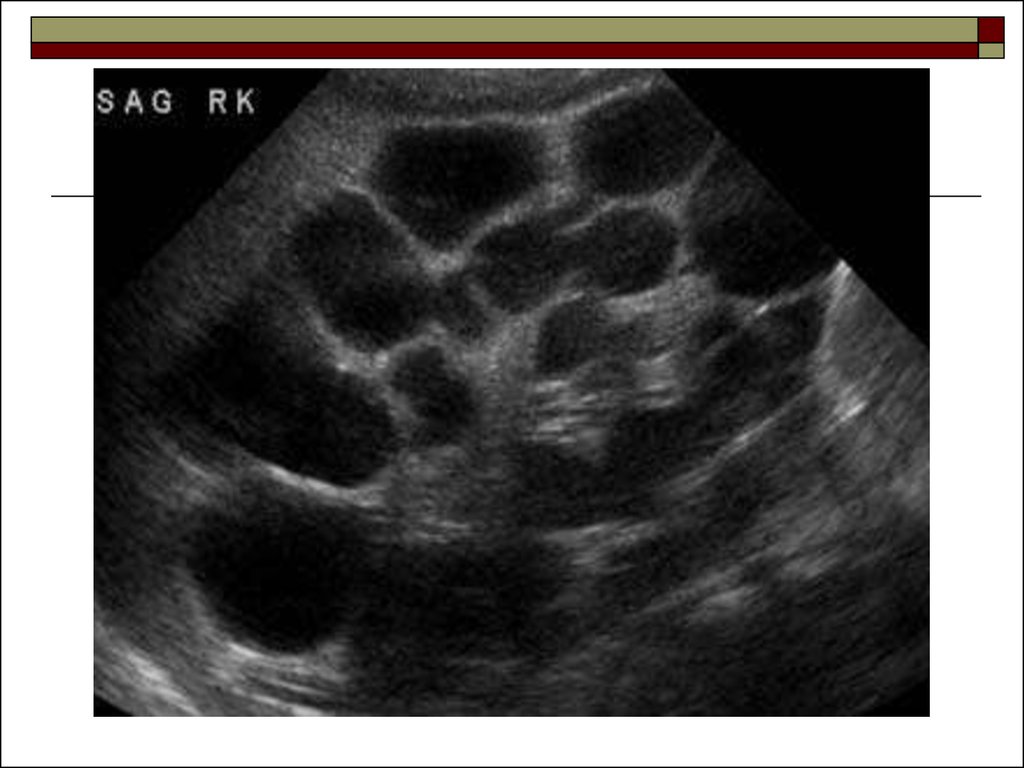
71.
Ultrasoundenlarged kidneys
cysts are seen as anechoic lesions (black) with
distal acoustic enhancement
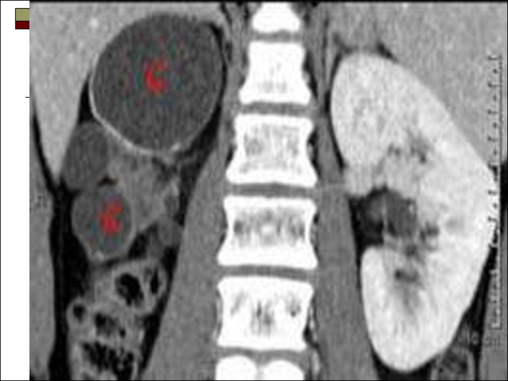
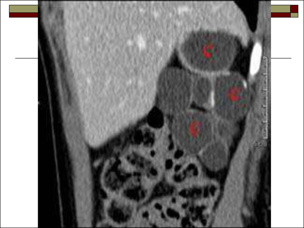
CT
cysts will be seen as multiple hypodense
lesions with density of fluid
72.
73.
74.
75.
76.
77.
78. Retrocaval ureter
Normally the right ureter lies anterolateral tothe inferior vena cava.
Occasionally the right ureter takes an aberrant
course running sharply medially and behind
the inferior vena cava and then courses
anterior to the vena cava and then drops
inferiorly into the pelvis.
It may be associated with hydronephrosis due
to its abnormal course.
79.
80.
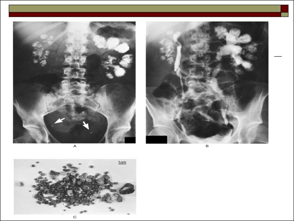
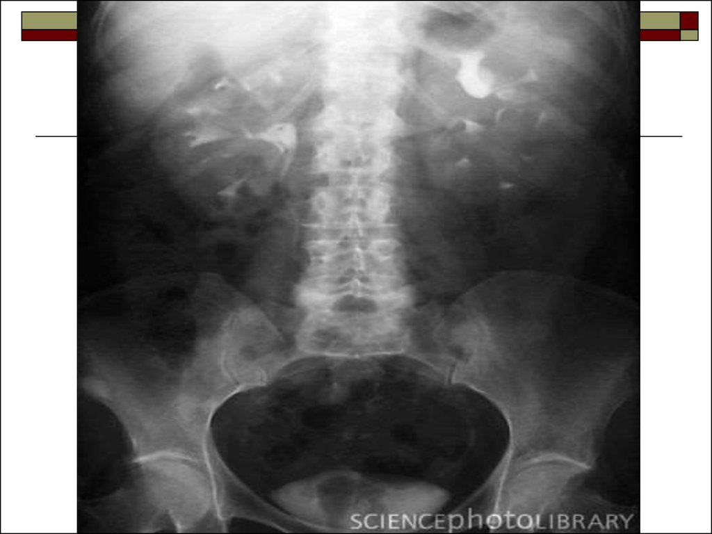
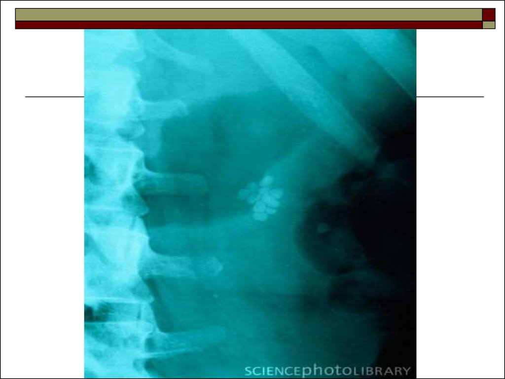
81. Urinary tract stones
Urinary tract stones are the stones within thecollecting system which are due to metabolic,
environmental, structural and genetic abnormalities.
Radio opaque stones:
calcium oxalate and phosphate stones
cysteine stones – they contain sulphur
struvite stones: this consists of magnesium
ammonium phosphate
82.
Radio lucent stones:uric acid stones
xanthine stones
Radiolucent stones are not visualized on xray, however, ultrasound and CT scan can
detect these radiolucent stones.
83.
UltrasoundStones will be seen as hyperechoic (bright) focus
within the collecting system with distal shadowing.
Dilatation of the collecting system may be present in
cases of obstruction.
Ultrasound is especially important in detecting
radiolucent stones not seen in IVU and plain x-ray.
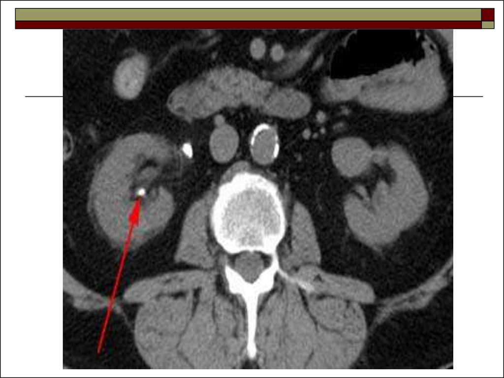
Ct scan
No enhanced CT scan is the modality of choice for
diagnosis calculus.
Advantages:
detection of multiple stones
other causes of abdominal pain which may mimic
renal colic
84.
85.
86.
87.
88. Hydronephrosis
Hydronephrosis is a dilatation of PCSsecondary to distal obstruction.
Causes
ureteric stones
ureteric stricture
pyeloureteric junction obstruction
bladder outlet obstruction
89.
IVUFindings may vary with the duration and degree of
the obstruction. Renal outline may be enlarged.
Crading
Grade1: minimal blunting of forniceal angle
Grade2: blunting of calyces with intact papillary
markings
Grade3: loss of papillary markings
Grade4: ballooning of the calyces
90.
Ultrasounddilatation of the collecting system will be seen
as hypoechogenicity (dark) within the (bright)
renal sinus
renal parenchyma may be thinned out in
severe hydronephrosis
91.
92.
93.
94. Hydroureter
Hydroureter is ureteric dilatation due to either obstructive ornon obstructive causes.
An absolute ureteral diameter exceeding 8 mm is considered
by some authors to represent a criterion for dilatation. In
general, asymmetry of ureteral caliber is a more significant
findings.
Early in its course, high-grade ureteral obstruction may be
associated with only minimal ureteral dilatation. More
chronic forms of obstruction and other chronic ureteral
conditions are typically associated with greater degrees of
ureteral dilatation.
No obstructive dilatation may occur as a result of high urine
flow, reflux, or inflammatory processes.
95.
CausesUreteric calculus
Ureteric stricture
Ureterocele
Congenital megaureter
Retroperitoneal tumor/Retroperitoneal fibrosis
Pelvic malignancies
96.
97.
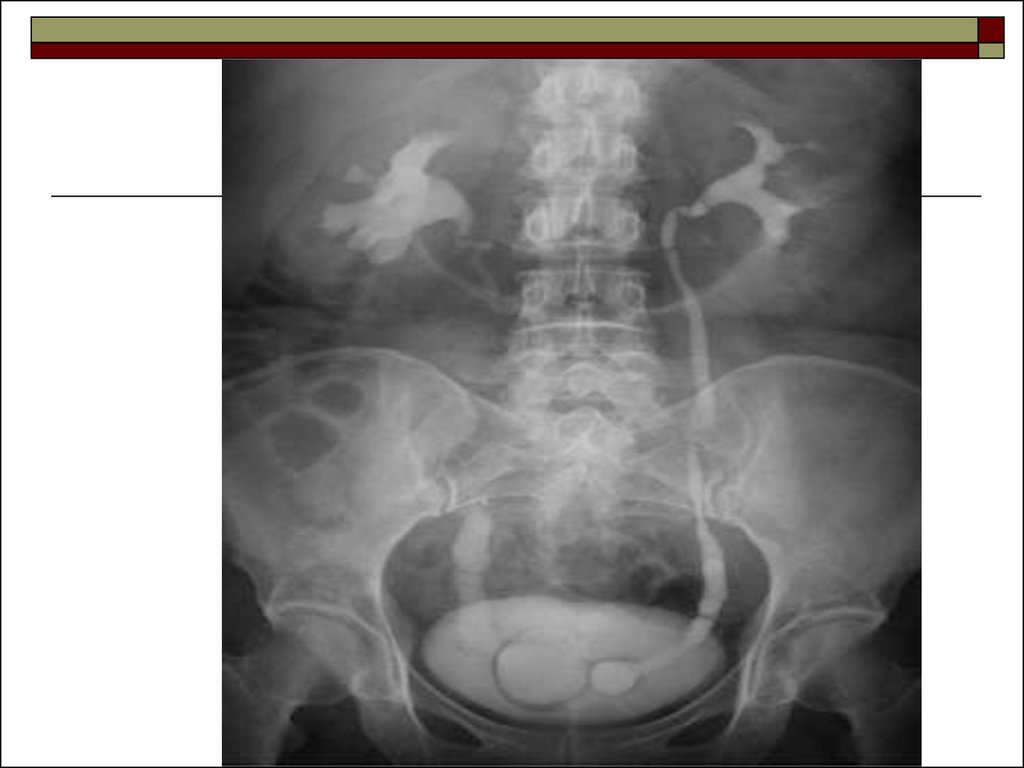
98. Ureterocele
Submucosal dilatation of the intramural distalureter which often protrudes into the bladder
lumen is called ureterocele.
IVU
Ureterocele can be seen as a contrast filled
structure with a thin smooth radiolucent wall
surrounded by contrast containing urine in the
bladder (cobra head appearance).
99.
100.
101.
102.
103. Primary megaureter
Primary megaureter is congenital abnormalmusculature of the distal ureter, leading to
focal failure of peristalsis.
Radiological signs
dilatation usually the distal third of the ureter
the calyces are normal
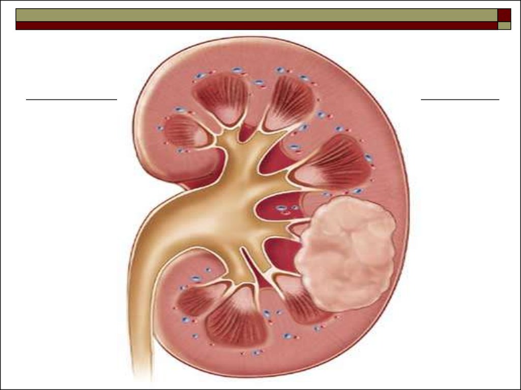
104. Renal cell carcinoma
Common age of presentation between 50 to70 years. Common urological malignancy in
adults, with a male: female ratio of 1:2.
Radiological imaging
Plain radiograph abdomen
soft tissue density mass in the renal fossa with
displaced bowel loops may be seen
105.
IVUdisplacement, compression and cut off of calyces,
change of axis of the kidney
enlargement of affected part of kidney with focal
bulge in renal contour
large tumor may displace entire kidney across
midline
upper pole tumor may cause caudal displacement of
calyces
large tumor mass obstructing the renal pelvis may
cause hydronephrosis
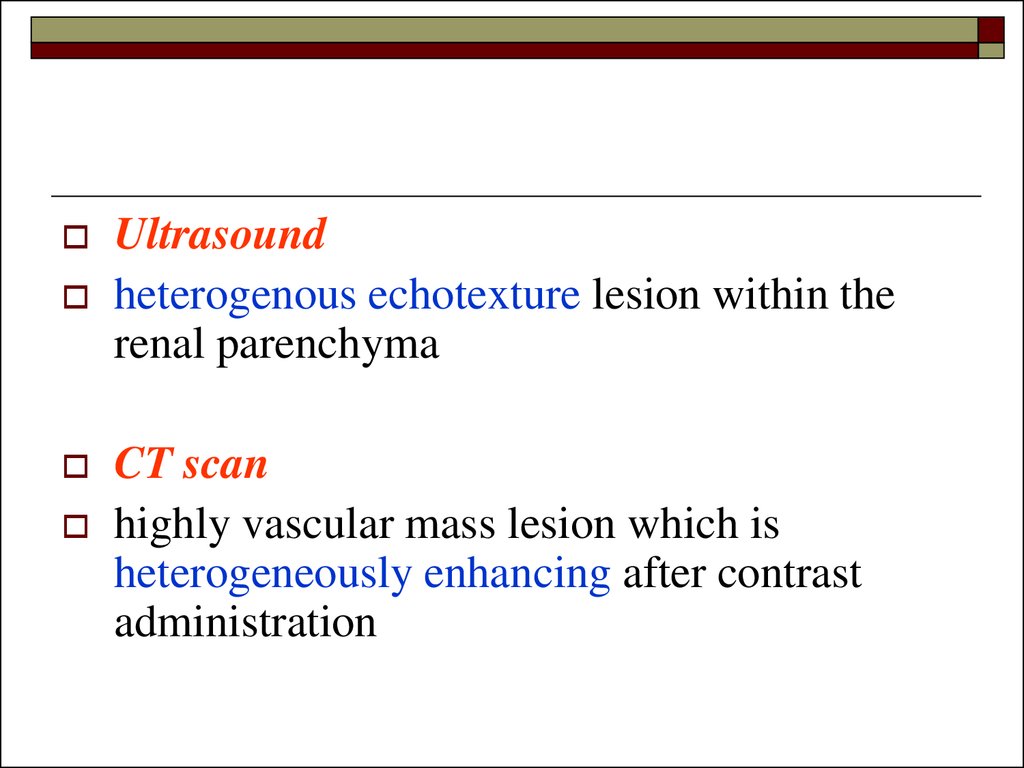
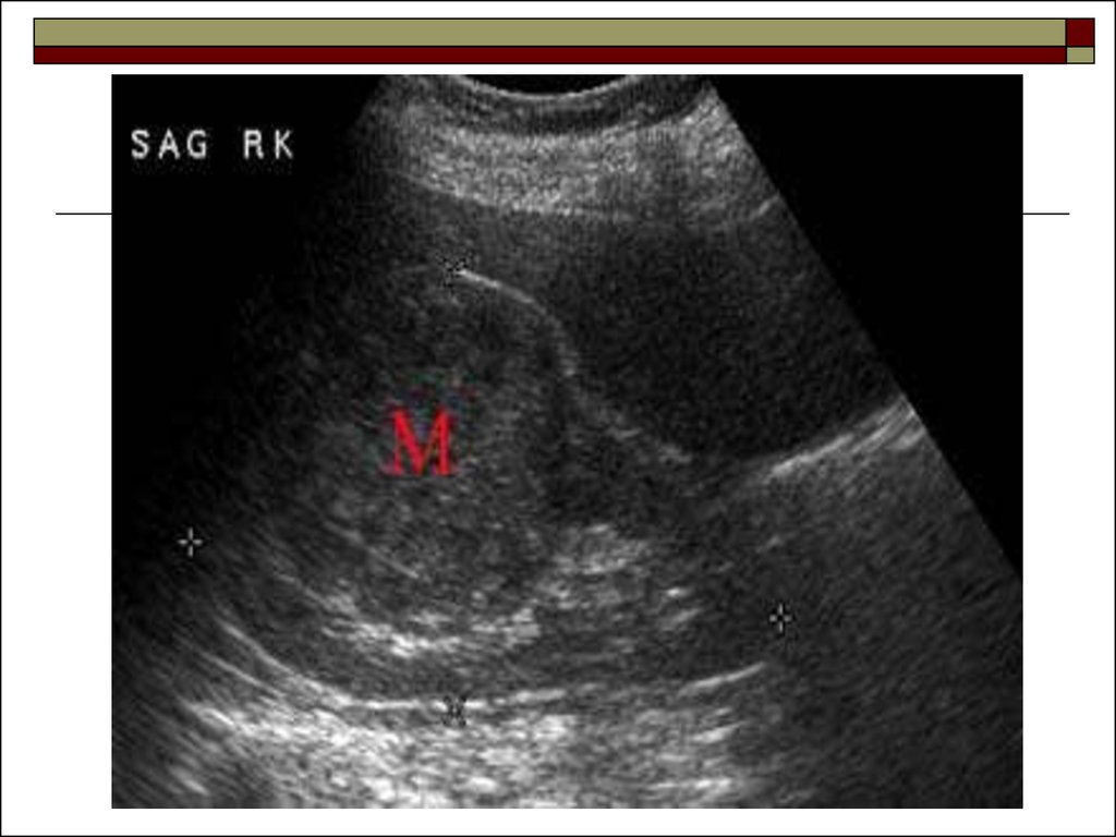
106.
Ultrasoundheterogenous echotexture lesion within the
renal parenchyma
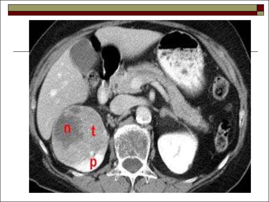
CT scan
highly vascular mass lesion which is
heterogeneously enhancing after contrast
administration
107.
108.
109.
110. Wilms tumor
commonest renal malignancy in childrenpresents mainly between 1 to 5 years of age
with peal incidence at 3 years
Radiological imaging
Plain radiograph
soft tissue mass in the renal area
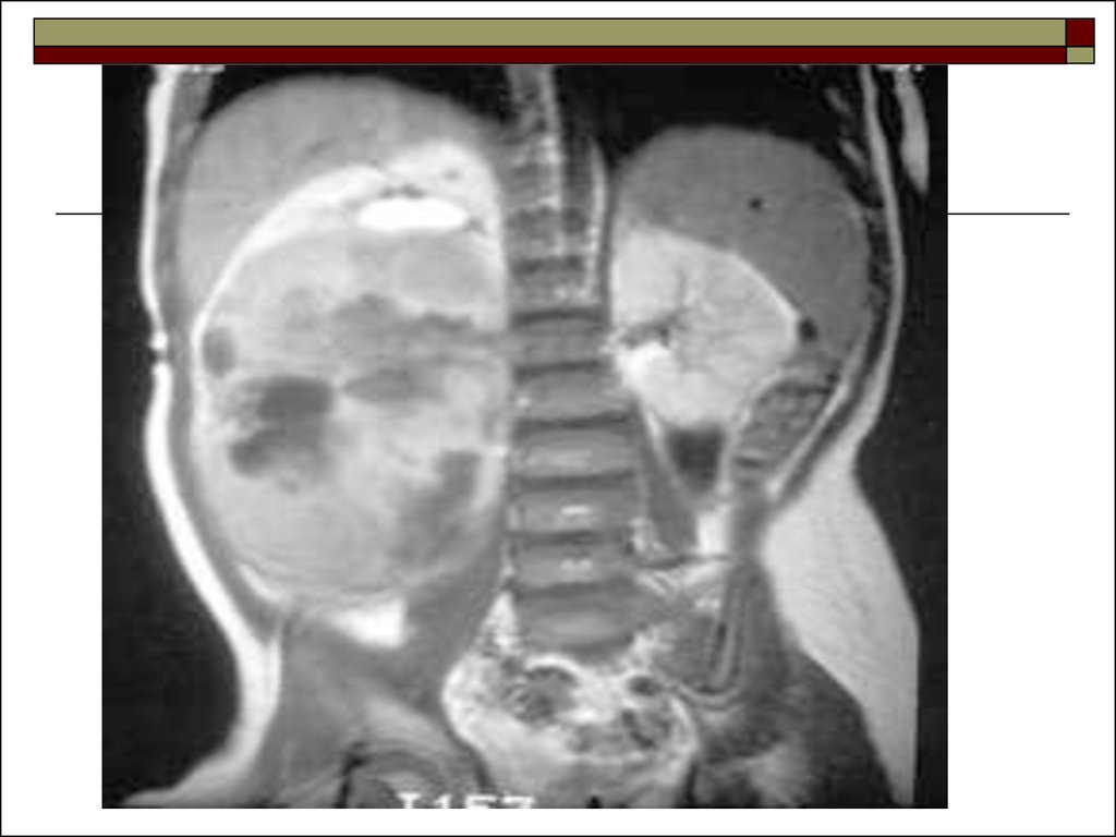
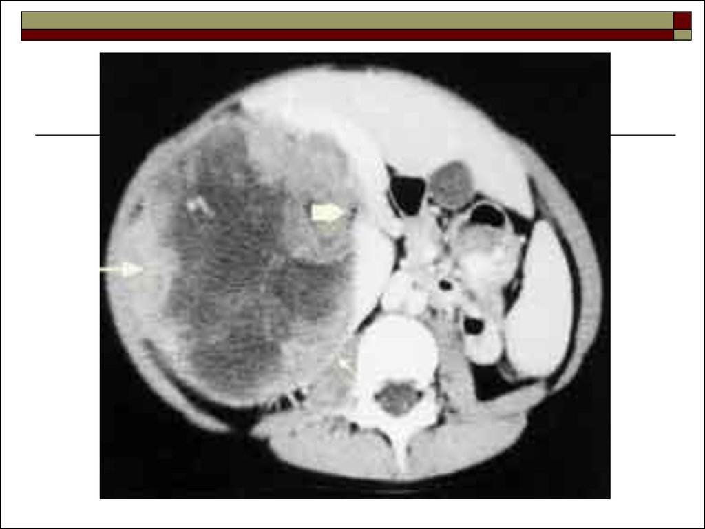
111.
IVUenlargement of affected part of the kidney
distortion of the PCS by a tumor
Ultrasound
heterogenous echotexture mass lesion arising from a
pole of the kidney
CT scan
well defined soft tissue density lesion which
enhances heterogeneously after contrast
administration arising from a pole of kidney
112.
113.
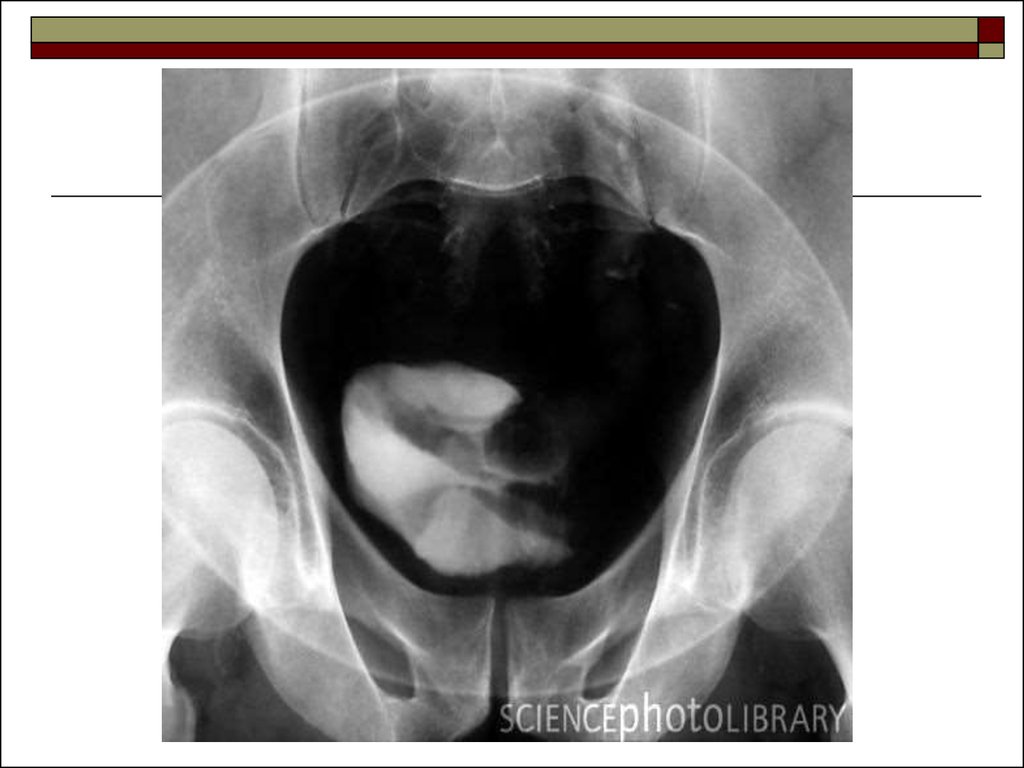
114. Diverticulum of bladder
It is outpouching of mucosa trough the wallsof the bladder.
Types:
congenital - due to weakness in the muscular
layers
acquired - this is usually secondary to distal
obstruction
115.
Imaging appearancesthe diverticulum may have wide neck or narrow neck
in the diverticulum with wide neck the diverticulum
gets filled with contrast while contrast enters bladder
and empties readily
in the narrow necked one, stasis of contrast for a
long period is noted; this type predisposes to urine
stasis, infection and stone formation
116.
117.
118.
119.
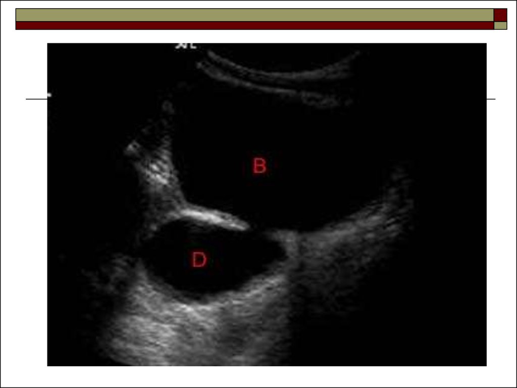
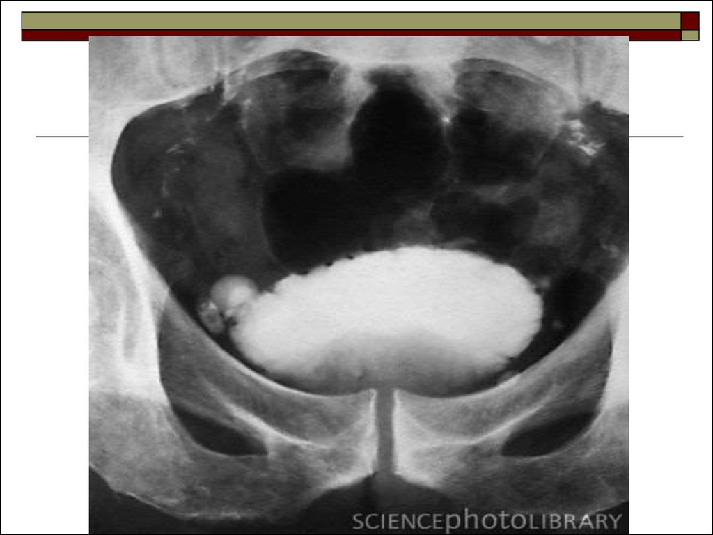
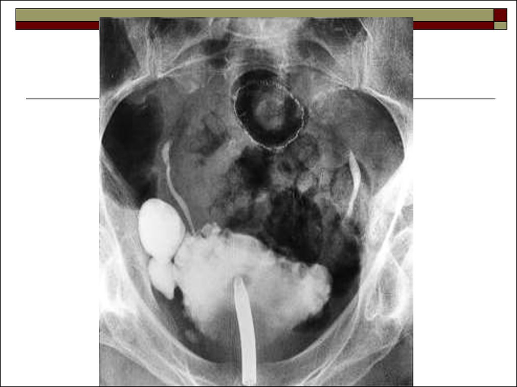
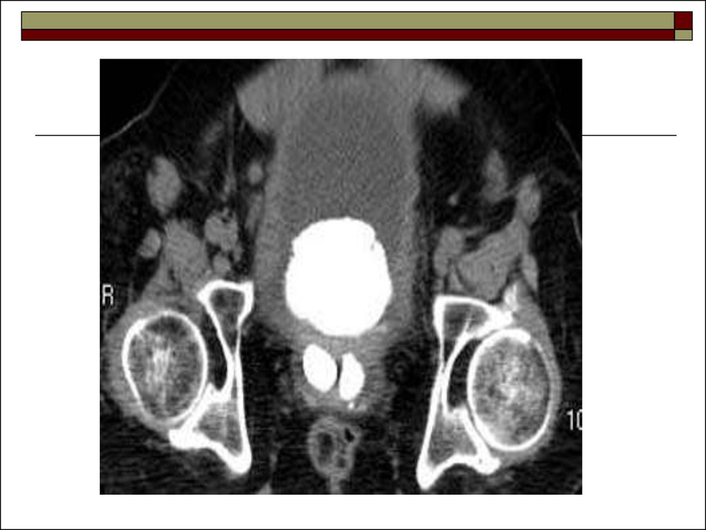
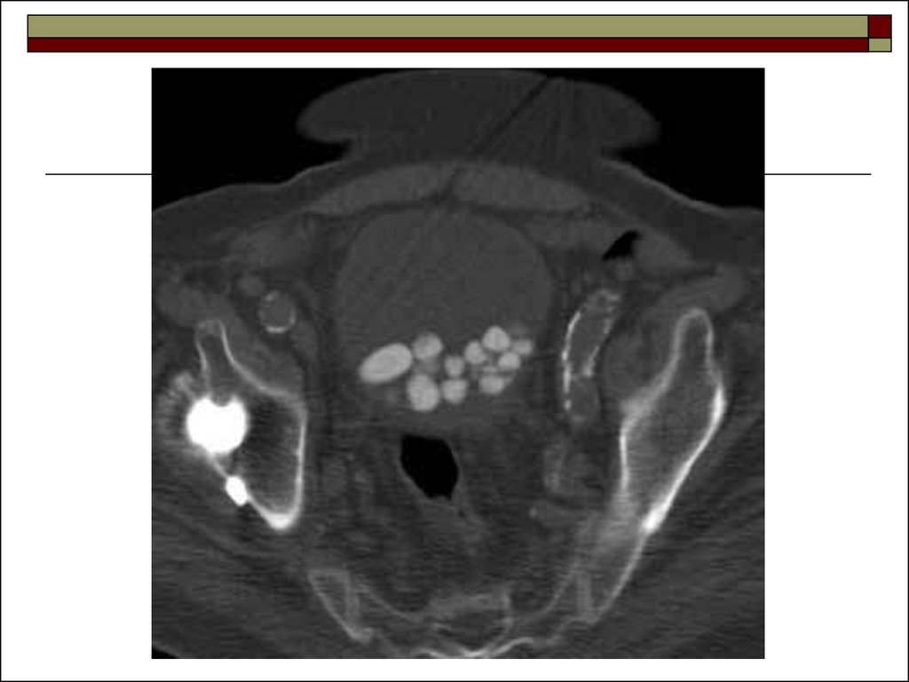
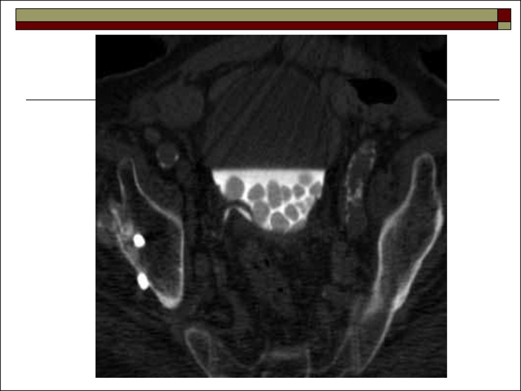
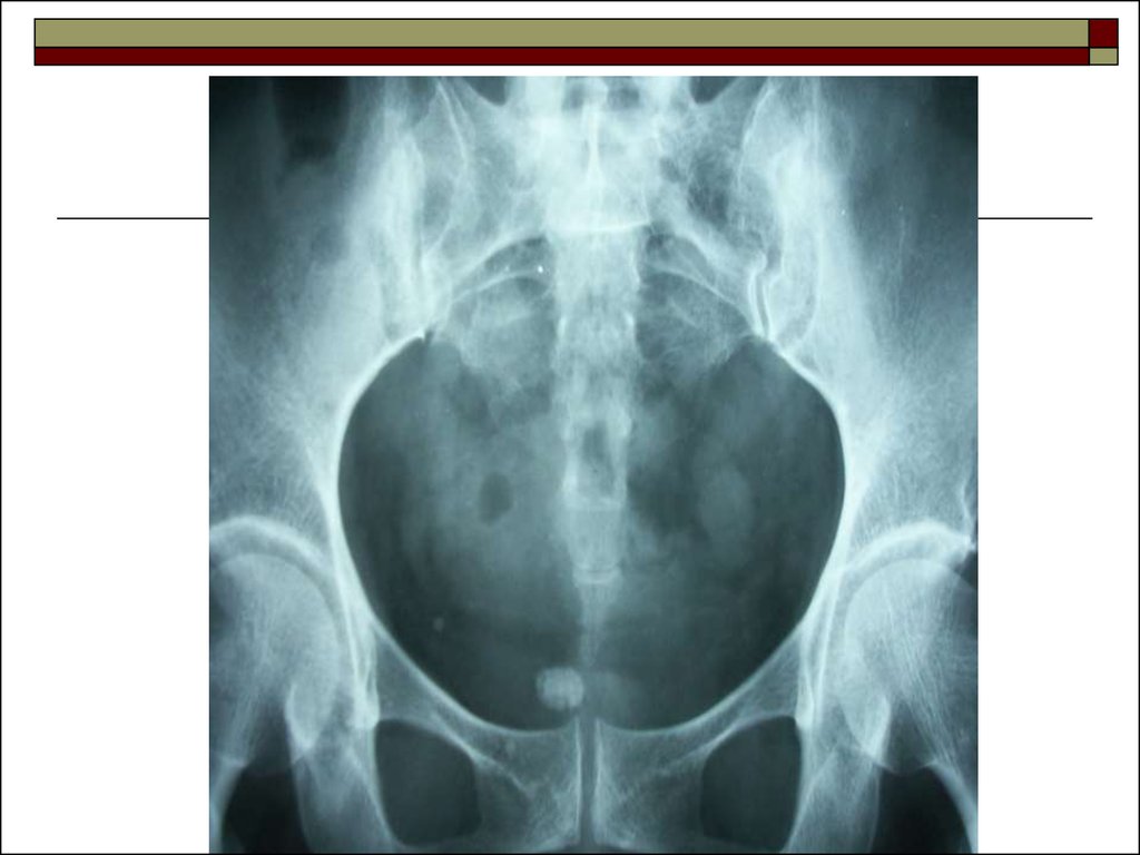
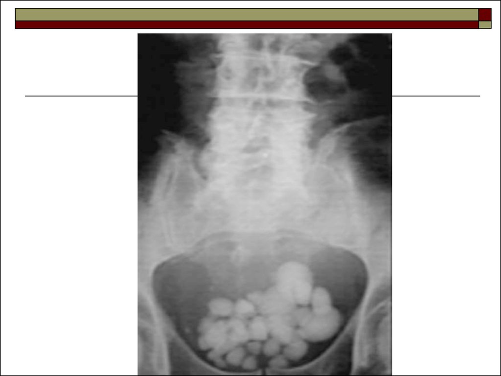
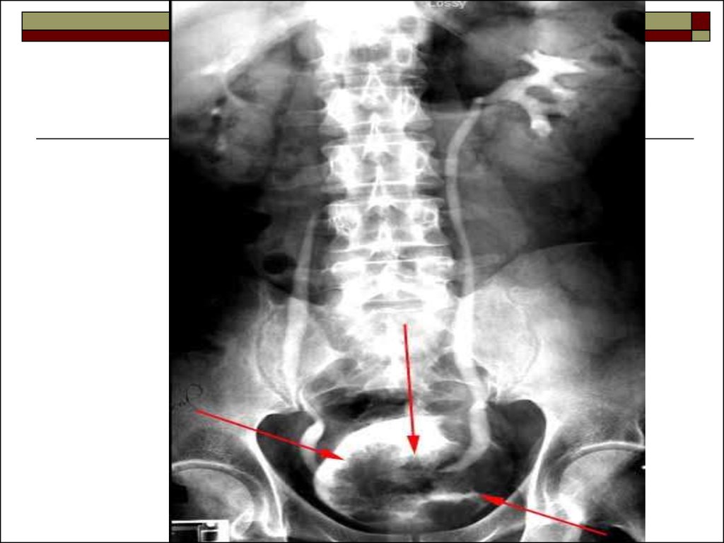
120. Bladder calculi
usually secondary to outflowobstruction/bladder diverticula or urinary tract
infections
it may occur in cases of hyperparathyroidism,
hyperuricemia or cystinuria
usually composed of triple phosphate and are
radio-opaque
121.
urinary bladder stones mimics phlebolith(stones in the venous wall) and should be
differentiated from it: phleboliths have central
lucency; bladder calculi do not have the
central lucency
ultrasound is modality of choice for diagnosis
of bladder calculi: calculi will be seen as
echogenic structure which show mobility
122.
123.
124.
125.
126.
127.
128. Bladder tumors
It commonly occurs in posterior and lateral wallsnear vesico-ureteric junction.
Types
epithelial tumors: almost 90% epithelial tumors are
malignant
nonepithelial tumor: 2.1 benign: papilloma,
leiomyoma, fibroma
2.2malignant
129.
Epithelial tumors:90%-transitional cell ca
1-10%-squamous cell ca
Clinical features
painless hematuria
Imaging
130.
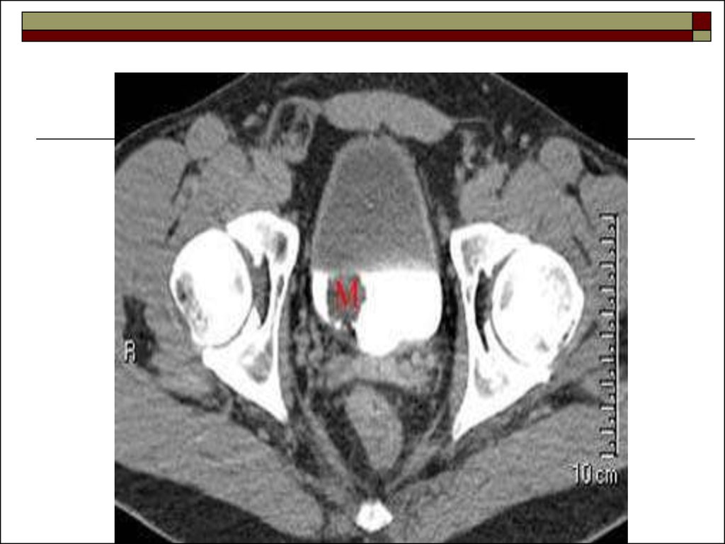
IVUfilling defect in the bladder
decreased capacity of bladder
may not detect small tumors
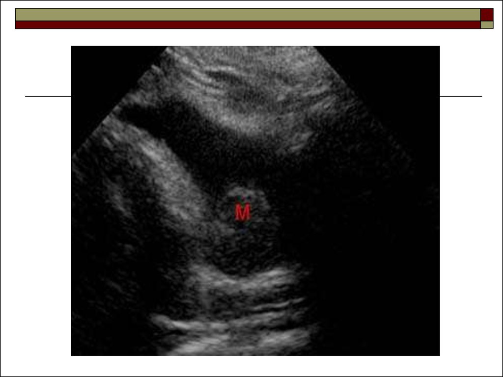
Ultrasound
focal irregular wall thickening
papillary mass protruding into the lumen of the
bladder
131.
132.
133.
134.
135.
136. Small smooth kidney
Unilateralischaemia due to renal artery stenosis
post obstructive atrophy
Bilateral
arterial hypertension
chronic glomerulonephritis
causes of unilateral small smooth kidney occurring
bilaterally
137. Large smooth kidney
Unilateralcompensatory hypertrophy
after trauma due to hematoma
gross hydronephrosis
Pyonephrosis
Bilateral
inflammatory: acute interstitial nephritis
neoplastic: leukemia, lymphoma
deposition of abnormal protein: amyloid, multiple myeloma