


")


 Медицина
Похожие презентации:


Drugs used in endocrine disorders
1.
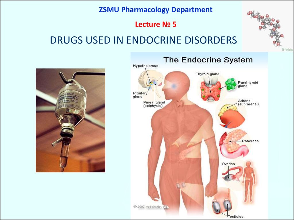
ZSMU Pharmacology DepartmentLecture № 5
DRUGS USED IN ENDOCRINE DISORDERS
2.
23.
Mechanism of actionOnce hormones reach a responsive cell, they bind with receptors
in the cell membrane ( protein hormones) or
inside the cell ( steroid and thyroid hormones).
Receptors may be increased (up-regulation) when there are
low levels of hormone or decreased (down-regulation) when
there are excessive amounts of hormone.
The hormone–receptor complex initiates intracellular reactions.
Many hormones act as a 1st messenger to the cell, and
the hormone–receptor complex activates a 2nd messenger.
The 2nd messenger then activates intracellular structures to
produce characteristic cellular functions and products.
4.
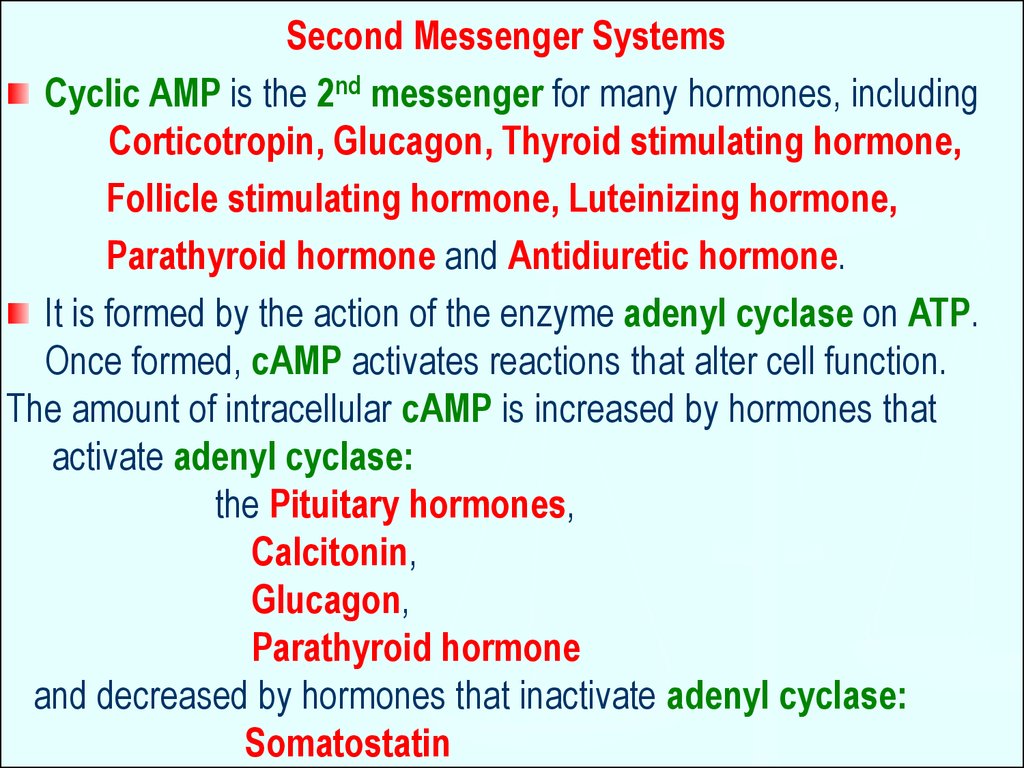
Second Messenger SystemsCyclic AMP is the 2nd messenger for many hormones, including
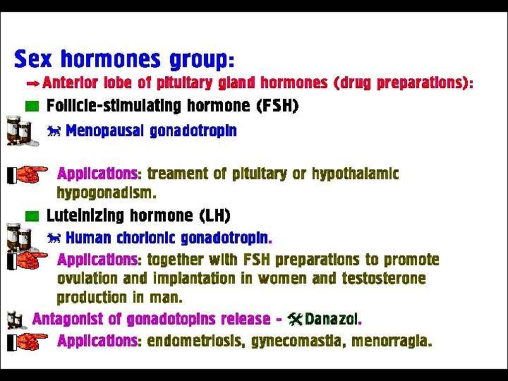
Corticotropin, Glucagon, Thyroid stimulating hormone,
Follicle stimulating hormone, Luteinizing hormone,
Parathyroid hormone and Antidiuretic hormone.
It is formed by the action of the enzyme adenyl cyclase on ATP.
Once formed, cAMP activates reactions that alter cell function.
The amount of intracellular cAMP is increased by hormones that
activate adenyl cyclase:
the Pituitary hormones,
Calcitonin,
Glucagon,
Parathyroid hormone
and decreased by hormones that inactivate adenyl cyclase:
Somatostatin
5.
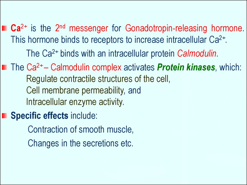
Ca2+ is the 2nd messenger for Gonadotropin-releasing hormone.This hormone binds to receptors to increase intracellular Ca2+.
The Ca2+ binds with an intracellular protein Calmodulin.
The Ca2+ – Calmodulin complex activates Protein kinases, which:
Regulate contractile structures of the cell,
Cell membrane permeability, and
Intracellular enzyme activity.
Specific effects include:
Contraction of smooth muscle,
Changes in the secretions etc.
6.
Some hormones activate cell membrane receptors and transformthem into phospholipase C, an enzyme that causes some of
the phospholipids in cell membranes to split into smaller molecules:
inositol triphosphate and diacylglycerol which act
as 2nd messengers to intracellular structures.
Inositol triphosphate mobilizes intracellular Ca2+ ions which fulfil
their functions as 2nd messengers.
Diacylglycerol activates an enzyme, protein kinase C that is
important in cell reproduction.
The lipid component of diacylglycerol is arachidonic acid the precursor for PGs, leukotrienes, and other local hormones
7.
Steroid Regulation of Protein SynthesisSteroid hormones are lipid soluble and cross cell membranes
easily.
Once inside the cell, the hormone molecules bind with specific
receptor proteins.
The hormone–receptor complex enters the nucleus of the cell
where it activates Gene Expression –
nucleic acids (DNA and RNA) and
the Genetic Code to synthesize
new proteins.
8.
89.
910.
1011.
1112.
1213.
Thyroid Hormones:Thyroxine (Tetraiodothyronine,T4 -contains 4 atoms of iodine)
Triiodothyronine (T3 - contains 3 atoms of iodine) is > potent and
has a more rapid onset but shorter duration of action
Calcitonin (a plasma Ca2+ lowering hormone).
L-Thyroxin (tab. 0.05 mg and 0.1 mg) is the drug of choice and
the standard replacement therapy for Hypothyroidism,
Endemic Goiter (a manifestation of iodine deficiency).
Triiodothyronine (tab. 0.02 mg and 0.05 mg) is the treatment of
choice for myxoedema coma, when its more rapid
action is required for emergency treatment.
Toxicity is related to thyroxine levels and manifests itself as
nervousness, heart palpitations and tachycardia,
intolerance to heat and unexplained weight loss.
14.
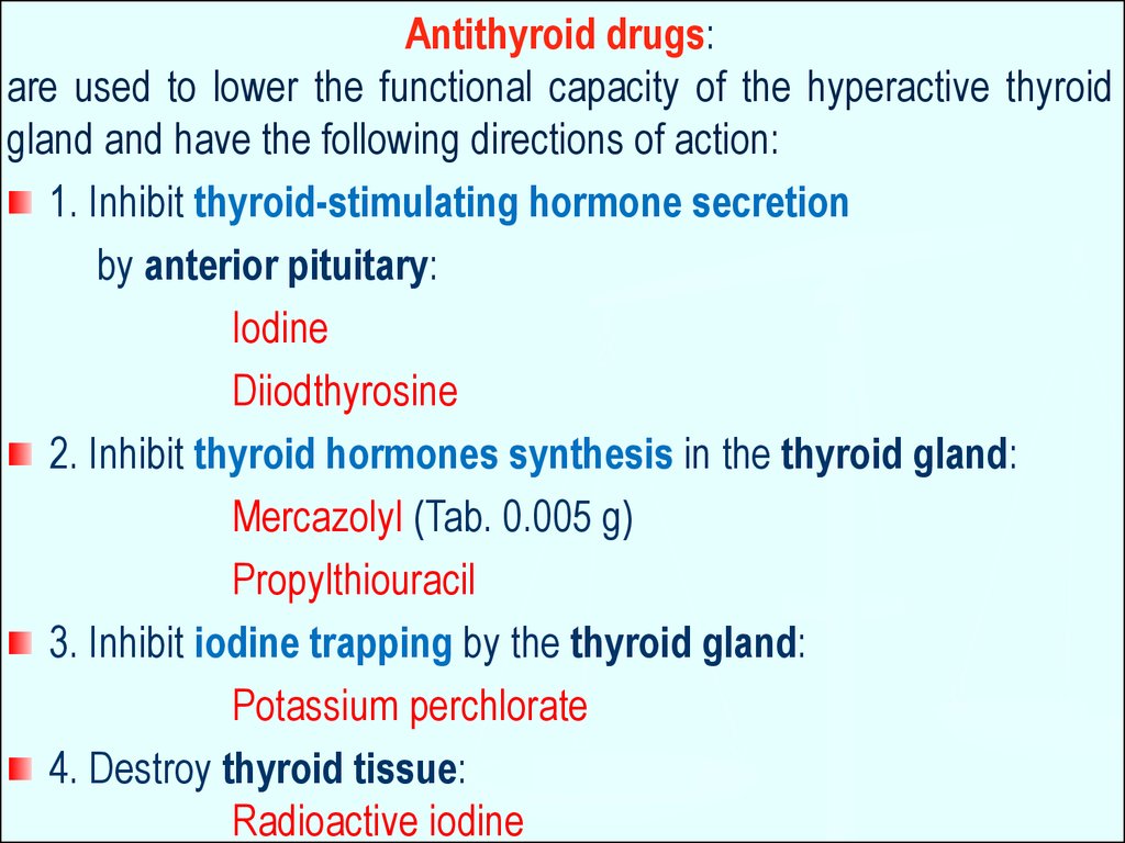
Antithyroid drugs:are used to lower the functional capacity of the hyperactive thyroid
gland and have the following directions of action:
1. Inhibit thyroid-stimulating hormone secretion
by anterior pituitary:
Iodine
Diiodthyrosine
2. Inhibit thyroid hormones synthesis in the thyroid gland:
Mercazolyl (Tab. 0.005 g)
Propylthiouracil
3. Inhibit iodine trapping by the thyroid gland:
Potassium perchlorate
4. Destroy thyroid tissue:
Radioactive iodine
15.
INSULIN PREPARATIONSA. Rapid Acting Insulin - max effect per 1-4 hours
short duration of action 4-8 hours
Regular Insulin - vial 5 and 10 ml – 40 U/ml
Insulin Lispro
Actrapid - vial 10 ml - 40 and 100 IU/ml SC or IV
B. Intermediate Acting Insulin - max effect per 6-12 hours
Intermediate duration of action 18- 24 hours
Semilente Insulin suspension
Lente Insulin: a mixture of 30% Semilente Insulin and
70% Ultralente Insulin
15
16.
C. Prolonged acting insulin:Ultralente Insulin
max effect per 12-18 hours
prolong duration of action 24-40 hours
Glucose
16
17.
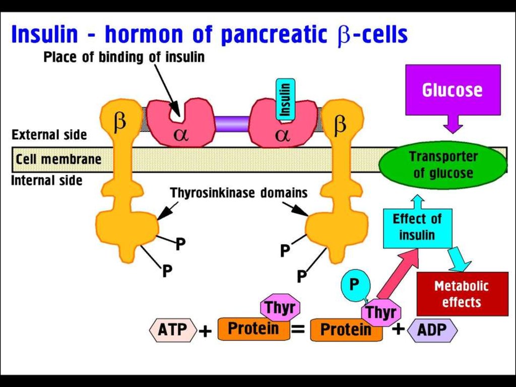
MECHANISM OF ACTION of INSULINInsulin binds to receptor on the surface of its target cells.
The receptor is a transmembrane glycoprotein complex consisting of
two α- and two β-subunits.
The α-subunits are entirely extracellular and each carries
an insulin-binding site,
the β-subunits are transmembrane proteins with
tyrosine kinase activity.
This activity is suppressed by the α-subunits, but
insulin binding causes a conformational change that activates
the tyrosine kinase activity of the β-subunits, which act
on each other and on other target proteins.
ATP levels rise and block K+ channels (KATP), leading to membrane
depolarization and an influx of Ca2+, which causes
pulsatile insulin exocytosis.
18.
1819.
1920.
Insulin is a fuel-storage hormone andaffects cell growth and differentiation.
Insulin ↓Blood Glucose by:
Glucose uptake into muscle and fat via
a transporter Glut-4
Glycogen synthesis
Glycogen breakdown
Gluconeogenesis
Adrenaline Blood Glucose by:
Inhibiting Insulin Release (via α2-Receptors)
Promoting Glycogenolysis (via β2-Receptors in
Striated Muscle and liver)
20
Somatostatin inhibits Insulin Release.
21.
Actrapid (vial 10 ml : 40 and 100 IU/ml for SC or IV) - is a fastacting human insulin produced in Saccharomyces cerevisiae
by recombinant DNA technology. It may be used
in combination with intermediate or long-acting insulin.
It is administered SC by injection in the abdominal wall, the thigh,
the gluteal region or the deltoid region.
Injection into a lifted skin fold minimizes the risk of unintended IM
injection. The needle should be kept under the skin
for at least 6 sec to make sure the entire dose is injected.
Injection sites should always be rotated within the same region in
order to reduce the risk of lipodystrophy.
SC injection into the abdominal wall ensures a faster absorption
than other injection sites. The duration of action will vary
according to the dose, injection site, blood flow, temperature
and level of physical activity.
22. Insulin Pens
2223.
2324. Insulin Inhaling Device (Exubera)
2425.
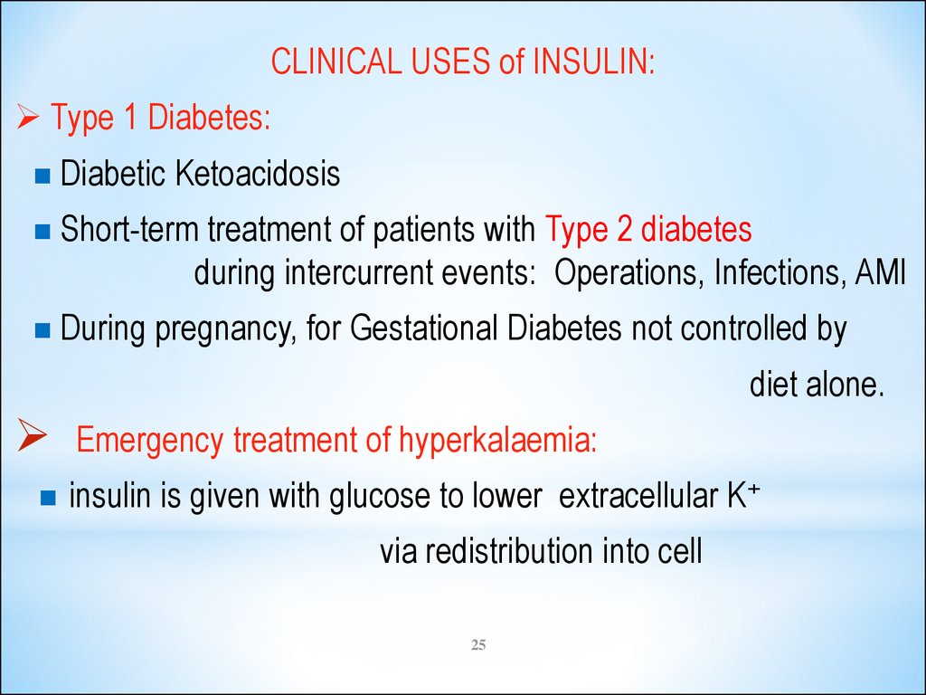
CLINICAL USES of INSULIN:Type 1 Diabetes:
Diabetic Ketoacidosis
Short-term treatment of patients with Type 2 diabetes
during intercurrent events: Operations, Infections, AMI
During pregnancy, for Gestational Diabetes not controlled by
diet alone.
Emergency treatment of hyperkalaemia:
insulin is given with glucose to lower extracellular K+
via redistribution into cell
25
26.
Oral (Synthetic) Hypoglycemic Agents:I. Stimulators of insulin release by beta cells:
1. Sulfonylurea derivatives:
I. Generation – moderate duration of action (8-24 hours):
Butamide (Tolbutamide)
II Generation – Long duration of action (24-60 hours):
Chlorpropamide (tab. 0.1 and 0.25 g)
Glibenclamide (tab.5 mg)
Glipizide (tab. 5 mg)
2. Meglitinides:
Repaglinide (tab. 1 mg)
Nateglinide (tab. 120 mg)
27.
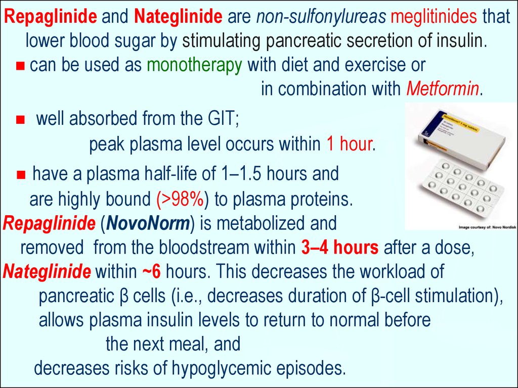
Repaglinide and Nateglinide are non-sulfonylureas meglitinides thatlower blood sugar by stimulating pancreatic secretion of insulin.
can be used as monotherapy with diet and exercise or
in combination with Metformin.
well absorbed from the GIT;
peak plasma level occurs within 1 hour.
have a plasma half-life of 1–1.5 hours and
are highly bound (>98%) to plasma proteins.
Repaglinide (NovoNorm) is metabolized and
removed from the bloodstream within 3–4 hours after a dose,
Nateglinide within ~6 hours. This decreases the workload of
pancreatic β cells (i.e., decreases duration of β-cell stimulation),
allows plasma insulin levels to return to normal before
the next meal, and
decreases risks of hypoglycemic episodes.
28.
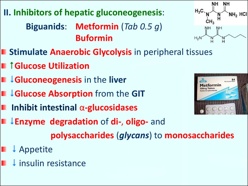
II. Inhibitors of hepatic gluconeogenesis:Biguanids: Metformin (Tab 0.5 g)
Buformin
Stimulate Anaerobic Glycolysis in peripheral tissues
Glucose Utilization
Gluconeogenesis in the liver
Glucose Absorption from the GIT
Inhibit intestinal α-glucosidases
Enzyme degradation of di-, oligo- and
polysaccharides (glycans) to monosaccharides
Appetite
insulin resistance
29.
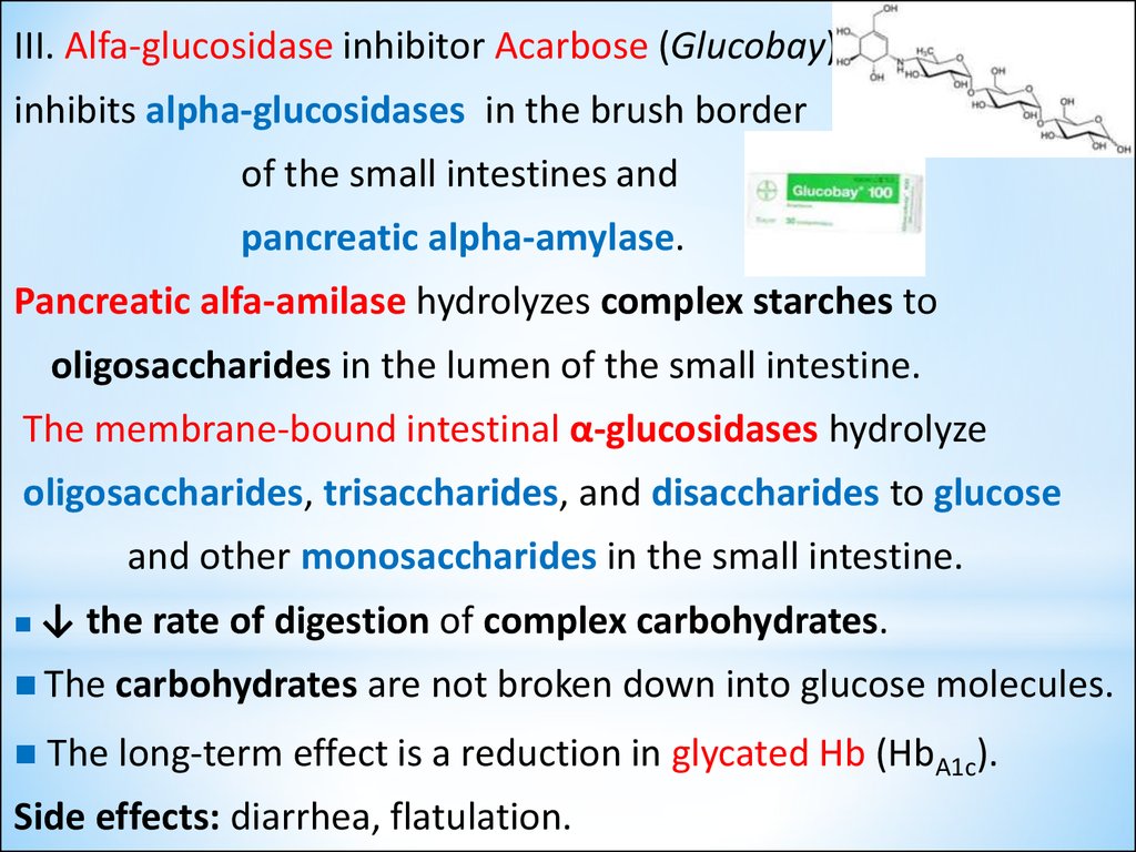
III. Alfa-glucosidase inhibitor Acarbose (Glucobay)inhibits alpha-glucosidases in the brush border
of the small intestines and
pancreatic alpha-amylase.
Pancreatic alfa-amilase hydrolyzes complex starches to
oligosaccharides in the lumen of the small intestine.
The membrane-bound intestinal α-glucosidases hydrolyze
oligosaccharides, trisaccharides, and disaccharides to glucose
and other monosaccharides in the small intestine.
↓ the rate of digestion of complex carbohydrates.
The carbohydrates
are not broken down into glucose molecules.
The long-term effect is a reduction in glycated Hb (HbA1c).
Side effects: diarrhea, flatulation.
30.
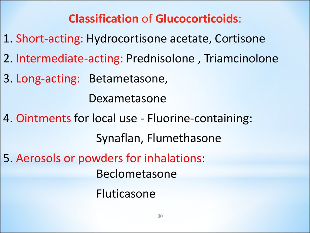
Classification of Glucocorticoids:1. Short-acting: Hydrocortisone acetate, Cortisone
2. Intermediate-acting: Prednisolone , Triamcinolone
3. Long-acting: Betametasone,
Dexametasone
4. Ointments for local use - Fluorine-containing:
Synaflan, Flumethasone
5. Aerosols or powders for inhalations:
Beclometasone
Fluticasone
30
31.
Action on mediators of inflammatoryand immune response:
GCs change Gene Expression:
Production of prostanoids owing to
Decreased Expression of COX-2
Generation of cytokines –
IL 1-6, IL-8, TNF- and cell adhesion factor –
through inhibition of transcription of
the relevant genes
Complement components in the plasma
Generation of induced NO
Histamine release from basophils
IgG production.
31
32.
Clinical uses of Glucocorticoids:1. Replacement therapy for patients with adrenal failure Addison’s disease
2. Anti-Inflammatory Immunosuppressive Therapy:
Asthma
Inflammatory conditions of skin, eye, ear or nose:
Eczema, Allergic Conjunctivitis or Rhinitis – topically
Hypersensitivity States: Severe Allergic Reactions – IV
3. In neoplastic diseases:
In
combination with Cytotoxic Drugs as a component of
Antiemetic Treatment in the treatment of Specific Malignancies
33. Adverse effects: Cushing’s syndrome:
Moon face, with red cheeksThin arms and legs: muscle wasting
BP, Itracranial Hypertension
Osteoporosis
Cataracts
Thinning of skin
Increased abdominal fat
Buffalo hump
Euphoria
Depression or emotional lability
Avascular necrosis of femoral head
Appetite, Obesity, Hyperglycemia
33
34.
3435. OESTROGENS
Natural:Estradiol - amp 0.1%-1 ml
Estriol - Tab 1 mg
Synthetic:
Ethinylestradiol - Tab 0,01 mg
Synoestrol Tab 1 mg, amp 0.1% - 1 ml
Mestranol
35
36. CLINICAL USEs of OESTROGENS:
Replacement therapy:- Primary Ovarian Failure
(e.g., Turner’s syndrome)
- Secondary Ovarian Failure
(Menopausal)
Contraception
Prostate and Breast Cancer
36
37. ANTIOESTROGENS
Clomiphene - Tab. 50 mgTamoxifen - Tab. 20 mg
Clomiphene:
Interfering with the Negative Feedback of
oestrogens on the hypothalamus and pituitary
=> Secretion of Gn-RH
Secretion of Gonadotropins
=> a Stimulation of Ovulation
Clinical use:
Infertility with Anovulatory Cycles
Breast Tumors
37
38. PROGESTOGENS
1. The naturally occurring hormone and itsderivatives:
Progesteron amp. 1% - 1 ml of oil solution
Oxyprogesterone Caproate amp. 25% - 1 ml
Pregnin Tab. 0.01
2. Testosterone derivatives:
Norethisterone Tab. 5 mg
Norgestrel
Desogestrel
Gestodene
38
39.
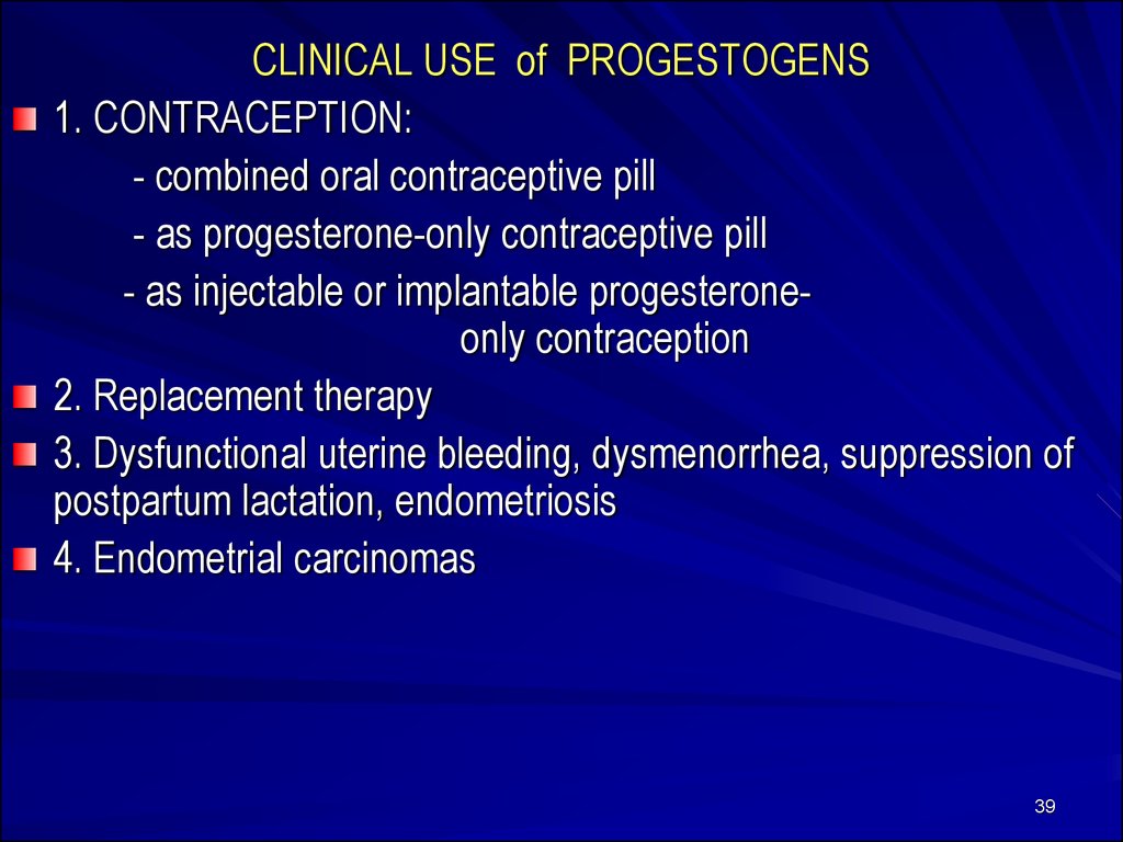
CLINICAL USE of PROGESTOGENS1. CONTRACEPTION:
- combined oral contraceptive pill
- as progesterone-only contraceptive pill
- as injectable or implantable progesteroneonly contraception
2. Replacement therapy
3. Dysfunctional uterine bleeding, dysmenorrhea, suppression of
postpartum lactation, endometriosis
4. Endometrial carcinomas
39