


 Медицина
Похожие презентации:


Hypoplastic left heart syndrome
1.
Crimea State Medical University,Simferopol
Biology Project
Teacher – Anna Zhukova
Made By - Mohammad Imran Sheikh
LA- 194 A
2.
3.
Hypoplastic left heart syndrome is a complex and rare heart defect present atbirth (congenital). The left side of the heart is critically underdeveloped in
hypoplastic left heart syndrome.
If your baby is born with hypoplastic left heart syndrome, the left side of the
heart can't effectively pump blood to the body. Instead, the right side of the
heart must pump blood to the lungs and to the rest of the body.
Medication to prevent closure of the connection (ductus arteriosus) between
the right and left sides, followed by either surgery or a heart transplant, is
necessary to treat hypoplastic left heart syndrome. With advances in care, the
outlook for babies born with hypoplastic left heart syndrome is better now than
in the past.
4.
Babies born with hypoplastic left heart syndrome usually are seriously ill soonafter birth. Hypoplastic left heart syndrome symptoms include:
Grayish-blue skin color (cyanosis)
Rapid, difficult breathing
Poor feeding
Cold hands and feet
Weak pulse
Being unusually drowsy or inactive
5.
At birth, the ductus arteriosus is still open, and there is higher than normalresistance to blood flow in the lungs. This allows for adequate oxygenation via
mixing between the atria and a normal appearance at birth. When the ductus begins
to close and pulmonary vascular resistance decreases, blood flow through the
ductus is restricted and flow to the lungs is increased.
In typical anatomy, the left side of the heart receives oxygen-rich blood from the
lungs and pumps it to the rest of the body. In people with HLHS, the aorta and left
ventricle are underdeveloped (beginning in utero), and the aortic
and mitral valves are either too small to allow sufficient blood flow or are atretic
(closed) altogether. As blood returns from the lungs to the left atrium, it cannot
be pumped to the rest of the body by the left ventricle. The neonate is reliant on
blood flowing through an atria septal defect to mix oxygenated and deoxygenated
blood, and on a patent ductus arteriosus to allow blood to reach the aorta and the
systemic circulation via the right ventricle. This is what defines HLHS as a "single
ventricle" defect.
6.
Hypoplastic left heart syndrome can be diagnosed prenatally or after birthvia echocardiography. Typical findings include a small left ventricle and
aorta, abnormalities of the mitral and aortic
valves, retrograde flow in the transverse arch
of the aorta, and left-to-right flow
between the atria. It is often
recognized during the second
Trimester of pregnancy, between 18
and 24 weeks’
gestation.
7.
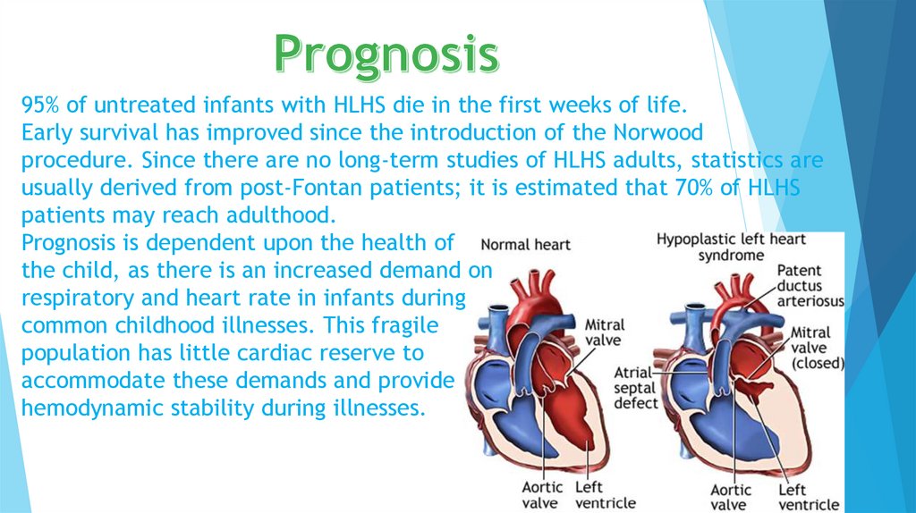
95% of untreated infants with HLHS die in the first weeks of life.Early survival has improved since the introduction of the Norwood
procedure. Since there are no long-term studies of HLHS adults, statistics are
usually derived from post-Fontan patients; it is estimated that 70% of HLHS
patients may reach adulthood.
Prognosis is dependent upon the health of
the child, as there is an increased demand on
respiratory and heart rate in infants during
common childhood illnesses. This fragile
population has little cardiac reserve to
accommodate these demands and provide
hemodynamic stability during illnesses.
8.
Double aortic arch is a relatively rare congenital cardiovascular malformation.DAA is an anomaly of the aortic arch in which two aortic arches form a
complete vascular ring that can compress the trachea and/or esophagus. Most
commonly there is a larger (dominant) right arch behind and a smaller
(hypoplastic) left aortic arch in front of the trachea/esophagus. The two arches
join to form the descending aorta which is usually on the left side (but may be
right-sided or in the midline). In some cases the
end of the smaller left aortic arch closes
(left atretic arch) and the vascular tissue
becomes a fibrous cord. Although in these cases a
complete ring of two patent aortic arches is not
present, the term ‘vascular ring’ is the accepted
generic term even in these anomalies.
9.
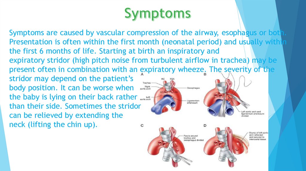
Symptoms are caused by vascular compression of the airway, esophagus or both.Presentation is often within the first month (neonatal period) and usually within
the first 6 months of life. Starting at birth an inspiratory and
expiratory stridor (high pitch noise from turbulent airflow in trachea) may be
present often in combination with an expiratory wheeze. The severity of the
stridor may depend on the patient’s
body position. It can be worse when
the baby is lying on their back rather
than their side. Sometimes the stridor
can be relieved by extending the
neck (lifting the chin up).
10.
Myocardial ischemia occurs when the blood flow through one or more of yourcoronary arteries is decreased. The low blood flow decreases the amount of
oxygen your heart muscle receives.
Myocardial ischemia can develop slowly as arteries become blocked over time.
Or it can occur quickly when an artery becomes blocked suddenly.
Conditions that can cause myocardial ischemia include:
1. Coronary artery disease (atherosclerosis). Plaques made up mostly of
cholesterol build up on your artery walls and restrict blood flow. Atherosclerosis
is the most common cause of myocardial ischemia.
11.
Surgical correction is indicated in all double aortic arch patients withobstructive symptoms (stridor, wheezing, pulmonary infections, poor
feeding with choking). If symptoms are absent a conservative approach
(watchful waiting) can be reasonable. Children with very mild symptoms may
outgrow their symptoms but need regular follow-up.
1. Anesthesia and intraoperative monitoring.
2. Open division of vascular ring.
3. Postoperative care.
12.
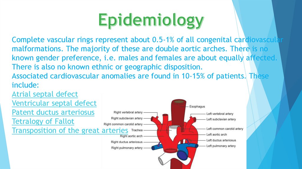
Complete vascular rings represent about 0.5-1% of all congenital cardiovascularmalformations. The majority of these are double aortic arches. There is no
known gender preference, i.e. males and females are about equally affected.
There is also no known ethnic or geographic disposition.
Associated cardiovascular anomalies are found in 10-15% of patients. These
include:
Atrial septal defect
Ventricular septal defect
Patent ductus arteriosus
Tetralogy of Fallot
Transposition of the great arteries
13.
Transposition of the great vessels (TGV) is a group ofCongenital heart defects involving an abnormal spatial
arrangement of any of the great vessels: superior and/or
inferior venae cavae, pulmonary artery, pulmonary veins,
and aorta. Congenital heart diseases involving only the
primary arteries (pulmonary artery and aorta)
belong to a sub-group called transposition
of the great arteries.
14.
Transposed vessels can present a large variety of atriovenous, ventriculoarterial and/or arteriovenous discordance. The effects may
range from a change in blood pressure to an interruption in circulation, depending
on the nature and degree of the misplacement and which vessels are involved.
Although "transposed" literally means "swapped", many types of TGV involve
vessels that are in abnormal positions, while not actually being swapped with each
other. The terms TGV and TGA are most commonly used in reference to dextroTGA – in which the arteries are in swapped positions; however, both terms are
also commonly used, though to a slightly lesser extent, in reference to levo-TGA –
in which both the arteries and the ventricles are swapped; while other defects in
this category are almost never referred to by either of these terms.
15.
On chest X-ray, transposition of the great vessels typicallyshows a cardio-mediastinal silhouette appearing as an
"egg on a string", wherein in which the enlarged heart
represents an egg on its side and the narrowed,
atrophic thymus of the superior mediastinum represents
the string.
X-ray showing characteristic finding
in case of Transposition of the great
vessels which is called egg on side
sign.
16.
For newborns with transposition, prostaglandins can be given to keepthe ductus arteriosus open which allows mixing of the otherwise isolated
pulmonary and systemic circuits. Thus oxygenated blood that recirculates
back to the lungs can mix with blood that circulates throughout the body.
The arterial switch operation is the definitive treatment for dextrotransposition. Rarely the arterial switch is not feasible due to
particular coronary artery anatomy and an atrial switch operation is
preferred.