


 Antibody Screen")


 Медицина
Похожие презентации:


 isoimmunization")


Rh Incompatibility and Disease
1. Rh Incompatibility and Disease
Name – Vanshul RanaGroup – 173B
International Medical Faculty
2. Rh Disease
• Occurs during pregnancy when there is anincompatibility between the blood types of the
mother and fetus
3. Blood Types
• A, B, O blood groups are specific types ofproteins found on the surface of RBC’s
• Also found in the cells and other body fluids
(saliva, semen, etc)
• O represents neither protein being present on
RBC
• Possible groups include: A, B, AB, or O
• A, B, O groups most important for transfusions
4. Rh Factor
• Proteins (antigens) occurring only on surfaceof RBC’s
• Rh + if proteins present
• Rh – if proteins absent
• A+, A-, B+, B-, AB+, AB-, O+, O• Most important for pregnancy
• Inheritance is Autosomal Dominant
• 15% Caucasian population is Rh-
5. Nomenclature
• Correct to say Rh(D) + or –• Rh blood system has other antigens: C, c, D, E,
e
• D is by far the most common and the only
preventable one
• Weak D (Du) also exists
• Also non Rhesus groups such as Kell, MNS,
Duffy (Fy) and Kidd (Jk) exist
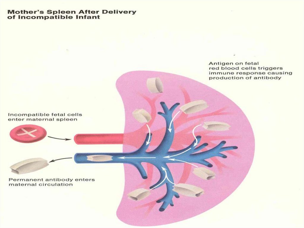
6. Why Does Rh Status Matter?
Fetal RBC cross to maternal circulationMaternal immune system recognizes foreign
antigens if fetus Rh + and mother Rh –
Antibodies are formed against fetal antigens
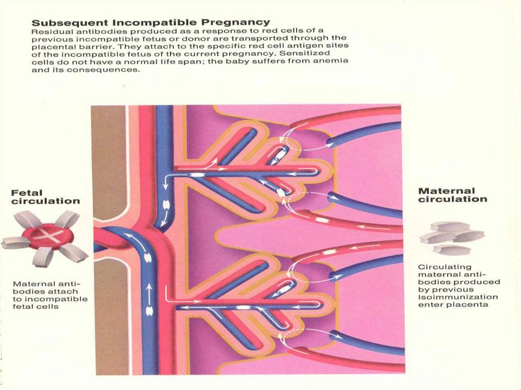
Subsequent pregnancy with Rh+ fetus,
immune system activated
and large amounts of Ab formed
IgG Ab cross placenta & attack fetal RBC
Fetal anemia, hydrops, etc
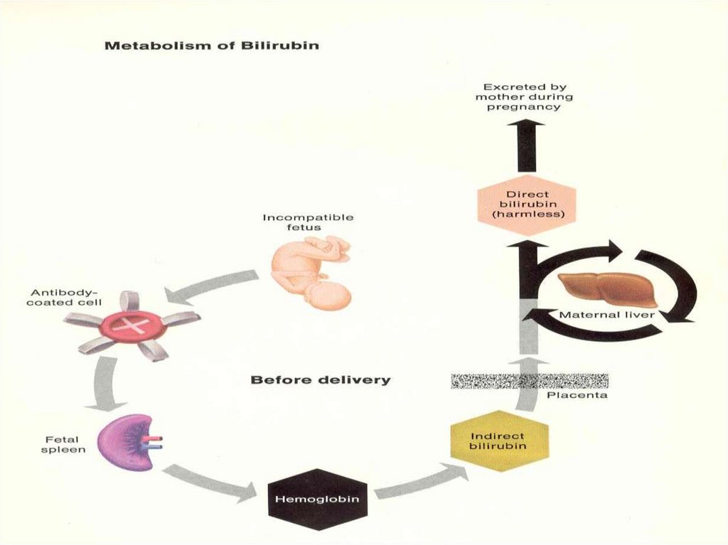
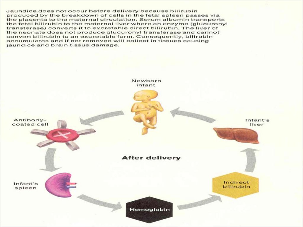
7.
8.
9.
10.
11.
12.
13. Pathophysiology
• Rh(D) antigen expressed by 30 d GA• Many cells pass between maternal & fetal circulation
including at least 0.1 ml blood in most deliveries but
generally not sufficient to activate immune response
• Rh antigen causes > response than most
• B lymphocyte clones recognizing foreign RBC antigen are
formed
14. Pathophysiology cont…
• Initial IgM followed by IgG in 2 wks- 6 mths• Memory B lymphocytes activate immune
response in subsequent pregnancy
• IgG Ab cross placenta and attach to fetal RBC’s
• Cells then sequestered by macrophages in
fetal spleen where they get hemolyzed
• Fetal anemia
15.
16.
17. Causes of RBC Transfer
abortion/ectopic
partial molar pregnancy
blighted ovum
antepartum bleeding
special procedures (amniocentesis, cordocentesis,
CVS)
external version
platelet transfusion
abdominal trauma
inadvertent transfusion Rh+ blood
postpartum (Rh+baby)
18. General Screening
ABO & Rh Ab @ 1st prenatal visit
@ 28 weeks
Postpartum
Antepartum bleeding and before giving any
immune globulin
• Neonatal bloods ABO, Rh, DAT
19. Gold Standard Test
• Indirect Coombs:-mix Rh(D)+ cells with maternal serum
-anti-Rh(D) Ab will adhere
-RBC’s then washed & suspended in Coombs
serum (antihuman globulin)
-RBC’s coated with Ab will be agglutinated
• Direct Coombs:
-mix infant’s RBC’s with Coombs serum
-maternal Ab present if cells agglutinate
20. + Rh(D) Antibody Screen
• Serial antibody titres q2-4 weeks• If titre ≥1:16 - amniocentesis or MCA dopplers
and more frequent titres (q1-2 wk)
• Critical titre – sig risk hydrops
• ** amnio can be devastating in this setting
• U/S for dating and monitoring
• Correct dates needed for determining
appropriate bili levels (delta OD450)
21. U/S Parameters
• Non Reliable Parameters:Placental thickness
Umbilical vein diameter
Hepatic size
Splenic size
Polyhydramnios
• Visualization of walls of fetal bowel from
small
amounts intraabdominal fluid may be
st
1 sign of impending hydrops
• U/S reliable for hydrops (ascites, pleural
effusions, skin edema) – Hgb < 70
22.
23. Amniocentesis
Critical titre/previous affected infantAvoid transplacental needle passage
Bilirubin correlates with fetal hemolysis
∆ optical density of amniotic fluid @ 450nm on
spectral absorption curve
• Data plotted on Liley curve
24.
25. Liley Curve
• Zone I – fetus very low risk of severe fetalanemia
• Zone II – mild to moderate fetal hemolysis
• Zone III – severe fetal anemia with high
probability of fetal death 7-10 days
• Liley good after 27 weeks
• 98% sensitive for detecting anemia in upper
zone 2/ zone 3
26. Middle Cerebral Artery Dopplers
• Measures peak velocity of blood flow• Anemic fetus preserves O2 delivery to brain by
increasing flow
• Sensitivity of detecting severe anemia when
MCA >1.5 MoM approaches 100%
• Not reliable > 35 weeks GA
27. Fetus at Risk
• Fetal anemia diagnosed by:• amniocentesis
• cordocentesis
• ultrasound
hydrops
middle cerebral artery Doppler
• Treatment:
• intravascular fetal transfusion
• preterm birth
28. Infant at Risk
• Diagnosis:• history of HDN antibodies?
• early jaundice < 24 hours
• cord DAT (“Coomb’s”) positive (due to HDN or ABO
antibodies)
• Treatment:
• Phototherapy
• Exchange or Direct blood transfusion
29.
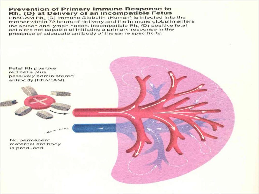
30. Prevention
• RhoGAM (120mcg or 300mcg)• Anti-D immune globulin
• Previously 16% Rh(D)- women became
alloimmunized after 2 pregnancies, 2% with
routine PP dose, and 0.1% with added dose @
28 wks