


 Медицина
Похожие презентации:

")
")


")
")


Туберкулез верхних дыхательных путей
1. Туберкулез верхних дыхательных путей
2.
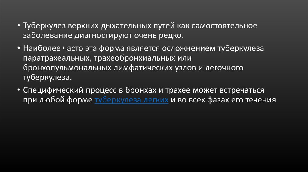
• Туберкулез верхних дыхательных путей как самостоятельноезаболевание диагностируют очень редко.
• Наиболее часто эта форма является осложнением туберкулеза
паратрахеальных, трахеобронхиальных или
бронхопульмональных лимфатических узлов и легочного
туберкулеза.
• Специфический процесс в бронхах и трахее может встречаться
при любой форме туберкулеза легких и во всех фазах его течения
3. Case report I
• A 48 year-old male presented with history of sore throat, occasionalfever, malaise for 1 year duration. He had been taking treatment in form
of various courses of antibiotics, analgesics, antihistamines since then
without any relief. He had no previous history of any serious illness,
chronic cough and other chest symptoms.
• While the general physical examination revealed normal findings, oral
examination showed enlarged right tonsil which was granular and
ulcerated. (Fig. 1a). It was firm on palpitation and did not bleed on
touch. There were no palpable neck nodes. Routine hematological
evaluation revealed a raised erythrocyte sedimentation rate (ESR) of
40 mm in first hour. Chest radiography was normal (Fig. 1b).
4. Случай 1
• 48-летний мужчина с историей боли в горле, прерывная лихорадка,недомогание на протяжении 1 года. Он принимал различных антибиотиков,
анальгетиков, антигистаминных препаратов с тех пор без каких-либо
помощи.
• У него не было каких-либо серьезных заболеваний, хронический кашель и
другие легочные симптомы.
• На осмотре обнаружено язва и зернистость правой миндалине. Плотная без
кровотечения. (Рис. 1а). Лимф. Узлов не увеличены. СОЭ высокая.
• Рентгенография грудной клетки без патологии (рис. 1b).
5.
a Granular tonsils, b chest X-ray (PA view) showing no abnormality, c after 1 month of ATT, d. after 6 month of ATT6.
• Biopsy was taken from ulcerated area of right tonsils and sent forhistopathological examination. Histology revealed granulomatous dense submucosal lymphoid infiltrate with multiple epithelioid histiocyte granulomas,
including multinucleate giant cells and caseous necrosis consistent with the
diagnosis of tuberculosis. Serological tests for acquired immunodeficiency
syndrome (AIDS) and syphilis were negative.
• Patient was managed with anti-tubercular treatment, with Rifampicin 450 mg,
ethambutol 800 mg, isoniazid 300 mg and pyrizinamide 1,500 mg for 2 months
and then a 4 month continuation phase of rifampicin 450 mg and isoniazid
300 mg. The patient had a good symptomatic response within the first month
of treatment (Fig. 1c) with total disappearance of ulcers and granular areas
after completion of treatment (Fig. 1d). On 1 year follow-up, he is free of
disease.
7. Case report II
• A 22 year old female presented with a history of odynophagia, haemoptysis,occasional fever and general malaise for 6 months. No history of cough,
previous chest infection or any other significant past illness was
given.Various empirical treatments consisting of antibiotics, antihistaminics,
analgesics etc. were tried at peripheral institutes but not to her satisfaction.
• On general physical examination, the patient was thin built and mildly
anaemic. There was no lymphadenopathy. Laboratory investigations
revealed Hb-8g% and ESR 50 mm in first hour, with rest of the parameters
being within normal limit. Chest X-ray shows no abnormality (Fig. 2b).
8.
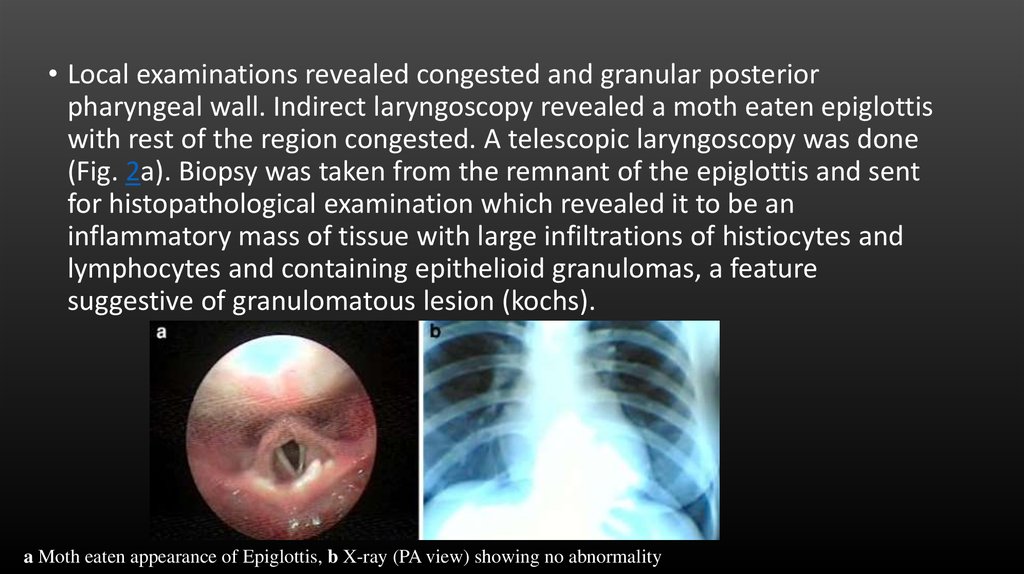
• Local examinations revealed congested and granular posteriorpharyngeal wall. Indirect laryngoscopy revealed a moth eaten epiglottis
with rest of the region congested. A telescopic laryngoscopy was done
(Fig. 2a). Biopsy was taken from the remnant of the epiglottis and sent
for histopathological examination which revealed it to be an
inflammatory mass of tissue with large infiltrations of histiocytes and
lymphocytes and containing epithelioid granulomas, a feature
suggestive of granulomatous lesion (kochs).
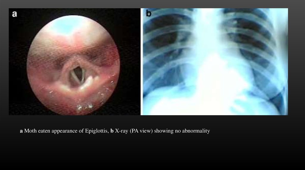
a Moth eaten appearance of Epiglottis, b X-ray (PA view) showing no abnormality
9.
a Moth eaten appearance of Epiglottis, b X-ray (PA view) showing no abnormality10. Case report III
• A 24 year old male student presented in the OPD with history of nasal block forthe past 6 month. He has no other difficulties. He had consulted a number of
times previously, and was treated as allergic rhinitis with steroid sprays, anti
histamines, antibiotics, analgesics etc., however with no improvement.
• On examinations, he was found to have no gross abnormalities within the nasal
cavities. Routine investigations revealed an abnormally high ESR of 86, with rest
of the parameters within normal limit. Nasal endoscopy revealed a smooth mass
in the nasopharynx (Fig. 3a). A ct scan of the nose and PNS was asked for,
whereby a report of a mass in nasopharynx with no extensions was given. The
mass was not vascular. (Fig. 3b). Punch biopsy was taken from the lesion and sent
for HPE. histopathological diagnosis was given to be granulomatous lesion,
probably tubercular. A repeat biopsy was done and chest x-ray was also done.
Chest x-ray came to be normal. (Fig. 3c). Repeat HPE report came as “fragments
of tissue lined with squamous and respiratory epithelium. The subepithelial tissue
shows lymphoid tissue with many epitheloid cell granuloma. Some of the
granulomas are confluent. Some show central necrosis. Stain for acid fast bacilli
are positive. Features consistant with tuberculosis.”
11.
a smooth mass innasopharynx,
b C T features of a mass
in nasopharynx,
c normal chest X-ray,
d mass after 1 month
of ATT, emass after
2 months of ATT, f mass
after 4 months of ATT
12. Case report III
• With all the above mentioned investigations, a diagnosis of primarytuberculosis of nasopharynx was reached and he was started on
standard dose of ATT. (4 drugs-rifampicin 450 mg, isoniazid 300 mg,
ethambutol 800 mg and pyrazinamide 1,500 mg for next 2 months
followed by rifampicin 450 mg and Isoniazid 300 mg for 4 months). He
showed remarkable regression of disease on follow-up at 1, 2 and
4 months. (Fig. 3d, e, f). At 6 months the mass completely regressed.
• The patient is under regular 6 monthly follow-up.
13. Conclusion
• Commonly URT primary TB can be misdiagnosed as traumatic oraphthous ulcer, haematological disorder, other granulomatous
disease, lymphoma or carcinoma. Diagnosis however must be based
on high degree of clinical suspicion backed by histopathological and
cytological identification of the bacilli. Hence, though uncommon,
primary tuberculosis of the upper respiratory tract should be borne in
mind, especially in a country like ours.
14. Reference
• 1. Chakravarti A, Pal S, Sahni JK. Primary tuberculosis of tonsil and posterior oropharyngeal wall. IndianJ Tuberc. 2008;55(1):48–50. [PubMed]
• 2. Usulu C, Oysu C, Ukluman B. Tuberculosis of the epiglottis, a case report. Eur Arch
Otolaryngol.2008;265(5):599–601. doi: 10.1007/s00405-007-0492-9. [PubMed] [Cross Ref]
• 3. Edizer DT, Karaman E, Mercan H, Alimoglu Y, Esen T, Cansiz H. Primary tuberculosis involving
epiglottis, a rare case report. Dysphagia. 2010;25(3):258–260. doi: 10.1007/s00455-009-92566. [PubMed][Cross Ref]
• 4. Savic D, Kosanovic H, Crejia D. Isolated tuberculosis of the nasopharynx. Srp Arh Celok
Lek.1961;89:99. [PubMed]
• 5. Martison FD. Primary tuberculosis of the nasopharynx in a nigerian. J Laryngol Otol. 1967;81:229–
234. doi: 10.1017/S0022215100066998. [PubMed] [Cross Ref]
• 6. Graff S. Die Bedentung des epipharynx fur die menschilidie. Pathologie Klin Wschr. 1936;27:953. doi:
10.1007/BF01777670. [Cross Ref]
• 7. Hollander AR. The nasopharynx: a study of 140 autopsy findings. Laryngoscope. 1946;56(6):282–
304.[PubMed]