


 Медицина
Похожие презентации:
")


")


")

Lung tuberculoma
1.
LUNG TUBERCULOMALecturer of the chair of phthisiopulmonology
of Saratov Medical University
Candidate of medical science Pankratova Liudmila
2.
Lung tuberculoma•Lung tuberculoma unites etiologically various
capsulated caseous foci of more than 1 cm in diameter
•The prevalence of tuberculoma among all forms of
pulmonary tuberculosis is
6-10 %.
3.
4.
THE LUNG TUBERCULOMA•The lung tuberculoma has the distinctive original
clinical and anatomical display of secondary form of
the pulmonary tuberculosis.
• It is characterized by the development of the dense
caseous focus (some time several focuses) in lungs, of
rounded forms, sharply outlined from surrounding
tissue by fibrotic capsule.
5.
CONDITIONS FOR FORMATION OF TUBERCULOMASPhysically active people.
Decreased virulence and pathogenicity of
Mycobacterium.
Increased resistance of the organism to Mycobacterium.
Social factors:
Male sex.
Age 20-40 years.
6.
The source of tuberculoma formationis mainly of two forms of pulmonary tuberculosis:
•infiltrative-pneumonic and focal.
•Besides this, tuberculoma forms from cavernous pulmonary
tuberculosis by means of filling the cavity with caseous
masses.
•Filled cavities refer to tuberculoma only conditionally, as the
filling of a cavity occurs mechanically, while tuberculomas are
an original phenomenon in lung tissue.
7.
PATHOMORPHOLOGICAL CLASSIFICATION OFTUBERCULOMAS.
Infiltrative-pneumonic
tuberculoma
Caseoma
Pseudotuberculoma
8.
INFILTRATIVE-PNEUMONICTUBERCULOMA
Presents as a round focus of pneumonia,
containing masses of clotty necrosis, clearly
limited from the surrounding lung tissue.
The capsule is weakly expressed.
Usually as a result of infiltrative tuberculosis.
9.
CASEOMABig focus of caseous pneumonia surrounded by a fresh
capsule.
Types:
1. Solitary homogenic caseoma (massive caseous focus
inside the capsule)
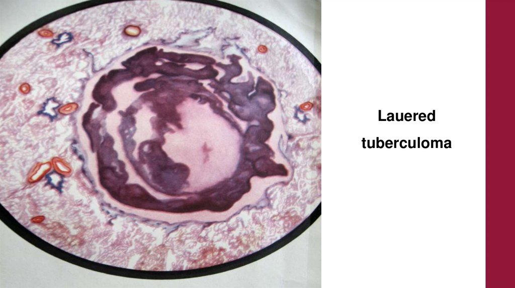
2. Solitary layered caseoma (alternation of layers of
caseous masses with layers of connective tissue).
3. Conglomerated caseoma (multiple caseous foci
surrounded by one capsule).
10.
Тuberculoma (solitary (homogeneous) and lauered)11.
Тuberculoma(solitary)
(homogeneous)
12.
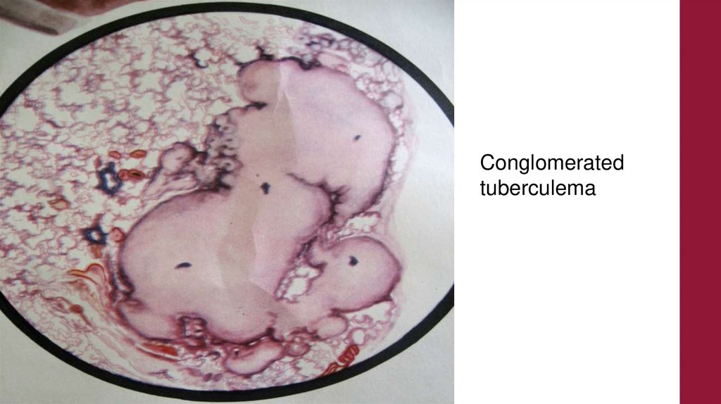
Conglomeratedtuberculema
13.
Laueredtuberculoma
14.
PSEUDOTUBERCULOMAOnly revealed in case of dynamic
observation of the patient and
histological examination of material
after operation.
15.
There are three clinical variants of tuberculomacourse:
1. progressing,
•described by occurrence of disintegration at some stage of
illness,
• perifocal inflammation around tuberculoma,
•bronchogenic dissemination in surrounding lung tissue.
16.
Variants of the tuberculema aggravation:•1) development of the perifocal inflammation;
•2) cavitation - discharge of the caseous
masses from a cavity, through draining
bronchus.
17.
2. stable –•absence of tuberculoma X-ray changes
•or rare aggravations without signs of tuberculoma
progressing;
18.
3. regressing tuberculomais characterized by its
•slow reduction in size,
•with subsequent formation of focus or group of foci,
induration field or combination of these changes.
19.
PREVALENCE OF TUBERCULOMA•The prevalence of tuberculoma among all forms of
pulmonary tuberculosis is 6-10 %.
•This tendency is explained by the fact that vast
infiltrative pneumonic processes, under treatment and
increased body resistance, become limited, condensed,
lose their aggravated course.
•However, the process does not heal completely and
precisely outlined dense formation remains.
20.
Clinical pattern•As tuberculoma itself is a parameter of high body
resistance, patients with this form of pulmonary
tuberculosis frequently are revealed accidentally, at
fluorography examinations,
•preventive examinations, and
•in presence of other diseases.
•Practically, patients have no complaints.
21.
Physical examination•At physical examination of a patient, there are no
pathological signs in lungs.
• Crackles are heard only at massive flare-up with
extensive infiltrative changes in lung tissue around
tuberculoma.
22.
CURRENT OF THE DISEASE1. Start of the disease:
Debut of the disease is asymptomatic.
The method of revealing tuberculomas is usually active i.e.
prophylactic fluorography.
2. Stable period:
Satisfactory condition of the patient.
No infringement of general work capacity.
Still asymptomatic.
Physical examination reveals no pathological findings.
23.
CURRENT OF THE DISEASE3. Period of progression:
Moderate expression of symptoms of
tuberculous intoxication.
Appearance of “chest” symptoms.
Physical examination reveals:
Dullness of percussion sounds.
Localised rales.
24.
CURRENT OF THE DISEASE4. Period of regression:
Reversal of symptoms.
The tuberculoma gradually decreases in
size, becomes indurated and deposition of
calcium crystals may also occur.
Carnification may also occur.
Conglomerated tuberculoma may fragment
into foci.
25.
Physical examination•At physical examination of a patient, there are no
pathological signs in lungs.
• Crackles are heard only at massive flare-up with
extensive infiltrative changes in lung tissue around
tuberculoma.
26.
X-ray picture of tuberculoma•X-ray image of tuberculoma looks like rounded
shadow with precise contours.
•Inside focus enlightenment could be observed due to
disintegration.
•Sometimes perifocal inflammation and small amount
of bronchogenic focuses, and calcification sites can be
defined.
27.
28.
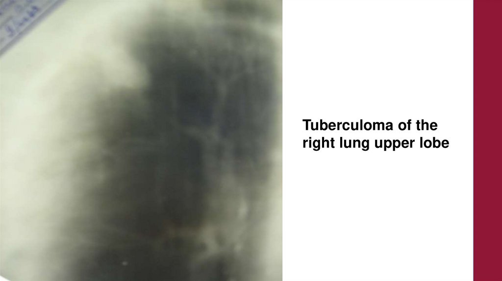
Tuberculomas of theright lung upper lobe
29.
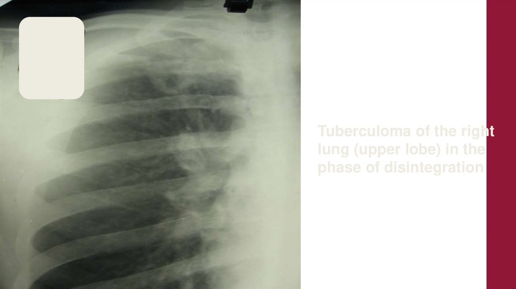
TUBERCULOMA IN THE PHASE OF DISINTERGRATIONCharacterised by eccentric locaiisation of semilunar shaped or beam-shaped zones of
enlightment around the medial edge of the
tuberculoma.
This is accompanied by communication of the
tuberculoma with the lung root due to formation
of broncho-vascular channels.
30.
31.
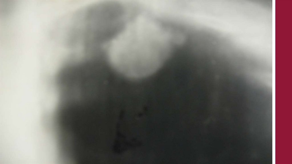
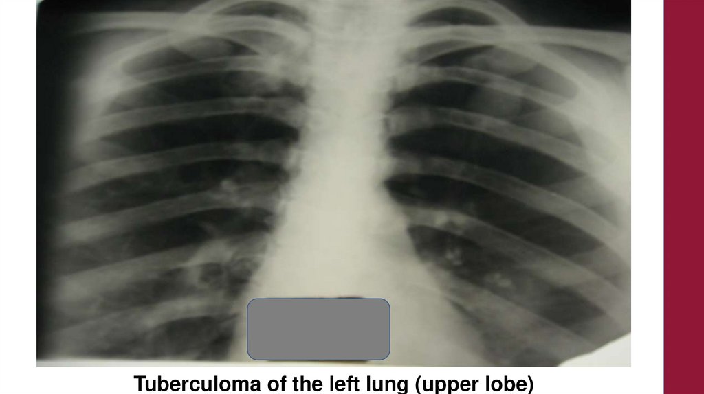
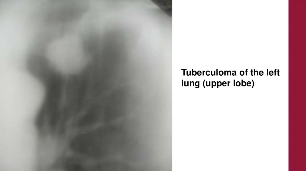
Tuberculoma of the left lung (upper lobe)32.
Tuberculoma of the leftlung (upper lobe)
33.
Tuberculoma of theright lung (upper lobe)
34.
Tuberculoma of the rightlung (upper lobe) in the
phase of disintegration
35.
36.
Tuberculoma of the leftlung in the phase of
disintegration
37.
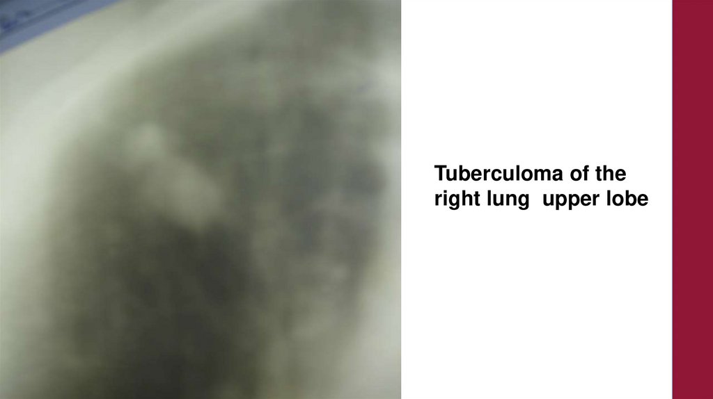
Tuberculoma of theright lung upper lobe
38.
Tuberculoma of theleft lung
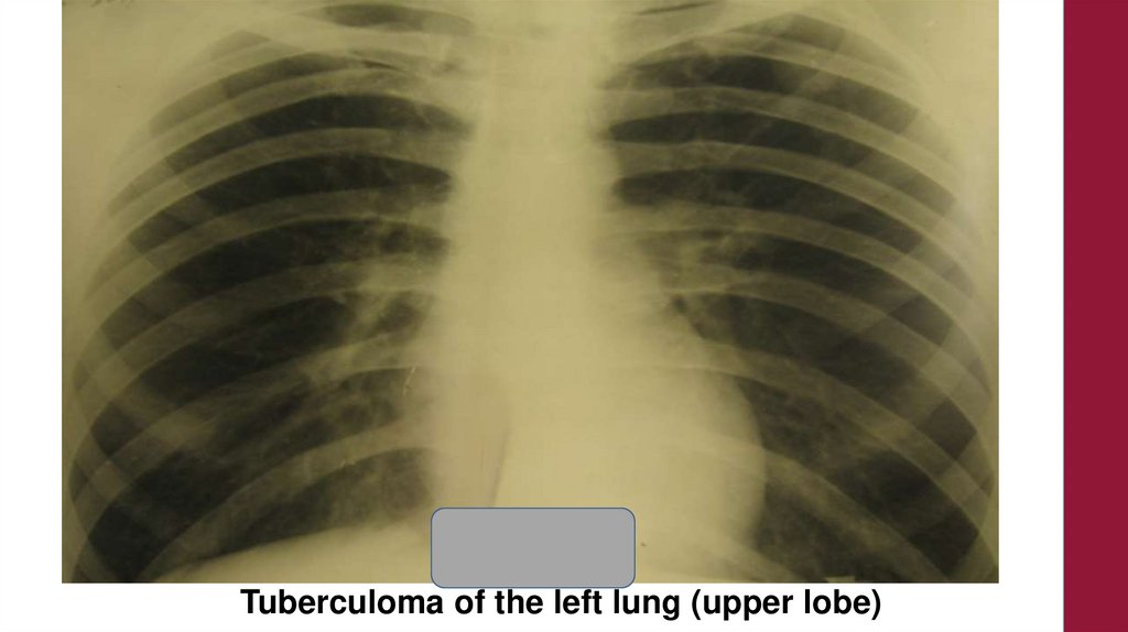
(upper lobe)
39.
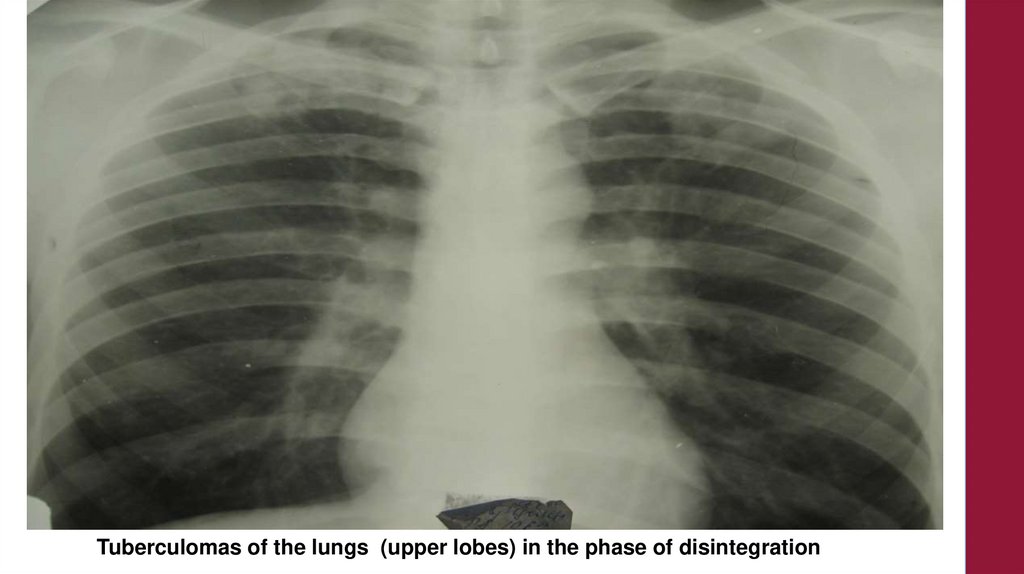
Tuberculomas of the lungs (upper lobes) in the phase of disintegration40.
Tuberculomas (multiple)of the lungs in the phase of disintegration41.
Tuberculoma of the left lung (upper lobe)42.
Tuberculoma of theright lung upper lobe
43.
Pat. I.A.A, June. Tuberculoma of the left lung (upper lobe)44.
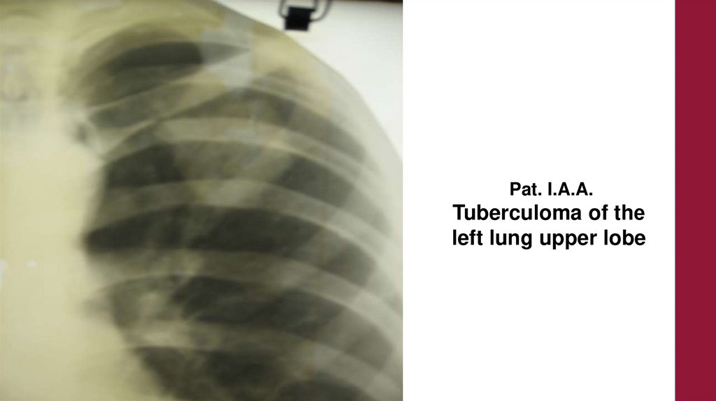
Pat. I.A.A.Tuberculoma of thе
left lung upper lobe
45.
Pat. I.A.A, October. Tuberculoma of the left lung In the phase of disintegration46.
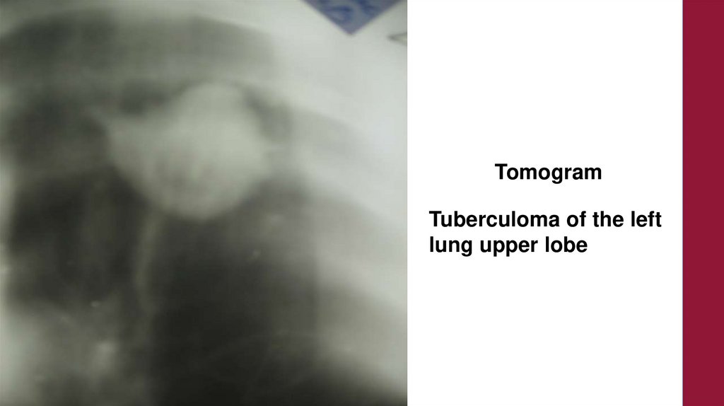
TomogramTuberculoma of the left
lung upper lobe
47.
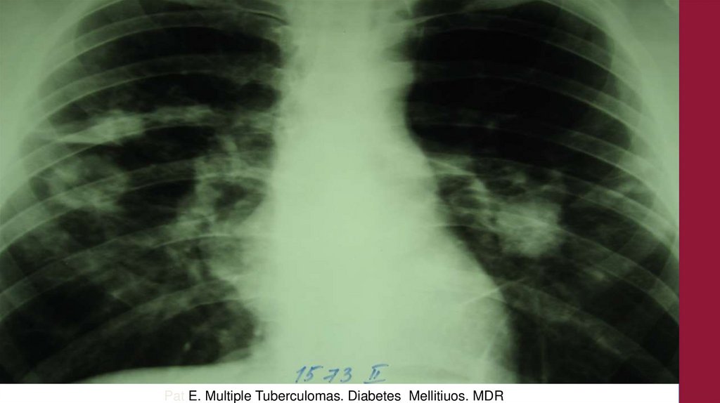
Pat E. Мultiple Tuberculomas. Diabetes Mellitiuos. MDR48.
Pat E Negative Dynamics Diabetes Mellitius. MDR49.
Tuberculoma of theright lung
(upper lobe)
Pat M.
(July)
50.
Pat M. March, ( 8 months later).51.
Pat M. March.Tomogram.
52.
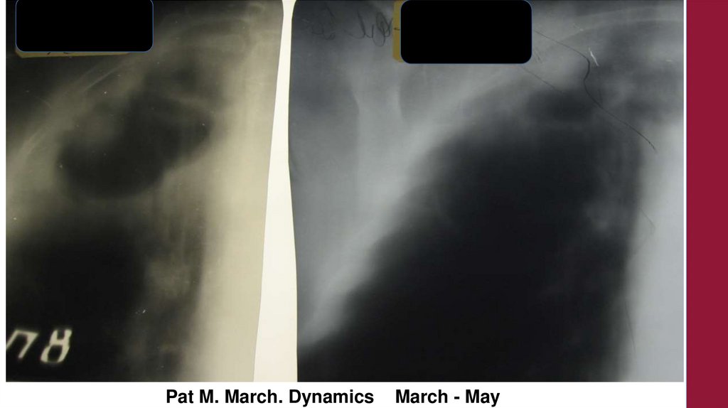
Pat M. March. DynamicsMarch - May
53.
Tuberculoma of the left lung (upper lobe) Pat. G., May.54.
LABORATORY FINDINGSGeneral blood analysis may reveal no significant
changes:
Lymphocytosis in 20% of the cases.
Ziel-Nelseen staining of sputum:
In the absence of disintegration the sputum is
Mycobacterium positive in 10-15% of the cases.
In the presence of disintegration the sputum is
Mycobacterium positive in 70% of the cases.
55.
BLOOD PICTUREBlood picture is also without peculiarities.
•Sometimes moderate elevation of ESR and
•moderate leukocytosis are observed at acute
stages.
56.
Mycobacterium tuberculosis•Mycobacterium tuberculosis is not found in sputum at
stable course of tuberculoma.
•Discharge of bacilli exists in tuberculoma at presence of
disintegration if there is connection with drainage of
bronchus.
57.
Tuberculin testsPatients with lung tuberculoma in most
cases positively react to tuberculin.
•Mantoux test is often hyperergic.
58.
TreatmentBefore the discovery of antituberculosis drugs, the
forecast of tuberculoma was bad.
•Tuberculoma gave massive flare-up with subsequent
transition in heavy forms of pulmonary tuberculosis.
•Now course of tuberculoma regresses or proceeds
chronically without aggravations among 80% of patients.
59.
Treatment•When tuberculoma is diagnosed the patient must
be hospitalized for long term treatment.
•Surgery is recommended if disintegration is
present in tuberculoma and the patient continues
to expectorate МВТ and there is no desirable
results to long therapy.
60.
TREATMENTGeneral principles of treatment of TB
patients but the antiTB drugs do not
penetrate into the tuberculoma.
Surgical treatment is more effective.
61.
Chemotherapy62.
Surgical treatment.•Usually operation is made with minimal removal of
lung tissue. It is segmental resection.
•Surgical treatment is used also in cases, when there is
no certainty that the patient has tuberculosis because
it is difficult to differentiate tuberculoma from other
lung diseases, especially tumor.
63.
Differential diagnostics64.
Differential diagnosticsX-ray picture of tuberculoma is isolated rounded focus in
lung tissue. It's typical for many diseases.
Practically patients more often have
•cancer of lung,
•benign tumors,
•pneumonia complicated by an abscess, and
•parasitic lung diseases
65.
Differential diagnosticsIt is necessary
•to collect detailed anamnesis,
•carefully examine all organs and systems of the patient to
differentiate one disease from another.
•X-ray examination is especially important.
•Sputum is investigated for МВТ, atypical cells and fungi.
•In some cases pneumonocentesis is made.
•The ex juvantibus treatment of tuberculosis is often used and if
the focus in lungs under the influence of specific treatment
decreases, it testifies its tubercular origin.
66.
Differential diagnosticsFor diagnosis of tuberculoma,
•Computer tomography
•bronchological examination with catheter biopsy and
•puncture of bifurcation lymph nodes has received high
development.
These techniques allow to put correct diagnosis almost in
90% of cases.
67.
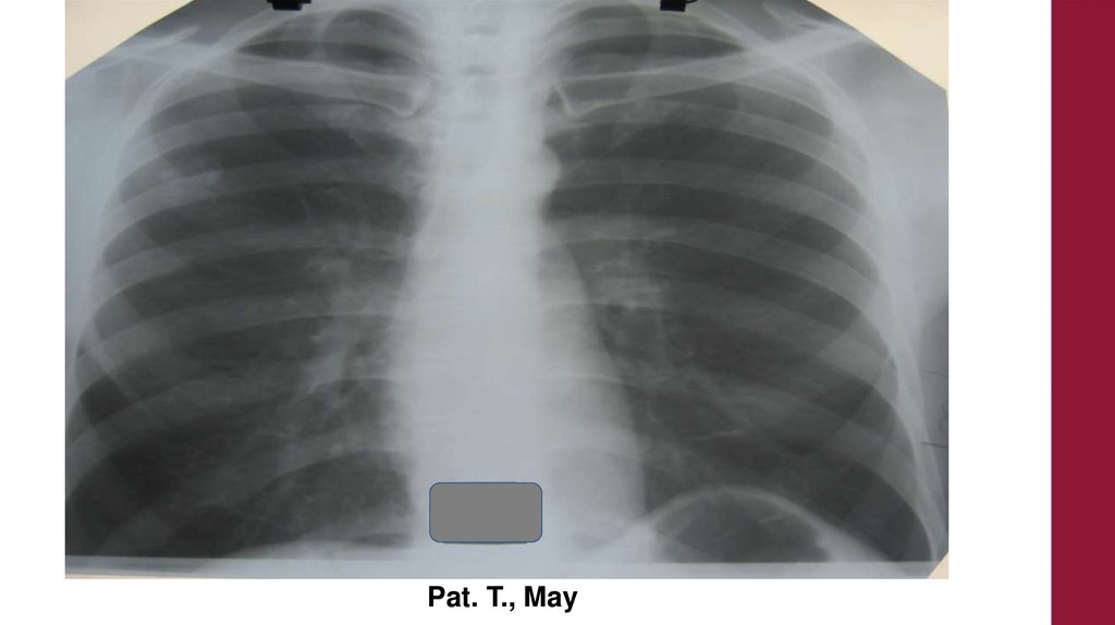
Pat. T., May68.
Pat. T., December69.
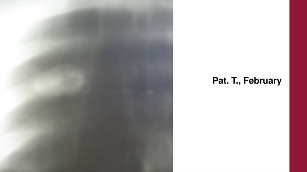
Pat. T., February70.
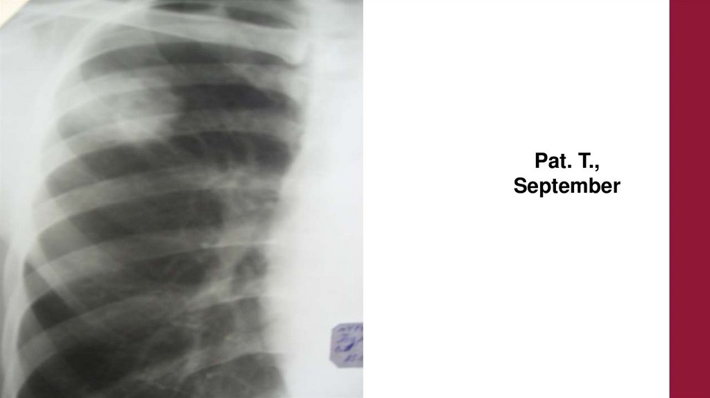
Pat. T.,September
71.
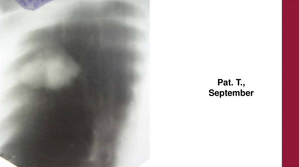
Pat. T.,September
72.
Pat. T.,September
73.
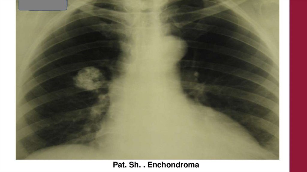
Benign tumor74.
Pat. L.,tomogram
75.
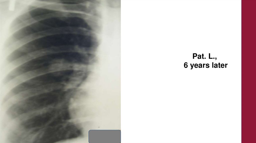
Pat. L.,6 years later
76.
Pat. L.,Tomogram
6 years later
77.
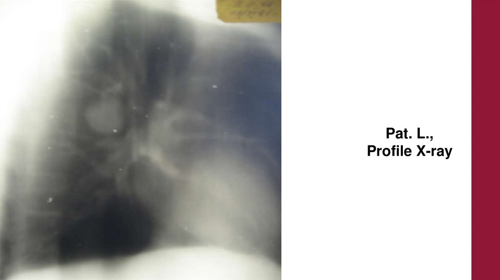
Pat. L.,Profile X-ray
78.
Pat. K.Echinococcus
79.
Pat. K.Echinococcus
Tomogram
80.
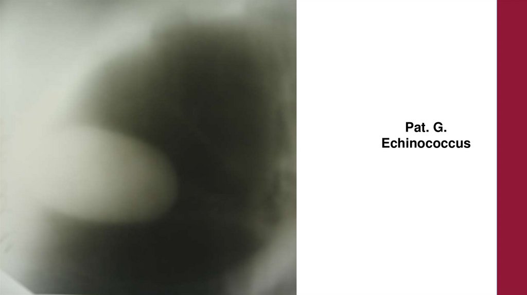
Pat. G.Echinococcus
81.
Pat. G.Echinococcus
82.
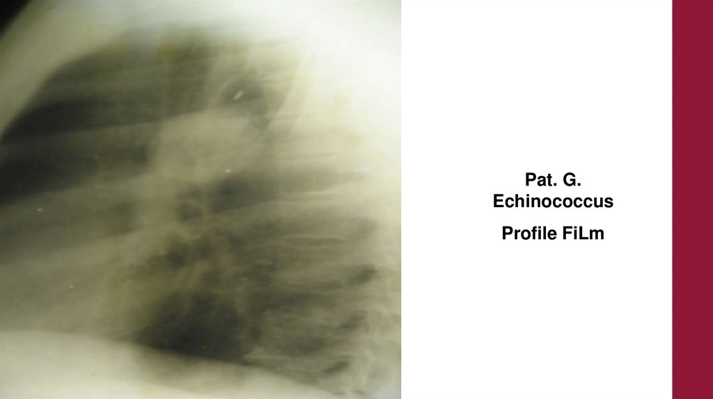
Pat. G.Echinococcus
Profile FiLm
83.
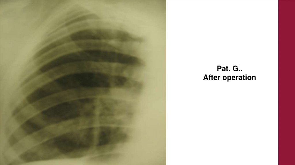
Pat. G..After operation
84.
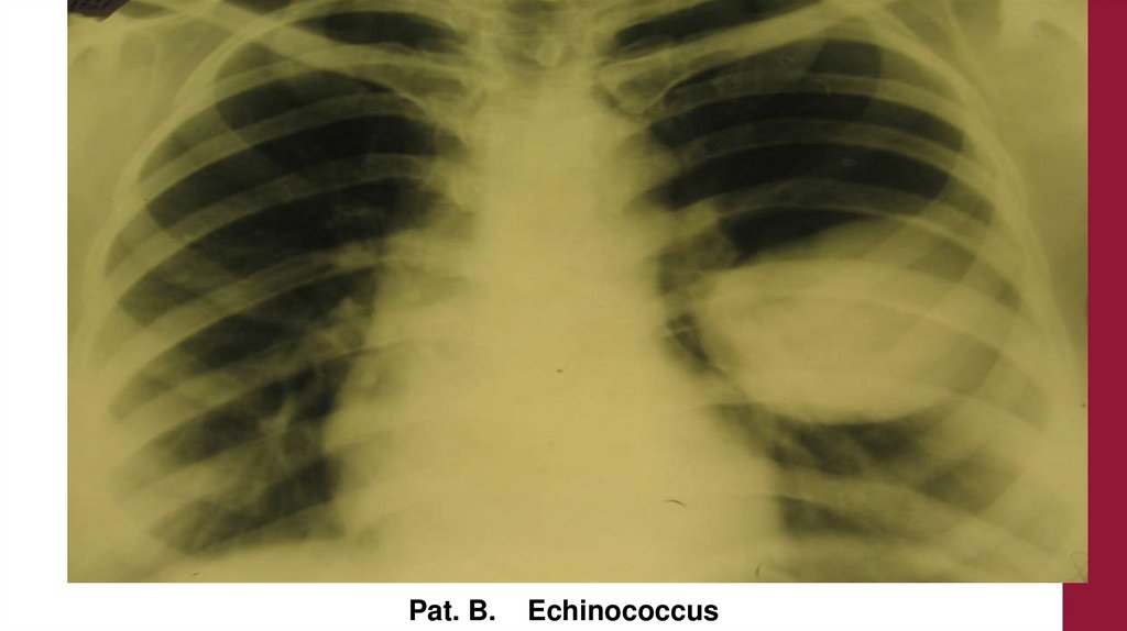
Pat. B.Echinococcus
85.
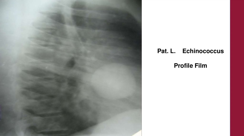
Pat. L.Echinococcus
Profile Film