


 Биология
Похожие презентации:


Anatomy of the brainstem
1.
Anatomy of the brainstem2.
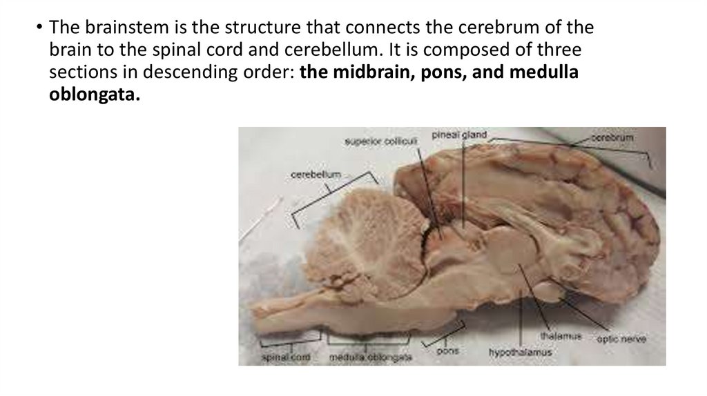
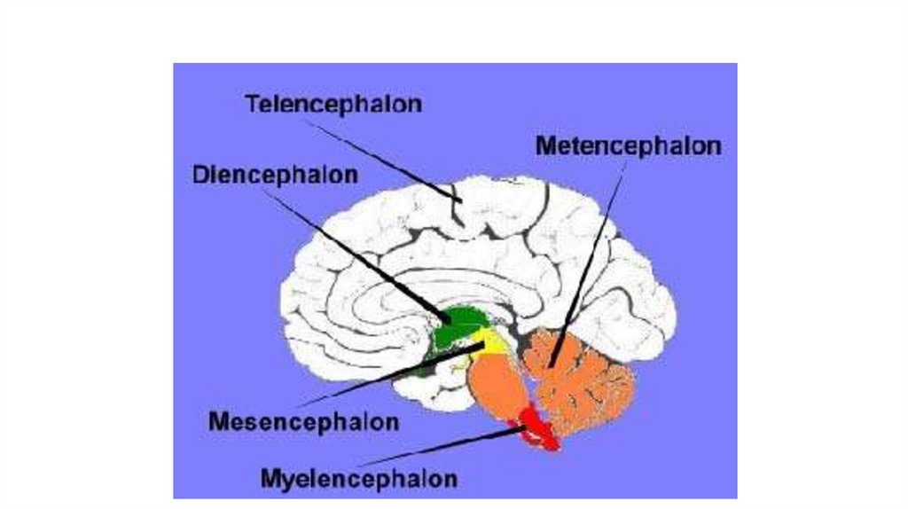
• The brainstem is the structure that connects the cerebrum of thebrain to the spinal cord and cerebellum. It is composed of three
sections in descending order: the midbrain, pons, and medulla
oblongata.
3.
• It is responsible for many vital functions of life:• such as breathing
• Consciousness
• Blood pressure
• Heart rate
• Sleep.
4.
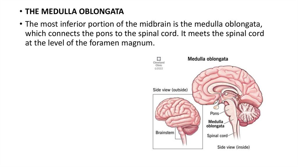
• THE MEDULLA OBLONGATA• The most inferior portion of the midbrain is the medulla oblongata,
which connects the pons to the spinal cord. It meets the spinal cord
at the level of the foramen magnum.
5.
• The anterior portion of the medulla oblongata contains the pyramids.The pyramids carry motor fibers from the precentral gyrus, or motor
cortex, to the grey matter of the spinal cord, where they synapse and
continue to the muscles of the body through the peripheral nervous
system. The pyramids contain a decussation caudally in which the
majority of the motor fibers contained cross to the contralateral side
of the body.
6.
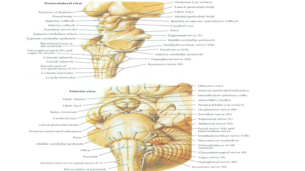
• Internal structure of the medulla oblongata• It contains the nuclei of cranial nerves from VIII to XII:
• Vestibulocochlear nerve (VIII) – medial and inferior vestibular nuclei,
anterior and posterior cochlear nuclei (all are sensory)
• Glossopharyngeal nerve (IX) – ambigus nucleus (motor), nucleus of tractus
solitarius (sensory), inferior salivatory nucleus (parasympathetic)
• Vagus nerve (X) - ambigus nucleus (motor), nucleus of tractus solitarius
(sensory), dorsal vagal nucleus (parasympathetic)
• Accessory nerve (XI) - accessory nucleus (motor)
• Hypoglossal nerve (XII) - hypoglossal nucleus (motor)
7.
8.
THE PONS• The pons connects the medulla oblongata inferiorly to the midbrain
superiorly. The anterior portion of the pons is convex and can be
easily seen as a visible distention when viewing the brainstem
anteriorly. The surface of the anterior distention contains the basilar
groove, which is where the basilar artery rests. The posterior pons is
connected to the cerebellum by the middle cerebellar peduncles,
which are the largest of the cerebellar peduncles. The posterior
portion of the pons forms the superior portion of the floor of the
fourth ventricle.
9.
• Internal structure of the pons• It contains the nuclei of cranial nerves from V to VIII:
• Trigeminal nerve (V) – motor nucleus (supply muscles of mastication), main
sensory nucleus (touch and pressure sensation), mesencephalic nucleus
(proprioceptions), spinal nucleus (pain and temperature sensation)
• Abducent nerve (VI) – nucleus of abducent nerve (supply rectus lateralis
muscle)
• Facial nerve (VII) – motor nucleus of facial nerve (supply facial muscles),
nucleus of tractus solitarius (sensory), inferior salivatory nucleus (supply
glands of the head)
• Vestibulocochlear nerve (VIII) – lateral and superior vestibular nuclei
(sensory)
10.
11.
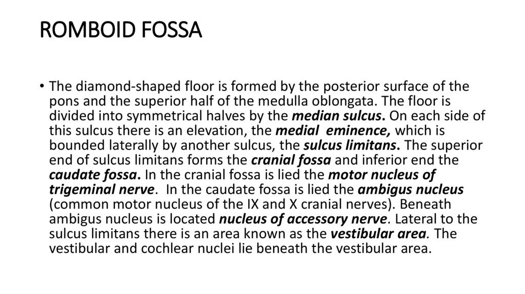
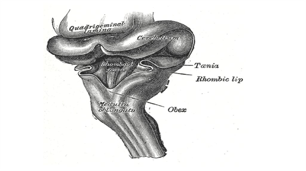
ROMBOID FOSSA• The diamond-shaped floor is formed by the posterior surface of the
pons and the superior half of the medulla oblongata. The floor is
divided into symmetrical halves by the median sulcus. On each side of
this sulcus there is an elevation, the medial eminence, which is
bounded laterally by another sulcus, the sulcus limitans. The superior
end of sulcus limitans forms the cranial fossa and inferior end the
caudate fossa. In the cranial fossa is lied the motor nucleus of
trigeminal nerve. In the caudate fossa is lied the ambigus nucleus
(common motor nucleus of the IX and X cranial nerves). Beneath
ambigus nucleus is located nucleus of accessory nerve. Lateral to the
sulcus limitans there is an area known as the vestibular area. The
vestibular and cochlear nuclei lie beneath the vestibular area.
12.
13.
• Laterally from the sulcus limitans and medially from the vestibulararea in the upper half are located vegetative nuclei of the facial nerve
– superior salivatory nucleus and glossopharyngeal nerve – inferior
salivatory nucleus; in the lower half is located solitary tract nucleus
(common sensory nucleus of the VII, IX and X cranial nerves).
14.
THE MIDBRAIN (MESENCEPHALON)• The midbrain serves as the connection between the pons and the
diencephalon. It also connects posteriorly to the cerebellum via the
superior cerebellar peduncles. The anterior part of the midbrain
contains the crus cerebri with the interpeduncular fossa located
between them. The crus cerebri carry motor cortical spinal fibers,
corticonuclear fibers, and pontine fiber tracts.
15.
16.
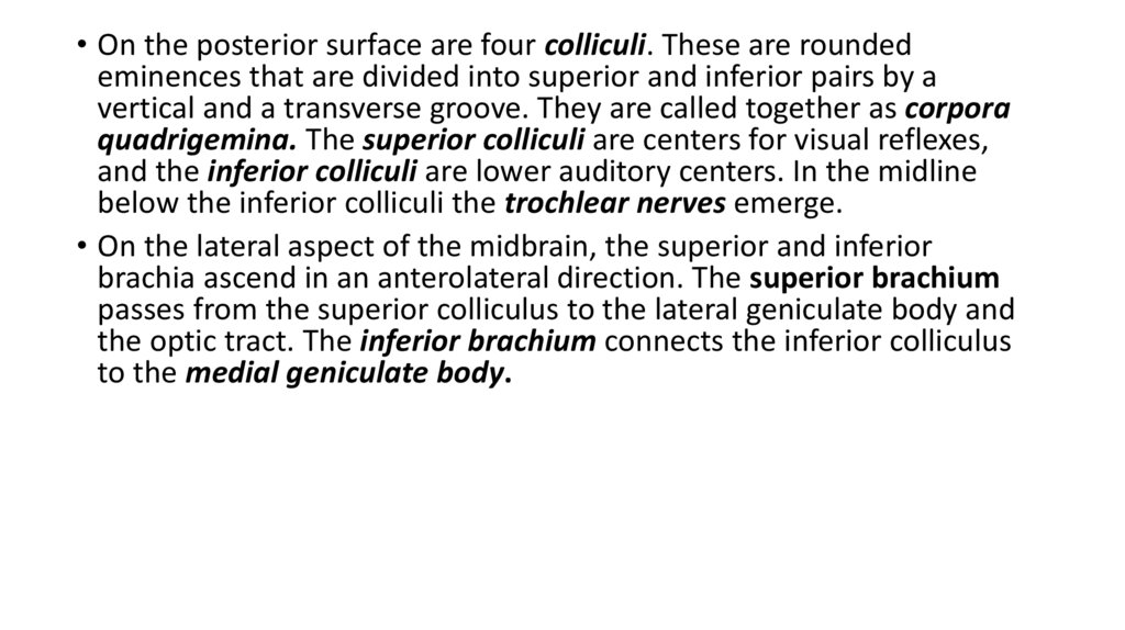
• On the posterior surface are four colliculi. These are roundedeminences that are divided into superior and inferior pairs by a
vertical and a transverse groove. They are called together as corpora
quadrigemina. The superior colliculi are centers for visual reflexes,
and the inferior colliculi are lower auditory centers. In the midline
below the inferior colliculi the trochlear nerves emerge.
• On the lateral aspect of the midbrain, the superior and inferior
brachia ascend in an anterolateral direction. The superior brachium
passes from the superior colliculus to the lateral geniculate body and
the optic tract. The inferior brachium connects the inferior colliculus
to the medial geniculate body.
17.
• Internal structure of the midbrain• It contains the nuclei of cranial nerves III and IV:
• Oculomotor nerve (III) – nucleus of oculomotor nerve (supply muscles
of eyeball), Edinger-Westphal nucleus (supply cilliary and sphincter
pupillae muscles)
• Trochlear nerve (IV) – nucleus of trochlear nerve (supply superior
oblique muscle)
18.
THE CEREBELLUM• The cerebellum is situated in the posterior cranial fossa and covered
superiorly by the tentorium cerebelli. It is the largest part of the hindbrain
and lies posterior to the fourth ventricle, the pons and the medulla
oblongata. It consists of two cerebellar hemispheres joined by a narrow
median vermis. The sagittal section of vermis is called arbor vitae cerebelli
or “tree of life”. The cerebellum is connected to the posterior aspect of the
brainstem by three symmetrical bundles of nerve fibers called the superior,
middle, and inferior cerebellar peduncles. The superior cerebellar
peduncles are connected with the midbrain, the middle cerebellar
peduncles are connected with the pons and the inferior cerebellar
peduncles are connected with the medulla oblongata. A deep horizontal
fissure that is found along the margin of the cerebellum separates the
superior from the inferior surfaces. On the inferior surface is situates
depression – vallecula cerebelli.
19.
20.
STRUCTURE OF THE CEREBELLUM• The cerebellum is composed of an outer covering of gray matter
called the cortex and inner white matter. The white matter of the
cerebellum has on section the appearance of small leaves of a plant
which correspond to each folium (gyri of cerebellum) covered on the
periphery by a cortex of grey matter. The gray matter of the cortex
may be divided into three layers: (1) an external layer, the molecular
layer; (2) a middle layer, the Purkinje cell layer; and (3) an internal
layer, the granular layer.