


")

")

 loop:")


")
:")


 Биология
Похожие презентации:


Cranio-cerebral nerves
1.
CRANIO-CEREBRAL NERVES2.
Cranial - cerebral nerves are nerveswalking away from a cerebrum or
included in him.
There are 12 pairs of cranio-cerebral
nerves, that pierce a skin, muscles,
organs of head and neck, and also the
row of organs is thoracal and abdominal
cavities.
3.
Distinguish:•motoriuss (III, IV, VI, XI and XII of
pair);
•mixed nerves (V, VII, IX and X of
pair)
containing
all
functional
explorers;
•nerves of sense-organs - I and II of
pair.
4.
olfactoryoculomotor
trochlear nerve
abducens
preddverno cochlear
hypoglossus
additional
visual
trifacial
facial
glossopharyngeal
vagus
5.
Motor cranial nerves6.
Classification of motoriussMotoriuss begin in the motive kernels of
barrel.
To mainly motive take the group of
oculomotoriuss: oculomotor (III), block
(IV), taking (VI), additional (XI),
innervating
sternal-clavicular-mammiform and trapezoidal muscles, subglossal
(XII), innervating muscles of language.
7.
OculomotoriusThis nerve is mainly motor, however, it also
contains parasympathetic fibers to smooth
muscle of the eyeball, sympathetic fibers and a
small number of sensory fibers.
A conglomerate of nuclei III pairs located in
the Central gray matter of the midbrain (at the
bottom of the IV ventricle, at the level of the
corpora quadrigemina).
8.
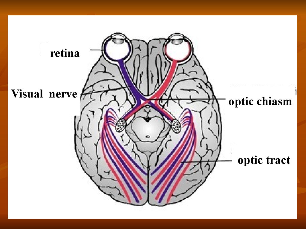
retinaVisual nerve
optic chiasm
optic tract
9.
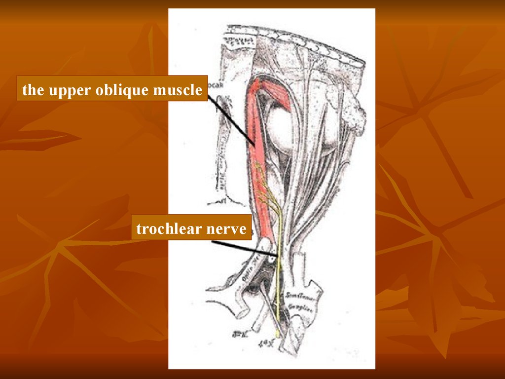
Trochlear nerve (IV pair)Этот нерв обеспечивает только верхнюю
косую мышцу, которая двигает
зрачок вперед-вниз и вбок. Все волокна
нерва переходят на противоположную
сторону тела между центральным ядром и
мышцей. Следовательно, дисфункция
одного блокового нерва будет
воздействовать на противоположную
мышцу.
10.
the upper oblique muscletrochlear nerve
11. Trochlear nerve
AnatomyTrochlear nerve emerges from the brain
stem, in the area of attachment of the
sail Rostral to the caudal hills corpora
quadrigemina. Together with the
trigeminal nerve it enters orbital cleft,
out there in the fossa and branches into
the dorsal oblique muscle of the eye.
12.
Pathology and clinical symptomsIsolated anomalies of the trochlear
nerve are rare in clinical practice and
difficult to diagnose. Cats that have
vertically oriented pupils, a small
dorsolateral rotation of the affected eye
may occur due to paralysis of the dorsal
oblique muscle of the eye.
13. Abducens nerve (VI pair)
Abducens nerve provides lateralrectus, which moves the pupil laterally.
Dysfunction of the nerve results in
strabismus is called convergent. In this
case, the nerve fibers don't cross midline
of the body, and dysfunction of one
abducens nerve only affects the muscle
located on the same side.
14.
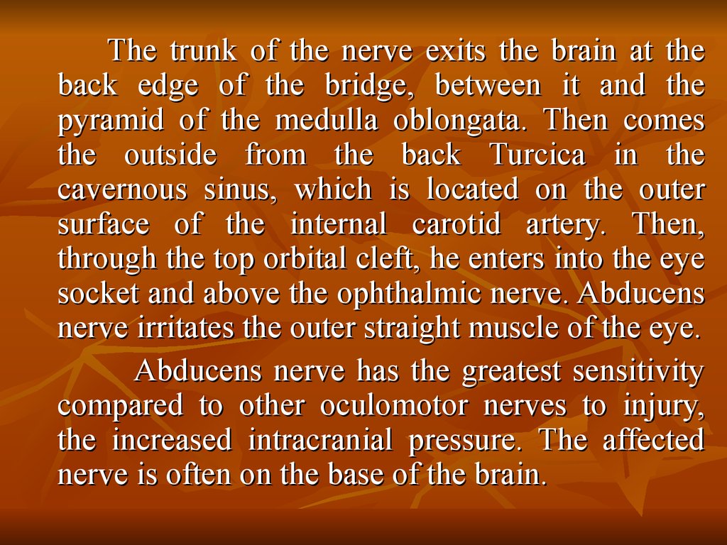
The trunk of the nerve exits the brain at theback edge of the bridge, between it and the
pyramid of the medulla oblongata. Then comes
the outside from the back Turcica in the
cavernous sinus, which is located on the outer
surface of the internal carotid artery. Then,
through the top orbital cleft, he enters into the eye
socket and above the ophthalmic nerve. Abducens
nerve irritates the outer straight muscle of the eye.
Abducens nerve has the greatest sensitivity
compared to other oculomotor nerves to injury,
the increased intracranial pressure. The affected
nerve is often on the base of the brain.
15.
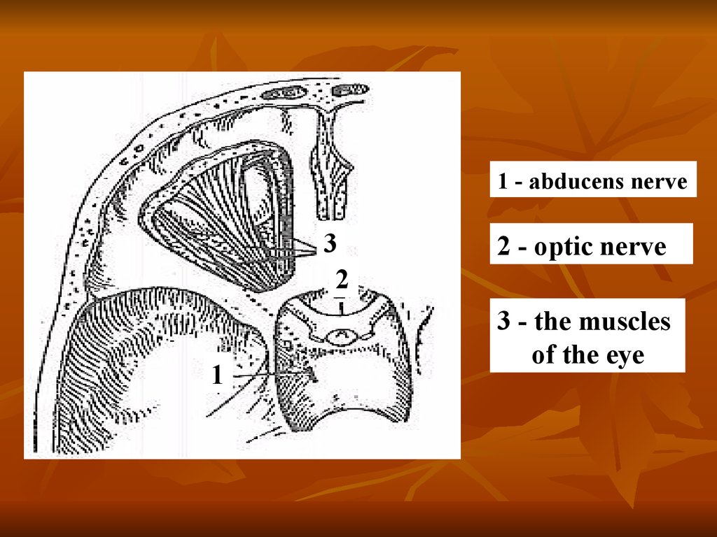
Abducens nerveAnatomy
Nucleus abducens nerve are
located on both sides of the median
sulcus in the caudal part of the bridge
near the medulla oblongata and beneath
the bottom of the IV cerebral ventricle.
Fiber abducens nerve through the
orbital gap enter the orbit and Innervate
the above muscles.
16.
1 - abducens nerve3
2
1
2 - optic nerve
3 - the muscles
of the eye
17. Hypoglossal nerve (XII pair)
Formed by processes of nerve cells of thesame nucleus, which is located in the medulla
oblongata. The nerve exits the skull through the
hypoglossal canal of the occipital nerve, innervates
muscles of the tongue and partly by some of the
muscles of the neck.
Hypoglossal nerve mainly caused by
gorkovatam connections with the opposite
hemisphere. Central motor neuron for muscles of
the tongue is the bottom portion of the precentral
gyrus.
18.
Hypoglossal nerve (XII pair)Anatomy
The neurons forming the hypoglossal nerve
originate from the hypoglossal nerve centre
in the medulla oblongata, at the level of the
fourth ventricle.
19. Hypoglossal nerve and cervical (hyoid) loop:
1 - hypoglossal nerve;2 - thyrohyoid branch;
3 - forward spine;
4 - dorsal root;
5 - cervical (hyoid) loop;
6 - speaking branch.
20.
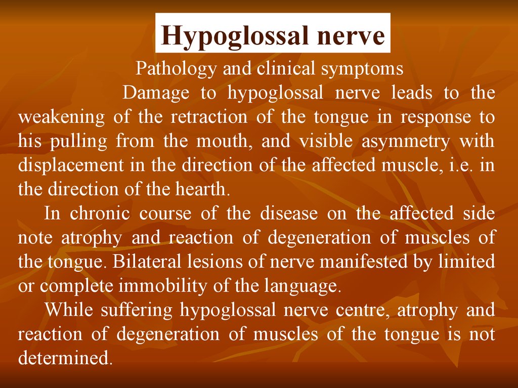
Hypoglossal nervePathology and clinical symptoms
Damage to hypoglossal nerve leads to the
weakening of the retraction of the tongue in response to
his pulling from the mouth, and visible asymmetry with
displacement in the direction of the affected muscle, i.e. in
the direction of the hearth.
In chronic course of the disease on the affected side
note atrophy and reaction of degeneration of muscles of
the tongue. Bilateral lesions of nerve manifested by limited
or complete immobility of the language.
While suffering hypoglossal nerve centre, atrophy and
reaction of degeneration of muscles of the tongue is not
determined.
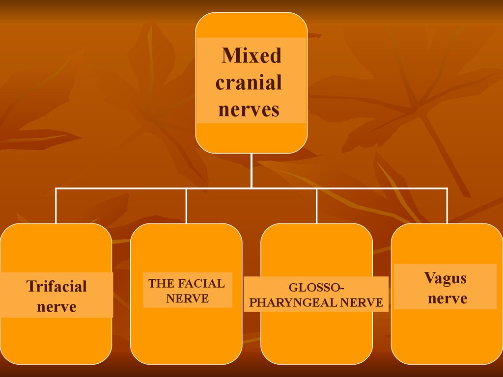
21. Mixed cranial nerves
22.
MixedСмешанные
cranialЧМН
nerves
Trifacial
Тройничный нерв
nerve
THE FACIAL
Лицевой
нерв
NERVE
GLOSSOЯзыкоглоточный нерв
PHARYNGEAL NERVE
Vagus
Блуждающий
nerve нерв
23. Trifacial nerve
Trigeminal nerve(from lat. nervus trigeminus)
V pair
of cranial nerves mixed
character.
Ternary nerve (shown in yellow)
24.
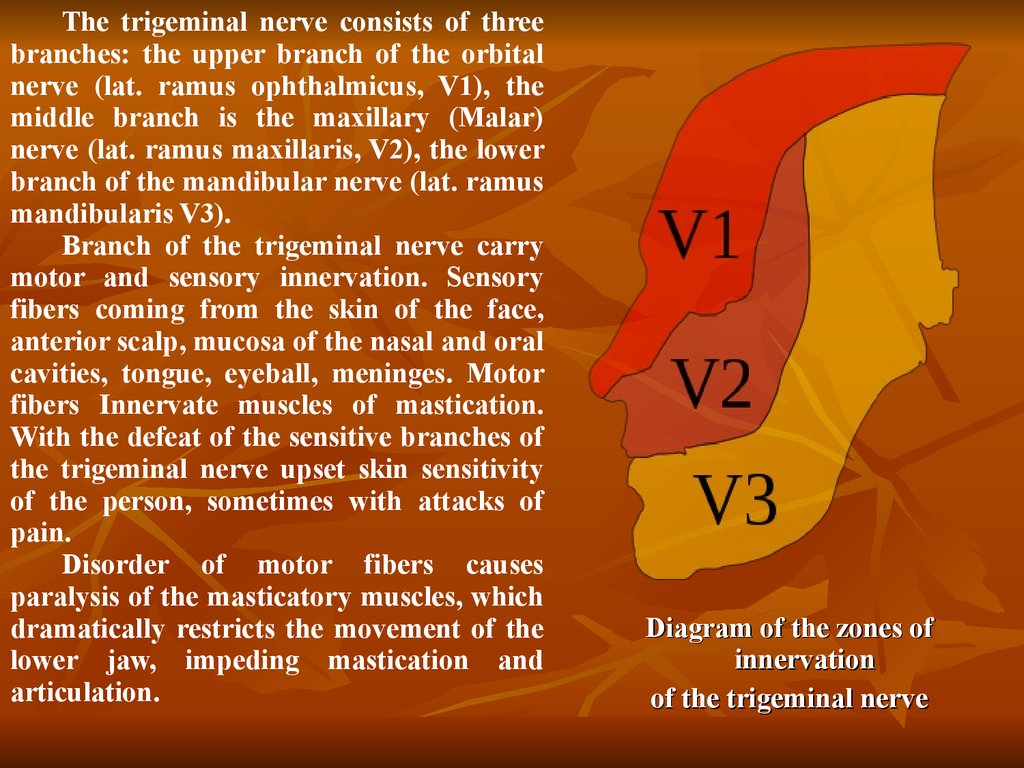
The trigeminal nerve consists of threebranches: the upper branch of the orbital
nerve (lat. ramus ophthalmicus, V1), the
middle branch is the maxillary (Malar)
nerve (lat. ramus maxillaris, V2), the lower
branch of the mandibular nerve (lat. ramus
mandibularis V3).
Branch of the trigeminal nerve carry
motor and sensory innervation. Sensory
fibers coming from the skin of the face,
anterior scalp, mucosa of the nasal and oral
cavities, tongue, eyeball, meninges. Motor
fibers Innervate muscles of mastication.
With the defeat of the sensitive branches of
the trigeminal nerve upset skin sensitivity
of the person, sometimes with attacks of
pain.
Disorder of motor fibers causes
paralysis of the masticatory muscles, which
dramatically restricts the movement of the
lower jaw, impeding mastication and
articulation.
Diagram of the zones of
innervation
of the trigeminal nerve
25. Trigeminal nerve
AnatomyThe nerve center of the trigeminal nerve is weakly expressed
anatomically, it is located in the lateral reticular formation at the level
of the Rostral legs of the cerebellum, dorsal to trapezoidal body.
Motor axons pass through the trigeminal ganglion and the foramen
ovale, are connected with the maxillary nerve tract and Innervate the
temporal, chewing, medial and lateral pterygoid muscles and the
Rostral part of the digastric.
Sensory pathways of the facial parts presented in the three branches.
The maxillary branch innervates the nose, the upper jaw; eye branch
provides the sensitivity of the eyeball and cornea; and the mandibular
branch is the nerve of General sensibility to the temporal region and
region of the lower jaw, and motor – to chewing muscles.
Each branch needs to be checked for sensitivity.
26. Pathology Disease affecting the sensory and motor functions of the trigeminal nerve: infectious diseases; injuries; tumors; vascular disease.
Neurological deficit is manifested in the decrease in muscle tone andinability to close the mouth. Bilateral trigeminal motor paralysis was
observed at rabies and idiopathic neuritis of the trigeminal nerve.
Bilateral damage causes paralysis of the muscles of the mouth, resulting
in lost the ability to close the mouth. Unilateral damage can lead to
decreased tone masticatory muscles, accompanied by atrophy of this muscle
group.
However, unilateral damage rarely have an impact on eating animals.
Sometimes, polyneuropathy can affect the trigeminal nerve, leading to
atrophy of the masticatory muscles.
The diagnosis can be confirmed by electromyography.
However, it should be noted that the most common cause of bilateral
atrophy of the masticatory muscles is myositis. In such cases it is necessary
to differentiate myositis and neuropathy.
27.
Regeneration of thetrigeminal nerve
Neurology
of the trigeminal nerve
28. The facial nerve
The facial nerve enters thetemporal bone through the
internal auditory hole. Deep in
the temporal bone it goes
through the facial canal (lat.
canalis facialis) and exits via the
stylomastoid hole, (lat. foramen
stylomastoideum),
and
then
divides into five branches.
Despite the fact that the facial
nerve runs through parotid gland
(lat. glandula parotidea), it does
not innervates it. This task is
performed
by
the
glossopharyngeal nerve.
29.
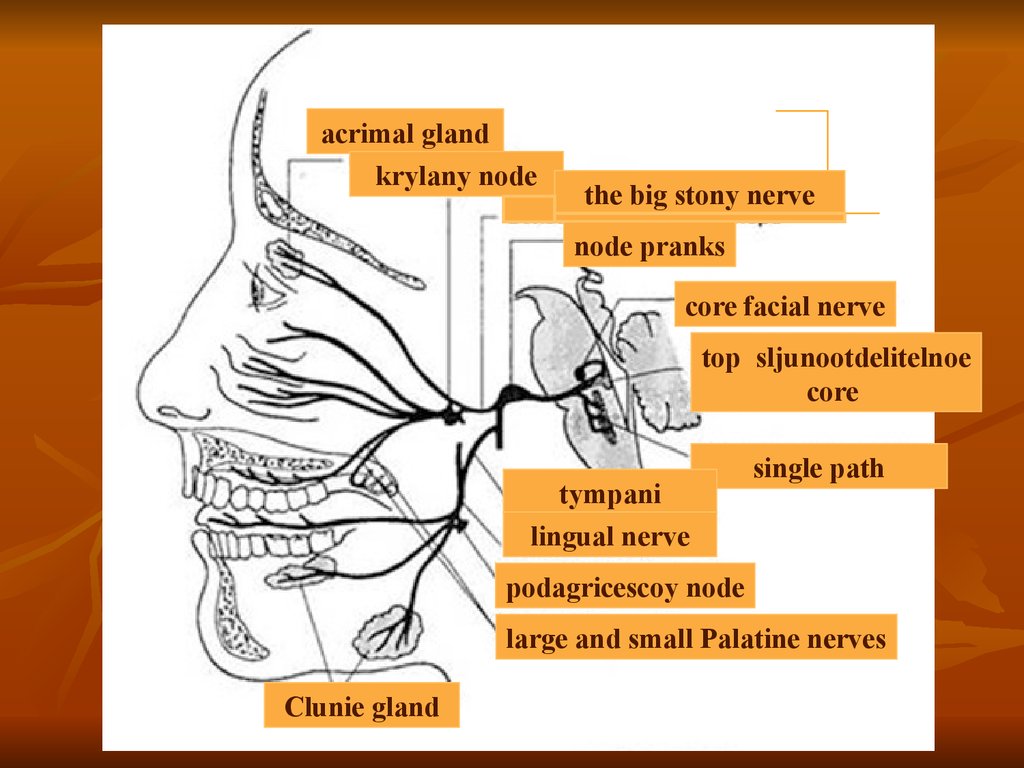
acrimal glandkrylany node
the big stony nerve
node pranks
core facial nerve
top sljunootdelitelnoe
core
tympani
lingual nerve
single path
podagricescoy node
large and small Palatine nerves
Clunie gland
30. The facial nerve (VII nerve)
AnatomyThe facial nerve is a mixed nerve, which unites the two
nerve: the facial and intermediate. The nucleus of the facial
nerve occur within the boundaries of the bridge of the
brain.
After leaving the brain stem in the furrow between the
Pons and medulla oblongata, and facial nerve enters the
internal auditory meatus and, passing through the facial
canal, exits via the stylomastoid hole and Innervate the
muscles of the ears, eyelids, nose, cheeks, lips, and the
caudal portion of the digastric
31. The location of the nuclei of the facial nerve and its root in the brainstem (Browse):
1 red nucleus, 2 — cellview water (cavity of themidbrain), 3 — the lamina quadrigemina, 4 —
pineal gland, 5 — srednedushevoj the path of the
trigeminal nerve, 6 — trochlear nerve,
7 — bridle front brain sails, 8 — motor trigeminal
nucleus, 9 is the knee of the facial nerve (loop n.
facialis covering - abducens nerve), 10 — the roof
of the IV ventricle or tent, 11 — plexus meninges
of the IV ventricle, 12 — the single way, the 13 gray wing (the nucleus of the vagus nerve), a 14 hypoglossal nerve, 15 is the Central channel, 16 —
spinal path of trigeminal nerve, 17 — accessory
nerve, an 18 - accessory nerve, 19 — hypoglossal
nerve, 20 — accessory nerve,
21 — vagus nerve, 22—, double -, 23 —
hypoglossal nerve, 24 — glossopharyngeal nerve,
the 25 — bottom - olive, 26 — sljunootdelitelnoe -,
27 — acoustic nerve 28 facial nerve, 29 —
abducens nerve, the 30 - facial nerve, 31 —
trigeminal nerve, 32 — varolii bridge, 33 — leg of
the cerebellum, 34 — oculomotor nerve
32. The divisions of the facial nerve
In the facial canal the nerve divides into severalbranches:
great stony nerve, which carries parasympathetic fibers to
pterygoid-Palatine site;
it emerges from the channel through the hole on the upper
surface of the pyramid;
drum string the mixed nerve departs from the facial nerve
via barrancominas the gap and goes forward and down to the
junction with the lingual nerve. The nerve contains the
afferent taste fibers from the anterior part of the tongue and
sljunootdelitelnye parasympathetic fibers to the sublingual
and submandibular salivary glands;
tremendou nerve - the motor nerve, innervates tremendous
muscle of the tympanic cavity.
33. Anatomo-topographic diagram of the structure of the facial nerve:
1 — the bottom of the IV ventricle, 2 —nucleus of the facial nerve, 3 —
stylomastoid hole, 4 — posterior
auricular muscle, 5 — occipital Vienna,
6 — posterior belly digastric, 7 —
chilopoda muscle, 8 — branches of the
facial nerve to the facial muscles and
subcutaneous muscle of the neck, 9 —
muscle, lowering the angle of the
mouth, 10 — mentalis, 11 — muscle,
lowering the lower lip, 12 — buccal
muscle, 13 — circular muscle of the
mouth, 14, 15 — muscle lifting the
upper lip 16 — the zygomatic muscle,
17 — the circular muscle of the eye, 18
muscle, the corrugator supercilium, 19
— frontal muscle, 20 — tympani, 21 —
lingual nerve, a 22 — Kralovny node,
23 — trigeminal site, 24 — internal
carotid artery, 25 — intermediate
nerve, 26 — the facial nerve, a 27 —
predverno-cochlear nerve
34.
Pathology and clinical symptomsClinical symptoms depend on the level of the lesion. For
example, if the damage is external to the facial canal, there
will be signs of paralysis of the facial muscles:
• inability to close the eye gap;
• paresis or paralysis of comissary lip on the affected side;
• impairment of movement of the ear on the damaged side;
• an asymmetric deviation of nasal mirrors to the healthy side,
as a result of muscle tone in the nose, not the greeters
counter;
• sometimes a small enlargement of the pupil, due to a decrease
in tone spherical eye muscle on the affected side. Facial
paralysis can be unilateral or bilateral and is not always
associated with a lesion of the facial muscles.
35. Facial nerve paresis
36. Diagnostic methods of neurology facial nerve
Clinical neurological examinationInstrumental methods
Electromyography
Doppler ultrasound with assessment of blood
circulation in vertebral-basilar pool
CT scan of the brain
MRI of the brain
37. Glossopharyngeal nerve
Glossopharyngeal nerve IXpair of cranial nerves (n.
glossophaгyngeus)
mixed
nerve
contains
motor,
sensory
and
parasympathetic (secretory)
fibers, has 4 cores, which are
located in the posterior part
of the medulla oblongata.
38. Symptoms
Slight unilateral paresis of the soft palate.Disorders of swallowing is usually mild.
The decrease in the secretion of the parotid
gland.
A decrease in the sensitivity of the
posterior pharyngeal wall and soft palate.
Loss of taste on the posterior third of the
tongue.
Can develop spasm glossopharyngeal
muscles of laringospasm
Increased salivation.
39.
With the defeat of motor nuclei ofthe vagus nerve disturbances of
swallowing, phonation, articulation,
breathing, and bulbar disorders. They
occur in bulbar paralysis, amyotrophic
lateral sclerosis, myelo-encephalitis
and other diseases.
40. Sensitive cranial nerves
Anatomy of the Chemoreceptors of the nasal mucosarecognize various odors and transmit sensory information
aksonam the olfactory nerve, which enters the cranial cavity
through the ethmoid bone and enters the olfactory bulb.
Pathology and clinical symptoms
Damage to the olfactory nerve are rare and difficult to
diagnose. The most common cause hyposmia is a chronic
rhinitis, which affects the olfactory cells of the nasal
mucosa. A tumor of the nasal cavity can also be the reason a
weak sense of smell.
Sometimes, the canine distemper virus can destroy as
neuroepithelial cells of the olfactory receptors of the nasal
mucosa and neurons in the olfactory bulb.