


 Английский язык
Похожие презентации:


Gestosis is a syndrome defined as violated
1.
2.
Gestosis is a syndrome defined as violatedadaptation of a woman to pregnancy.
Gestosis arises only in connection with
pregnancy, is etiologically linked to fetal egg
development, is characterized by various
symptoms, complicates the course of
pregnancy and usually disappears right after
or in some time after the end of pregnancy.
3.
Classification⚫ Early gestoses (1-st 3 months)
⚫ Late gestosis (after 20 weeks)
⚫ Rare forms of gestosis
4.
Aetiopathogenesis (theories)⚫ toxemic
⚫ allergic, immune
⚫ corticovisceral
⚫ hormonal
⚫ neurogenic
⚫ psycogenic
⚫ genetic
5.
The concept of "early gestosis" exists only in the practice ofobstetricians
gynecologists
of
the
ICU.
In the obstetric practice of foreign countries, this concept
does not exist, where these states are regarded as "minor"
complications of pregnancy, or "unpleasant symptoms
during pregnancy."
6.
1. Early gestosis, often occurs - vomiting of pregnantwomen
and
ptyalismus
(hypersalivation)
2. Early gestosis, rarely occurs - dermatoses of pregnant
women, cholestatic hepatosis of pregnant women, acute
fatty hepatosis of pregnant women, chorea of pregnant
women, osteomalacia during pregnancy.(
7.
Classification of vomiting:⚫ mild -3-5 times a day on an empty stomach of after
meals, reduced appetite
⚫ moderate- 10 times a day irrespective of food intake,
weight loss, weakness, apathy, electrolyte imbalance
⚫ severe – more than 10 times a day, no food is hold,
weight loss, low grade fever, icteric skin, acetonuria
with oliguria, tachycardia, hypotention,
hyperbilirubinemia,potassium reduction,
hypoproteinemia, hematocrit increase
8.
Differential diagnosis of vomiting in pregnantwomen should be carried out with the following
diseases:
⚫ food toxicoinfection
⚫ gastritis
⚫ Pancreatitis
⚫ Pyelonephritis
⚫ Cholelithiasis
⚫ viral hepatitis
⚫ Appendicitis
⚫ Meningitis
⚫ brain tumors
9.
Treatment⚫ Psychotherapy: electrical sleep ,laser reflexotherapy,
acupuncture, sedatives or tranquilizes (diazepam, seduxen)
⚫ Antiemetic agents – droperidol, aminazine, etapirazin,
cerucal
⚫ Water-electrolytic balance correction
⚫ Mild: dietary correction - fractional (5-6 times a day), balanced
nutrition, plenty of drinks, vitamin therapy.
⚫ In case of vomiting of pregnant women, moderate and severe,
hospitalization and prescription of drug therapy are indicated.
10.
Hypersalivation(ptyalismus)
The
amount of saliva with hypersalivation can reach 1.0 l
per day. Salivation does not cause severe irregularities in
the body, causes maceration of the skin and mucous
membrane of the lips.
In order to reduce the secretion of the salivary glands,
intramuscular administration of atropine is prescribed in 0.5 ml
0.1%
solution
2
times
a
day.
atropine is prescribed in 0.5 ml 0.1% solution 2 times a day. It is
advisable to rinse the mouth with infusion of sage, mint,
chamomile, oak bark and other means that have astringent
properties.
11.
Rare forms of gestosis⚫ Dermatoses
⚫ Chorea
⚫ Itching
⚫ Osteomalation
12.
Rare forms of⚫ dermatoses of pregnant women
gestosis
13.
Rare forms ofgestosis
⚫ pemphigoid of pregnant women
14.
Rare forms of⚫ Cholestatic hepatosis of pregnant women
gestosis
⚫
Acute fatty hepatosis of pregnant women
15.
⚫ Chorea (tetany) occurs in connection with a violation ofcalcium metabolism, due to the hypofunction of the
parathyroid glands.
Clinically, it manifests convulsive uncoordinated muscle
twitching of the upper, lower extremities, sometimes the
face, very rarely the larynx or the stomach.
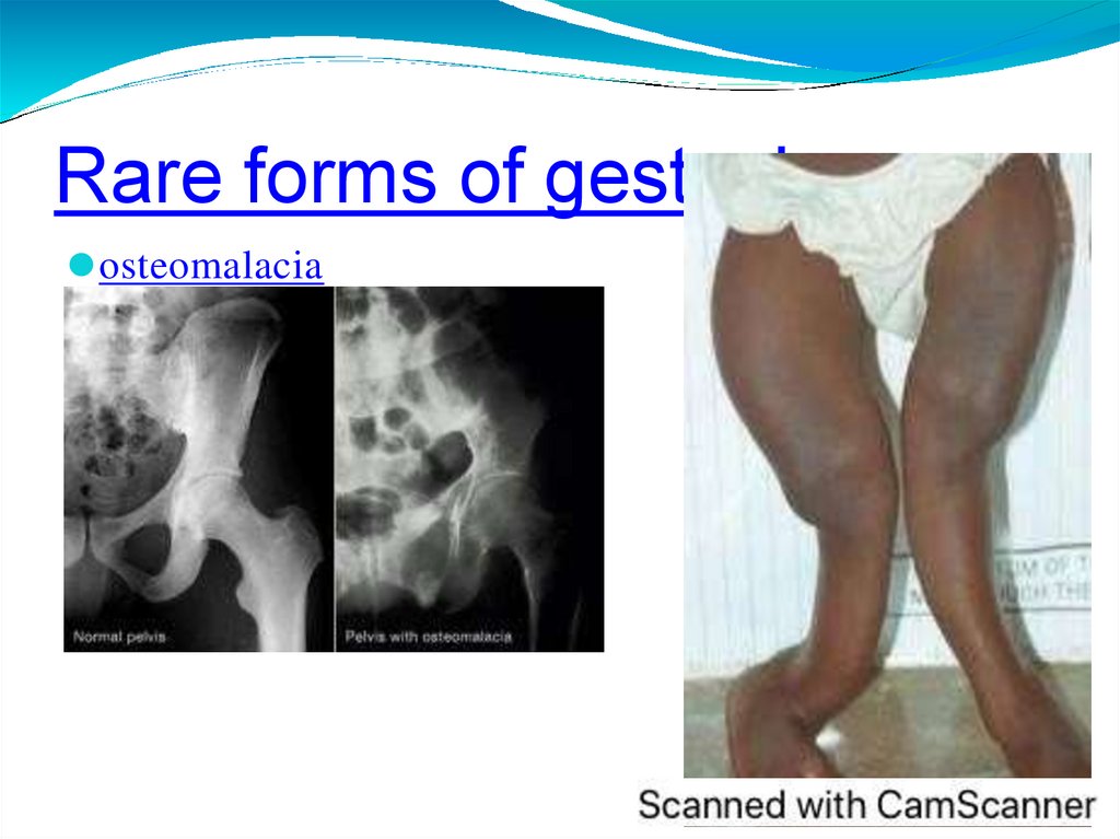
16.
Rare forms of gestosis⚫ osteomalacia
17.
Gestosis in modern obstetrics⚫ Late gestosis of pregnant women (LGP) is a
symptom complex of polyhedral and
polysystemic insufficiency that occurs during
pregnancy.
⚫ The frequency of LGP varies from 7% to 16%
among all pregnant women. In the structure
of mortality of pregnant women, parturient
women and mothers, LG takes one of the first
places.
18.
⚫ GESTOSIS is not an independent disease.⚫ this is a clinical manifestation of the
failure of the adaptation mechanisms of
the maternal organism to adequately
meet the needs of the fetus that develops.
This failure is realized through a varying
degree of expressiveness of perfusiondiffusion insufficiency in the motherplacenta-fetus system.
19.
Aetiology of gestosis⚫ The etiology of preeclampsia is not fully understood.
⚫ There are about 30 different theories. But the definition
of preeclampsia as a disease of adaptation is most
consistent with the ideas about it. Of particular
importance is immunological changes during
pregnancy.
20.
Aetiology and Pathogenesis⚫ Basic etiology is abnormal placentation : failure of
trophoblast invasion
⚫ Failure of second wave of endovascular trophoblast
migration resulting in reduction of blood supply to
fetoplacental unit.
⚫ 2 main things we should remember :
Endothelial Dysfunction due to oxidative stress and
inflammatory mediators, Vasospasm due to
imbalance b/w vasodilators(PGI2, NO) &
vasoconstrictors (TxA2, angiotensin 2, endothelin).
21.
Aetiology and Pathogenesis22.
Aetiology and Pathogenesis⚫ Abnormal trophoblast invasion – failure of the
trophoblast invasion to myometrial segments of the
spiral arteries.
⚫ Spiral arteries retain their muscular walls and prevent
the development of the high blood flow and increase
impedance in the utero-placental circulation.
⚫ Diffuse vasospasm caused by reduced sensitivity to
vasodilators (nitrous oxide and prostacyclin) and
enhanced sensitivity to vasoconstrictors (angiotensin).
23.
Lategestosis
24.
Patholog⚫ Diminished plasma volume
y
⚫ Increase extracellular fluid
⚫ Vasospasm
⚫ Disseminated intravascular coagulation in very
severe cases ischaemic changes in various organs,
when the disease is severe. General vasospasm lead to
changes of reologic properties of blood: appear stasis,
acedosis and formation of polyorganic insufficiency.
Hypovolemy lead to decrease of protein in plasma, as
a result-disorders of leaver functions, low of osmotic
pressure and liquid part of blood go away in
itraorganic space, then oedema, centralisation of
blood flow, increase of BP
25.
Changes in organswith preeclampsia:
⚫ Cardiovascular system: general vasospasm, increased
peripheral vascular resistance, hypovolemia.
⚫ Hematological changes: activation of platelets,
followed by consumption coagulopathy, decreased
plasma volume, increased blood viscosity,
hemoconcentration.
⚫ Urinary System: proteinuria, reduced glomerular
filtration rate, decreased excretion of uric acid.
⚫ Liver: necrosis, subcapsular hematoma.
⚫ CNS: brain edema, intracranial hemorrhage.
26.
Risk factors for1.Extragenital pathology: kidney, liver, hypertension,
preeclampsia:
chronic pulmonary and bronchial diseases, heart disease,
diabetes, obesity and other manifestations of
endocrinopathy.
2. Obstetric risk factors:
⚫ Family history of arterial hypertension (family history);
⚫ Availability of pre-eclampsia in previous pregnancy;
⚫ Age pregnancy (up to 19 years and over 30 years);
⚫ Polyhydramnios, multiple pregnancy;
⚫ Anemia of pregnant;
⚫ Izosensitization by Rh-factor and AB0 system.
⚫ 3. Social and domestic factors:
⚫ Bad habits;
⚫ Occupational hazard;
⚫ Unbalanced diet.
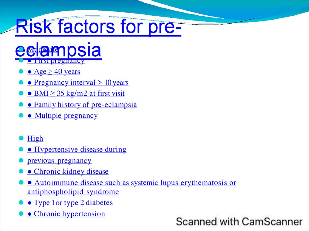
27.
Risk factors for preModerateeclampsia
⚫
⚫ ● First pregnancy
⚫ ● Age ≥ 40 years
⚫ ● Pregnancy interval > 10 years
⚫ ● BMI ≥ 35 kg/m2 at first visit
⚫ ● Family history of pre-eclampsia
⚫ ● Multiple pregnancy
⚫ High
⚫ ● Hypertensive disease during
⚫ previous pregnancy
⚫ ● Chronic kidney disease
⚫ ● Autoimmune disease such as systemic lupus erythematosis or
antiphospholipid syndrome
⚫ ● Type 1or type 2 diabetes
⚫ ● Chronic hypertension
28.
Late gestosisclassification
1. Gestational gypertension- appeared after 20 weeks of
pregnancy and is not accompanied by proteinuria up to
delivery
⚫ transient - normalisation of BP during 12 weeks after
delivery
⚫ chronic - continues during 12 weeks after delivery
2. Proteinuria during pregnancy- protein content of 0.3
g/l
3. Edema during pregnancy-local or generalized,
pathologic weight gain
29.
Pre-eclampsia –hypertension, which appeared after 20weeks of pregnancy with proteinuria, with/without
edema
4. Mild - diastolic pressure 90-99 mm Hg,proteinuria
<0.3 g/L
5. Moderate-diastolic pressure 100-109 mm
Hg,proteinuria 0.3-5 g/L ,edema of the face, hands, s.t.
headache
6. Severe –diastolic pressure >=110 mm Hg,
proteinuria> 5.0 g/L ,generalized edema ,
headache,visual impairment ,hyperreflexia, pain in the
epigastrium , oliguria (<500
ml/day).thrombocytopenia.
7. Eclampsia (any term)- convulsive attack in the
pregnant with pre-eclampsia
30.
Clinical⚫ The classic triad of symptoms of preeclampsia (edema,
manifestations:
proteinuria, hypertension), described in 1913 by the
German obstetrician Tsangmeister.
⚫ Headache, blurred vision, epigastric pain and right
upper quadrant are the clinical manifestations of
severe forms of preeclampsia.
31.
Edemas
32.
Proteinuria
33.
Diagnostic criteria for the severityof preeclampsia / eclampsia
Diagnosis
Diast.BP Proteinu
mm.Hg
ria,
g/day
Gestational
hypertension
or mild
preeclampsia
90-99
<0,3
Moderate
preeclampsia
100-109
0,3-5,0
Other signs
–
Swelling on the
face, hands
Sometimes
headache
34.
Diagnostic criteria for the severityof preeclampsia / eclamp
Diagnosis
Diast.BP Proteinuria,
mm.Hg
g/day
Other signs
Severe
preeclampsia
≥110
>5
Generalized swelling, severe
headache
Visual impairment
Epigaster pain or / and in
the right hypochondrium
Hyperreflexia
Oliguria (<500 ml / day)
Thrombocytopenia
Eclampsia
≥90
≤0,3
Convulsive seizure (one or
more)
35.
Additional clinical and laboratory criteriafor pre-eclampsia
Mild Preeclampsia
Moderate
preeclampsia
Severe
preeclampsia
Uric acid, mmol /
L
< 0,35
0,35-0,45
> 0,45
urea, mmol / L
< 4,5
4,5–8,0
>8
Creatinine, µmol / L
< 75
75–120
> 120 or
oliguria
Thrombocytes · 109 /L
> 150
80–150
< 80
Signs
36.
Mildpreeclampsia
Indications
for
hospitalization:
the appearance of at least one sign of
moderate preeclampsia, a violation of the
fetus.
In the case of a stable state of a woman
within the criteria of mild pre-eclampsia, the
management of pregnancy is expectant.
Сhildbirth is conducted according to
obstetric situation.
37.
ModeratePlanned
hospitalization of the pregnant woman in the
preeclampsia
hospital.
Initial laboratory examination: complete blood count,
hematocrit, platelet count, coagulogram, ALT and
AST, blood group and Rh factor (in the absence of
accurate information), urinalysis, determination of
daily proteinuria, creatinine, urea, plasma uric acid,
electrolytes (sodium and potassium), assessment of
the
condition
of
the
fetus.
Guard mode - half-bed mode, limiting physical and
mental stress.
38.
Moderatepreeclampsia
Delivery
The method of delivery at any time of gestation is
determined by the readiness of the birth canal and
the condition of the fetus. With the ineffectiveness
of the preparation of the birth canal with
prostaglandins a cesarean section have to
performed.
If the cervix is mature enough, labor induction is
performed and labor is carried out through the
birth canal.
39.
The clinical diagnosis of severepreeclampsia is also based on the classic
symptoms:
⚫ Hypertension - displays the degree of
vasospasm and is the basis of the
diagnosis.
⚫
Weight gain and swelling; An increase in the
body weight of a pregnant woman that is more
than 900.0 grams per month can be the first sign
of preeclampsia.
⚫ A headache in the forehead and occiput,
resistant to analgesics, may indicate a swelling of
the brain, often preceded by cramps.
40.
Features of severe pre⚫ Severe hypertension and proteinuria or mild or moderateeclampsia
hypertension and proteinuria with at least one of:
1.
2.
3.
4.
5.
6.
7.
8.
9.
severe headache
problems with vision such as blurring or flashing
severe pain just below ribs or vomiting
papilloedema
signs of clonus(≥ 3 beats)
Liver tenderness
HELLP syndrome
platelet count falls to < 100 x 109/litre
abnormal liver enzymes(ALT or AST rises to > 70
iu/litre).
41.
The clinical diagnosis of severepreeclampsia is also based on the
classic
symptoms:
⚫ Pain in the epigastrium or in the upper right
⚫
⚫
abdomen of the abdomen as a sign of edema or
hemorrhage in the liver. This symptom is a
symptom of severe preeclampsia, may be a
precursor to seizures.
Visual impairment - from the flashing of the flies
and the grid in front of the eyes to complete
blindness. They are associated with vasospasm,
ischemia and petechial hemorrhages in the cerebral
cortex, as well as spasm of retinal arterioles, its
ischemia and edema preceding retinal detachment.
Sensation of nasal congestion (perivascular
edema).
42.
Severe preeclampsia. Tactics ofconducting.
A pregnant woman is hospitalized in the
anesthesiology and intensive care unit of a level III
hospital to assess the risk of pregnancy for the
mother and fetus and to choose a method of delivery
within 24 hours. An individual ward with intensive
round-the-clock supervision of medical personnel is
distinguished. Immediate consultation with a
therapist, neuropathologist, oculist.
43.
Severemanagement tactics.
preeclampsia.
Treatment.
Security mode (strict bed).
In gestational periods up to 34 weeks, corticosteroids
for the prevention of RDS.
Antihypertensive therapy.
Infusion therapy
Monitoring the state of the pregnant
44.
Severepreeclampsia.
Management
Delivery is carried out taking into account the obstetric
tactics
situation.
Preference is given to childbirth through the natural birth
canal
with
adequate
anesthesia.
When birth canal is ready-amniotomy has been spent with
subsequent
labor
induction.
When the cervix is unavailable and there is no effect from the
preparation with prostaglandins, or in the case of progression
of hypertension, the threat of a convulsive seizure,
deterioration of the fetus, delivery is performed by cesarean
section.
45.
Pre - eclampsia when complicated withconvulsions and/or coma is called
eclampsia.
46.
Cause of convulsion : Cerebralirritation may be provoked by
⚫ Anoxia-spasm
of the cerebral vessels
following hypertension -> increased
cerebral vascular resistance ->fall in cerebral
oxygen consumption -> anoxia
⚫ Cerebral oedema – may contribute to
irritation
⚫ Cerebral dysrhythmia – increases following
anoxia or oedema
47.
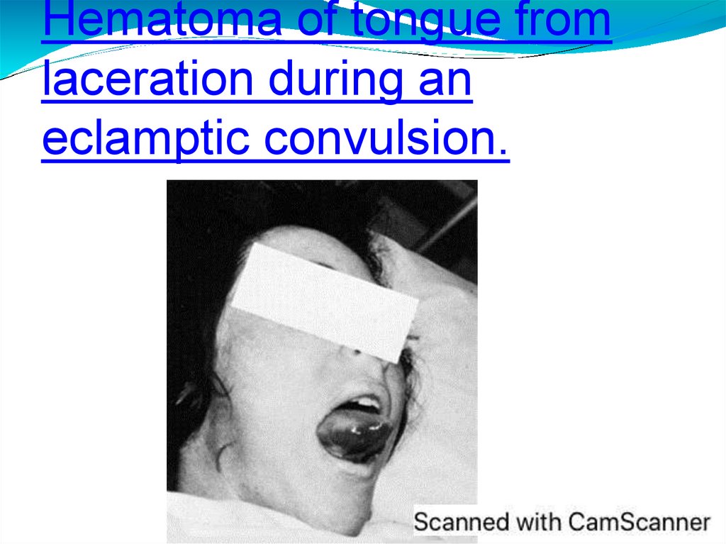
Hematoma of tongue fromlaceration during an
eclamptic convulsion.
48.
⚫ Headaches, pain under the breasts, blurredvision may be harbingers of seizures.
⚫Others can go after the first convulsions;
they can be from 10 to 100 or more in severe
cases of eclamptic status.
⚫ Death can occur from massive bleeding in
the brain. With cerebral hemorrhages,
hemiplegia may develops.
⚫In the absence of adequate treatment, an
eclamptic coma develops.
⚫ Loss of consciousness may be sudden
without the onset of convulsions “eclampsia
without eclampsia”. A differential diagnosis
of eclampsia with encephalitis, meningitis,
aneurysm, rupture of the cerebral vessels,
and hysteria should be made.
49.
Eclamptic seizure⚫ The patient may have 1or more seizures.
⚫ Seizures generally last 60-75 seconds.
⚫ The patient's face initially may become
distorted, with protrusion of the eyes.
⚫ The patient may begin foaming at the
mouth.
⚫ Respiration ceases for the duration of the
seizure.
50.
Eclamptic convulsions or fits:The fitsare epileptiform and consist of four
stages. stage: The patient become unconscious. There
⚫ Premonitory
is twitching of the muscles of the face, tongue and limbs. Eye
balls roll or turned to one side and become fixed. (30 sec.)
⚫ Tonic stage: The hole body goes into a tonic spasm – the
trunk-opisthotonus, limbs are flexed and hands clenched.
Respiration ceases and tongue protrudes between the teeth.
Cyanosis. Eye balls become fixed (30 sec.)
⚫ Clonic stage :All the voluntary muscles undergo contraction
and relaxation .Biting of the tongue occurs. Breathing is
stertorous and blood stained frothy secretions fill the mouth;
cyanosis gradually disappears (1-4 min)
⚫ Stage of coma
51.
⚫ A coma or a period of unconsciousness follows phase 2.⚫ Unconsciousness lasts for a variable period.
⚫ Following the coma phase, the patient may regain some
consciousness.
⚫ The patient may become combative and very agitated.
⚫ The patient has no recollection of the seizure.
⚫ A period of hyperventilation occurs after the tonic-clonic
seizure. This compensates for the respiratory and lactic
acidosis that develops during the apneic phase.
52.
⚫⚫
⚫
⚫
⚫
First aid for the development of
seizures and coma.
The patient is placed on a flat surface, avoiding damage.
They release the airways, open the mouth with a spoon or
spatula, draw the tongue forward and, if possible, aspirate
the contents of the oral cavity.
With renewed spontaneous respiration, oxygen is supplied.
With prolonged apnea, assisted ventilation immediately
begins.
With the discontinued cardiac activity, in parallel with the
АLV, a closed heart massage is performed and all methods
of cardiovascular resuscitation are performed.
To stop seizures in / in except for 20 ml of a 25% solution of
magnesium sulphate, 0.002 g of sibazone is injected and
administration is repeated after 10 minutes. 0.01g.
53.
⚫ Magnesium sulphate is considered to be thedrug of choice for anticonvulsant therapy. It
has anticonvulsant and sedative effects and
does not cause a marked suppression of
consciousness, which makes it possible to
remove the problem of differentiation
between the depth of drug allergies by
neuroleptics, sedatives and narcotic drugs.
Magnesium sulfate is also a diuretic and
hypotensive effect, and also reduces
intracranial pressure. The dose depends on
the woman and blood pressure levels.
54.
Complications of Eclampsia⚫ Tongue biting,
⚫ Head trauma
⚫ Broken bones
⚫ Pulmonary edema from aspiration pneumonitis or
heart failure
⚫ Death from massive cerebral hemorrhage
⚫ Hemiplegia from sublethal hemorrhage
⚫ Blindness from retinal detachment or occipital lobe
ischemia & edema
⚫ Rarely eclampsia followed by psychosis
55.
HELLP⚫ H (hemolis) - microangiopathic hemolytic anemiasyndrome:
⚫ EL (elevated liver ferments) – the increase of liver
enzymes concentration in blood plasma
⚫ LP (low platelet quantity) – the decrease of
thrombocytes level
56.
HELLP Syndrome• Hemolysis
• Elevated Liver enzymes
• Low Platelets
• < 36 wks
• Malaise (90%), epigastric pain (90%), N/V (50%)
• Self-limiting
• Multi-system failure
57.
⚫ The disease most often occurs during pregnancy, at 35weeks. In 10% of cases in a period of less than 27
weeks, and in 31% - in the first week after birth.
58.
CLINICAL SIGNS:- HEADACHE, VOMITATION, PAIN IN THE
abdomen , MOSTLY IN THE Right upper quadrant
(RUQ);
- INCREASING JAUNDANTS, HEPATIC INSUFFICIENCY;
- CONVULSIONS, COMA;
- UNCERTAKABLE LIVER TEAR AND
INTRAABDOMINAL BLEEDING;
- COAGULOPATHY, BLEEDING IN THE
POSTBIRTH PERIOD;
- FREQUENTLY TOTAL PONRP + MASSIVE
BLEEDING;
- Hepatic and renal insufficiency.
59.
HELLP⚫ Epigastric or right upper quadrant pain in a woman with
syndrome
preeclampsia often represents hepatic involvement. This is called
‘Pre eclamptic Angina’ . The pain responds poorly to analgesia
but both the pain and associated increases in liver enzymes
(AST, ALT) may subside (albeit temporarily) after blood pressure
lowering, particularly with vasodilators.
⚫ Thrombocytopenia is the commonest hematologic abnormality
seen in preeclampsia; the lower limit of the normal platelet
count in pregnancy is approximately 140x109/L but the risk of
spontaneous bleeding is not significantly increased until the
count falls below 50 x 109/L. Even so, there are concerns with
central neuraxial anesthesia and analgesic techniques at higher
levels (50-75 x 109/L), and surgical bleeding may be increased
even with moderate thrombocytopenia
60.
HELLP• Hemostasis is not problematic unless PLT <
syndrome
40,000
• Rate of fall in PLT count is important
• Regional anesthesia - contraindicated fall is
sudden
• PLT count normal within 72 hrs of delivery
• Thrombocytopenia may persist for longer periods.
• Definitive cure is delivery
61.
CHANGES IN THE LIVER WITHHELLP
syndrome
62.
Preeclampsia: Definition⚫ Hypertension
⚫ > 140/90
⚫ relative no longer considered diagnostic
⚫ Proteinuria
⚫ > 300 mg/24 hours or 1or 2+ on urine dipstick
⚫ Proteinuria is defined as urinary excretion 0.3 g
protein or greater in a 24-hour+2 or greater on urine
dip specimen
⚫ Edema (non-dependent)
⚫ ‘dry’ pre-eclampsia has been recognised hence the
exclusion of oedema from the diagnostic criteria.
63.
Severe Preeclampsia :Criteria (one or more)⚫ Blood Pressure: >160 systolic, >110 diastolic
⚫ Proteinurea: >5gm in 24 hours, over 3+ urine dip
⚫ Oligurea: less than 400ml in 24 hours
⚫ CNS: Visual changes, headache, scotomata, mental
status change
⚫ Pulmonary Edema
⚫ Epigastric or RUQ Pain: Usually indicates liver
involvement
⚫ Impaired Liver Function tests
⚫ Thrombocytopenia: <100,000
⚫ Intrauterine Growth Restriction: With or without
abnormal doppler assessment
⚫ Oligohydramnios
64.
Physical Findings⚫ Blood Pressure
inPreeclampsia
⚫ Proteinurea
⚫ Retinal vasospasm or Retinal edema
⚫ Right upper quadrant (RUQ) abdominal tenderness stems from
liver swelling and capsular stretch
⚫ Brisk, or hyperactive, reflexes are common during pregnancy,
but clonus is a sign of neuromuscular irritability that raises
concern.
⚫ Among pregnant women, 30% have some lower extremity edema
as part of their normal pregnancy. However, a sudden change in
dependent edema, edema in nondependent areas such as the
face and hands, or rapid weight gain suggests a pathologic
process and warrants further evaluation
65.
Laboratory Tests :- Uric⚫ Decreased renal urate excretion in
acid
preeclampsia
⚫ Serum uric acid exceeding 5.9 at 24 wk
(PPV 33%)
⚫ Not useful in differentiating GHT from
preeclampsia
⚫ usually (> 6mg/dl)
66.
Coagulation activation⚫ Thrombocytopenia and platelet dysfunction
⚫ Increased destruction cause platelet volumes increase
(younger platelet)
⚫ Preeclampsia : PAI-1increase increased relative to PAI2 because of endothelial cell dysfunction
67.
Instrumental Investigations⚫ Uterine artery doppler
⚫ Fetal Sonography
⚫
Fetal size
⚫ Amniotic fluid volume
⚫ Fetal and maternal dopplers
⚫ CT Scan (Brain)
⚫ ECG
⚫ Doppler sonography
⚫ Cerebral angiography
68.
⚫ Differential diagnosis :⚫ epilepsy , encephalitis , meningitis , cerebral tumor ,
cysticercosis , ruptured cerebral aneurysm
69.
Gross liver specimen from a woman with preeclampsia whodied from severe acidosis and liver failure. Periportal
hemorrhagic necrosis was seen microscopically.
70.
Computed tomographic scan of liver showing a subcapsularhematoma with a peripheral hyperdense rim corresponding to
more recent hemorrhage.
71.
Magnetic resonance imaging in a 22-year-old woman witheclampsia who had cortical blindness for 96 hours. A highsignal lesion (arrow) is apparent in the left occipital lobe.
72.
At postnatal review(6–8 weeks after
1. Offer medical review.
birth)
2. Offer specialist referral if antihypertensive treatment
still needed.
3. Repeat platelet count, transaminases and serum
creatinine measurements if indicated.
4. Carry out a urinary reagent-strip test. If proteinuria ≥
1+:
⚫ – offer further review at 3 months to assess kidney
function
⚫ – consider offering referral for specialist kidney
assessment.
73.
Antihypertensive Medications inManagement of HSIP
74.
⚫ Three short-acting antihypertensive agents⚫ hydralazine
⚫ short-acting [sublingual or orally administered]
nifedipine
⚫ labetalol
→ commonly used control acute very high blood
pressure in women with severe hypertension in pregnancy
who may require emergency cesarean section and often
receive magnesium sulfate
75.
⚫ First line drugs include⚫ methyldopa
⚫ labetolol
⚫ Second line agents are
⚫ hydralazine,
⚫ nifedipine
⚫ prazosin
76.
Hydralazin5-10 mg every 20 minutes IV
e⚫⚫ Dose:
Onset: 10-20 minutes
⚫ Duration: 3-8 hours
⚫ Side effects: headache, flushing, tachycardia, lupus
like symptoms
⚫ Mechanism: peripheral vasodilator
⚫ First Choice for severe PIH
77.
Labetalol⚫ Dose:
⚫ IV: 20mg, then 40, then 80 every 20 minutes, for a
total of 220mg
⚫ Oral 100 mg bid to be increased up to 200 mg qid.
maximum 2400mg daily)
⚫ Onset: 1-2 minutes
⚫ Duration: 6-16 hours
⚫ Side effects: hypotension
⚫ Mechanism: Alpha and Beta blocker
⚫ Used extensively in pregnancy. Has favorable effects.
(
78.
Nifedipine⚫ Dose: 10 mg po, not sublingual
⚫ Onset: 5-10 minutes
⚫ Duration: 4-8 hours
⚫ Side effects: chest pain, headache, tachycardia
⚫ Mechanism: CA channel block
⚫ Required by midwives and nurses in the absence of a
doctor
79.
Clonidine⚫ Dose: 1mg po
⚫ Onset: 10-20 minutes
⚫ Duration: 4-6 hours
⚫ Side effects: unpredictable, avoid rapid withdrawal
⚫ Mechanism: Alpha agonist, works centrally
80.
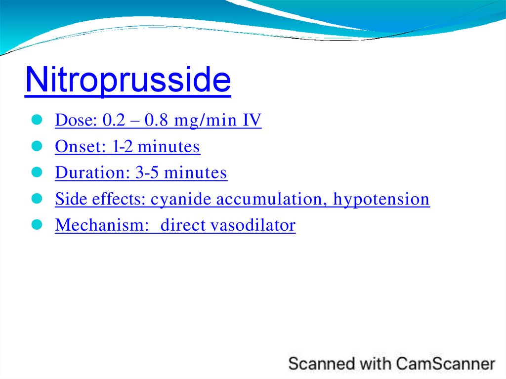
Nitroprusside⚫ Dose: 0.2 – 0.8 mg/min IV
⚫ Onset: 1-2 minutes
⚫ Duration: 3-5 minutes
⚫ Side effects: cyanide accumulation, hypotension
⚫ Mechanism: direct vasodilator
81.
Alpha-methyl Dopa⚫ The most commonly used and presumably the safest
with pregnancy.
⚫ The usual dose starts with 250mg tds to be increased
up to 2 grams per day.
⚫ It blocks the adrenaline release at post synaptic sites.
82.
Antenatal care and fetal monitoring83.
If previous severe eclampsia /pre-eclampsia needing birthbefore 34 weeks pre-eclampsia OR with baby’s birth weight
< 10th centile OR intrauterine death OR placental
abruption , Carry out
⚫ ultrasound fetal growth and amniotic fluid volume
assessment + umbilical artery
⚫ doppler velocimetry.
Start at 28–30 weeks, or at least 2 weeks before previous
⚫ gestational age of onset of hypertensive disorder if earlier
than 28 weeks.
Repeat 4 weeks later.
84.
⚫ If at least two moderate risk factors or at least one highrisk factor for pre-eclampsia,
⚫ Advise woman to take aspirin 75 mg/day from 12 weeks
until birth.
⚫ If fetal activity abnormal, carry out cardiotocography.
85.
Management in variousperiods of pregnancy
86.
Antenatal care87.
Mild hypertension (BP 140/90–149/99 mmHg)
Do not treat hypertension.
2. Measure BP at least 4 times a day.
3. Test kidney function, electrolytes, FBC,
transaminases, bilirubin 2 times a week.
1.
88.
Moderate hypertension(BP 150/100–159/109 mmHg)
⚫ Treat with first-line oral labetalol to keep BP < 150/80–
100 mmHg.
⚫ Measure BP at least 4 times a day.
⚫ Test kidney function, electrolytes, FBC, transaminases,
bilirubin 3 times a week.
89.
Timing of birthBefore 34 weeks
1. Manage conservatively (do not plan same-day delivery of
2.
baby).
Consultant obstetric staff to:
⚫ – document maternal (biochemical, haematological and clinical)
and fetal indications for elective birth before 34 weeks
⚫ – write plan for antenatal fetal monitoring.
2.
Offer birth (after discussion with neonatal and anaesthetic
teams and, if required, course of corticosteroids completed) if:
⚫ – severe refractory hypertension
⚫ – maternal or fetal clinical indication develops as defined in plan.
90.
Mild and moderate hypertension(140/90–159/109 mmHg)
Measure BP hourly.
2. Continue antenatal hypertensive treatment.
3. Carry out haematological and biochemical
monitoring according to criteria from antenatal
period, even if regional analgesia being considered.
4. Do not routinely limit duration of second stage of
labour if BP stable
1.
91.
Management ofsevere hypertension
(Eclampsia)
92.
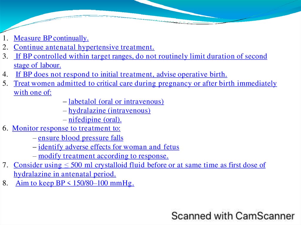
1. Measure BP continually.2. Continue antenatal hypertensive treatment.
3. If BP controlled within target ranges, do not routinely limit duration of second
stage of labour.
4. If BP does not respond to initial treatment, advise operative birth.
5. Treat women admitted to critical care during pregnancy or after birth immediately
with one of:
– labetalol (oral or intravenous)
– hydralazine (intravenous)
– nifedipine (oral).
6. Monitor response to treatment to:
– ensure blood pressure falls
– identify adverse effects for woman and fetus
– modify treatment according to response.
7. Consider using ≤ 500 ml crystalloid fluid before or at same time as first dose of
hydralazine in antenatal period.
8. Aim to keep BP < 150/80–100 mmHg.
93.
Anticonvulsants94.
⚫ Give intravenous magnesium sulphate if woman withsevere hypertension or severe pre-eclampsia has or
previously had eclamptic fit.
⚫ Consider giving intravenous magnesium sulphate if
birth planned within 24 hours in woman with severe
pre-eclampsia.
⚫ Do not use diazepam, phenytoin or lytic cocktail as
alternatives to magnesium sulphate* in women with
eclampsia.
95.
Regimen for magnesium sulphate96.
Loading dose4 g given intravenously slowly over 5-10 minutes,
followed by infusion of 1g/hour for 24 hours.
OR
10gm deep IM 5gm in each buttock
Maintenance dose
5gm deep IM, 2.5gm in each buttock 4hrly.
Continue 24hrs after last convulsion or delivery
(Further dose of 2–4 g given over 5 minutes if
recurrent seizures.)
97.
Corticosteroids98.
For fetal lung maturation⚫ If birth likely within 7 days in woman with pre-
eclampsia:
1. give 2 doses betamethasone 12 mg intramuscularly
24 hours apart between 24 and 34 weeks
2. consider giving 2 doses betamethasone 12 mg
intramuscularly 24 hours apart at 35–36 weeks.
⚫ For HELLP syndrome
Do not use dexamethasone or betamethasone to treat
HELLP syndrome
99.
⚫ Caesarean section versus induction of labourChoose mode of birth according to clinical
circumstances and woman’s preference.
100.
Diuretics & hyperosmotic agents101.
⚫ Diuretics : deplete intravascular volume , compromiseplacental perfusion , limited used to pulmonary edema
⚫ Hyperosmotic agents e.g. Mannitol , Isosorbide :-
leaks of agents through capillaries into lungs & brain
promote accumulation of edema
102.
103.
Antenatal management⚫ If the platelet count is sufficiently low to present a
hazard for operative delivery, a platelet transfusion
should be considered