


 Биология
Похожие презентации:


The cytoskeleton: microfilaments essential. Cell biology
1.
Lectures 21 and 22:The Cytoskeleton:
Microfilaments
Essential
Cell Biology
Third Edition
Chapter 17
2.
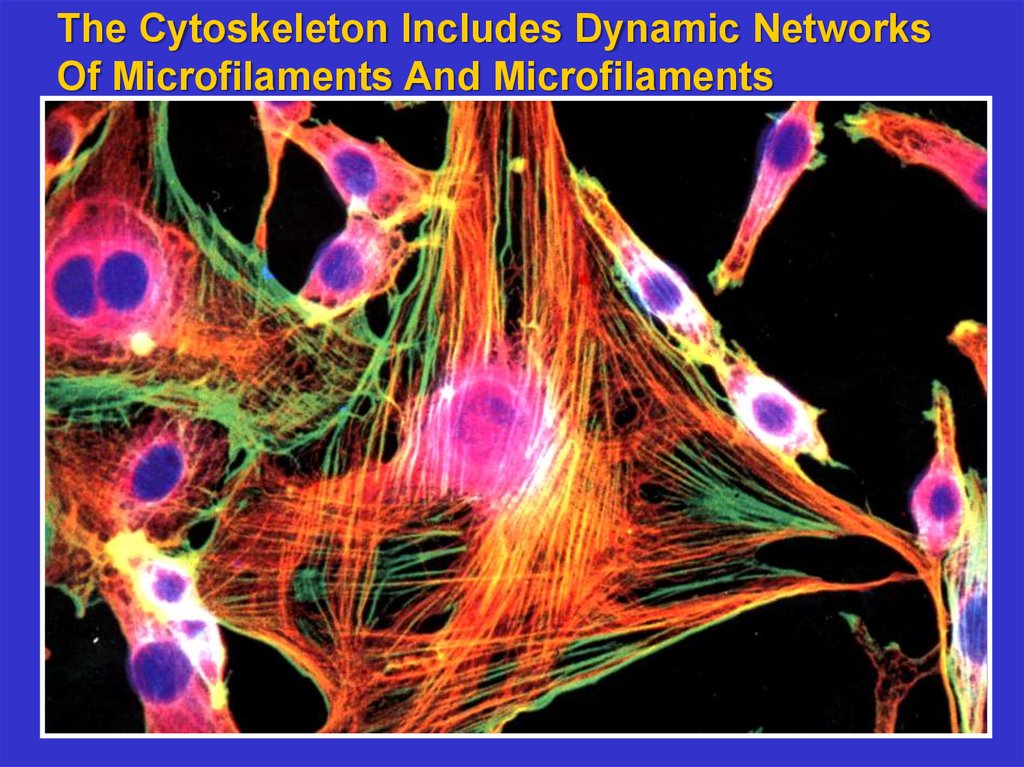
The Cytoskeleton Includes Dynamic NetworksOf Microfilaments And Microfilaments
3.
Microfilaments composed of actin are oftenfound at the cortex of cells. They also form
stress fibers that give shape to the cell. They
are highly dynamic forming networks that
serve as the basis for cellular motility.
4.
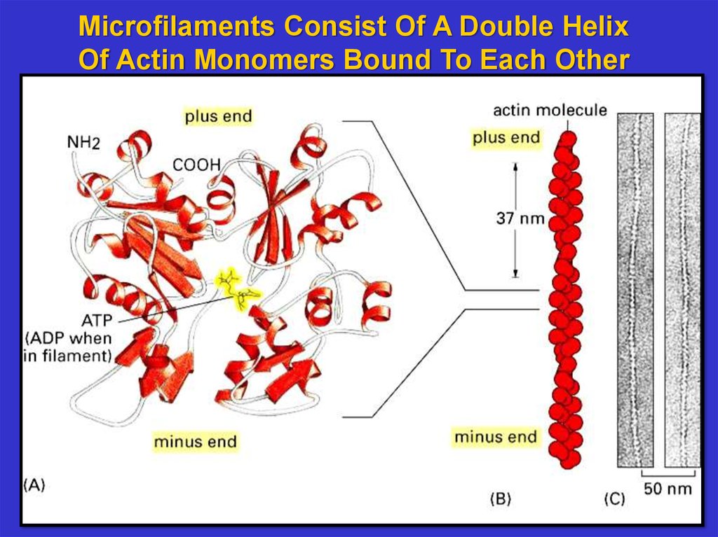
Microfilaments Consist Of A Double HelixOf Actin Monomers Bound To Each Other
5.
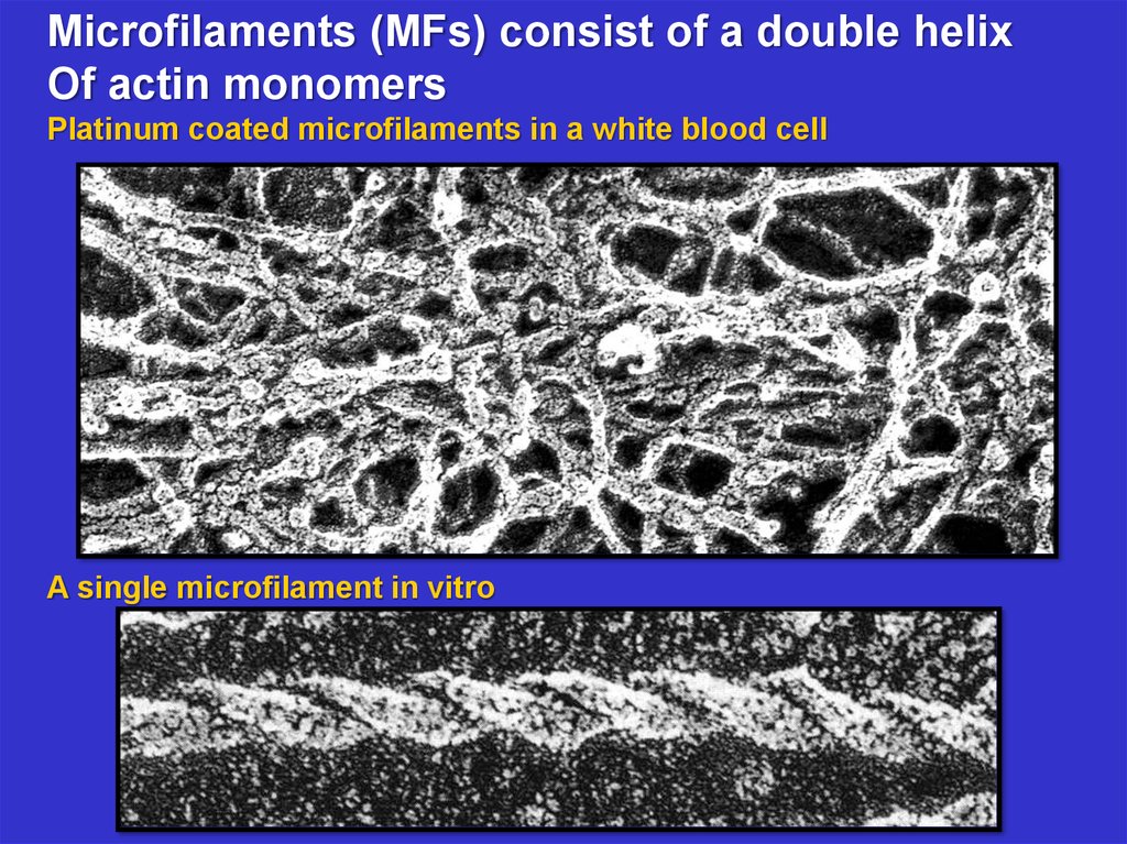
Microfilaments (MFs) consist of a double helixOf actin monomers
Platinum coated microfilaments in a white blood cell
A single microfilament in vitro
6.
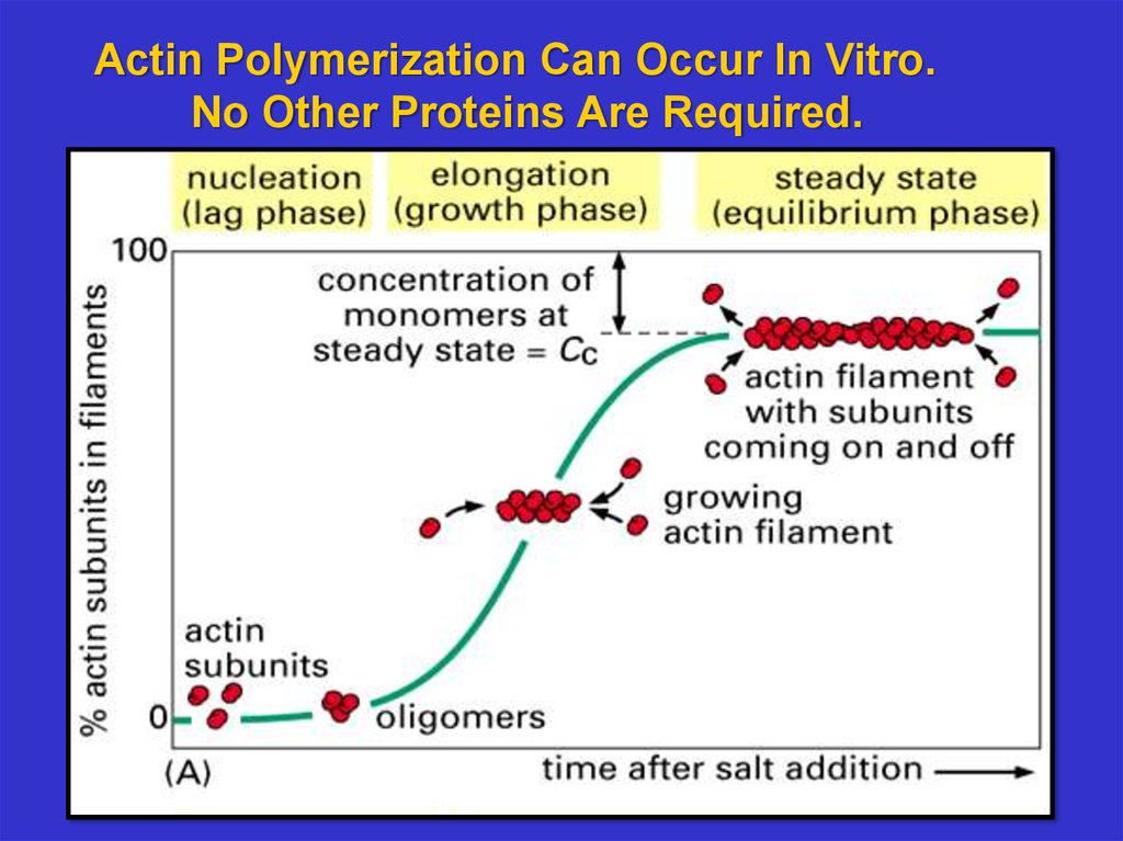
Actin Polymerization Can Occur In Vitro.No Other Proteins Are Required.
7.
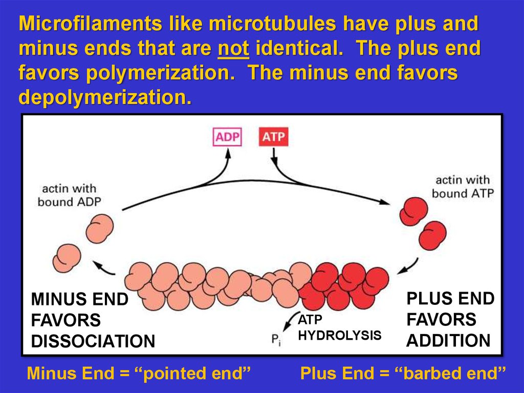
Microfilaments like microtubules have plus andminus ends that are not identical. The plus end
favors polymerization. The minus end favors
depolymerization.
MINUS END
FAVORS
DISSOCIATION
Minus End = “pointed end”
ATP
HYDROLYSIS
PLUS END
FAVORS
ADDITION
Plus End = “barbed end”
8.
Animation: Regulation of Actin Polymerization andDepolymerization inside cells using Profilin and Cofilin
Profilin binds to actin monomers (G-actin) to aid
polymerization; cofilin binds to microfilaments (f-actin) to
sever them and allow faster depolymerization
9.
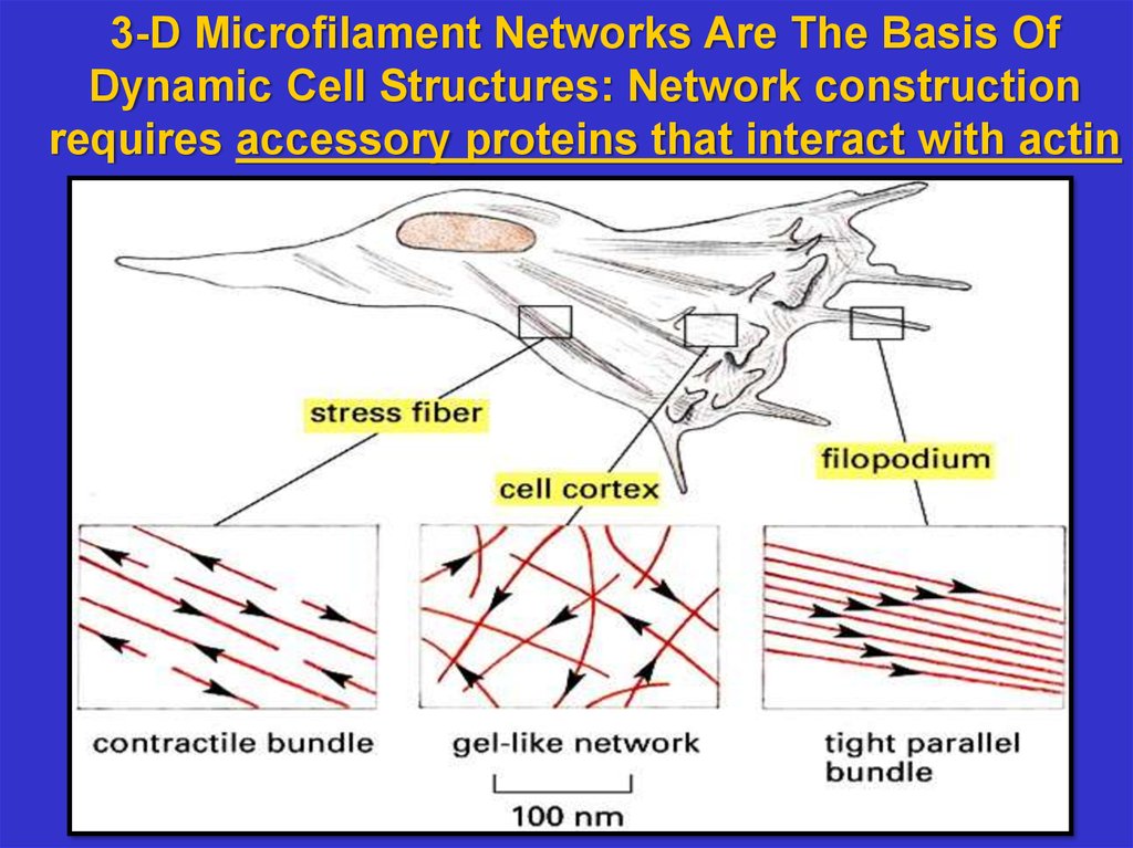
3-D Microfilament Networks Are The Basis OfDynamic Cell Structures: Network construction
requires accessory proteins that interact with actin
10.
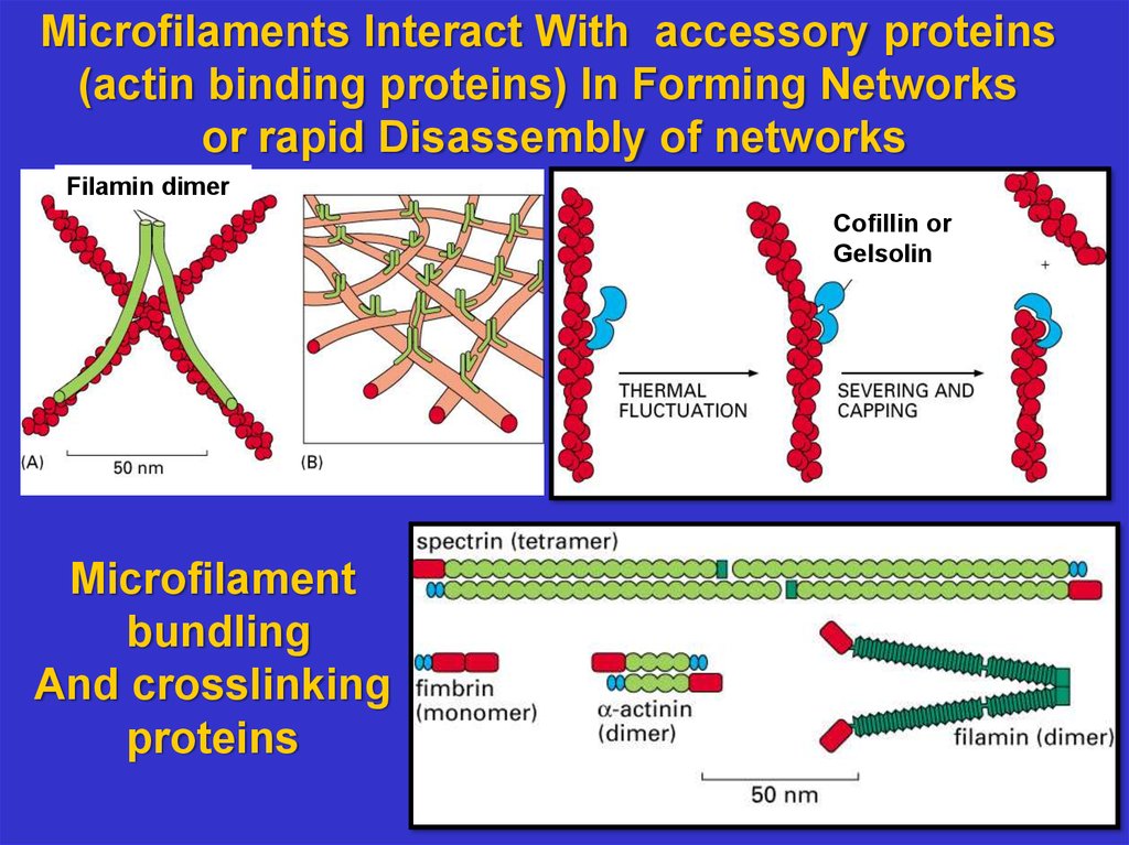
Microfilaments Interact With accessory proteins(actin binding proteins) In Forming Networks
or rapid Disassembly of networks
Filamin dimer
Cofillin or
Gelsolin
Microfilament
bundling
And crosslinking
proteins
11.
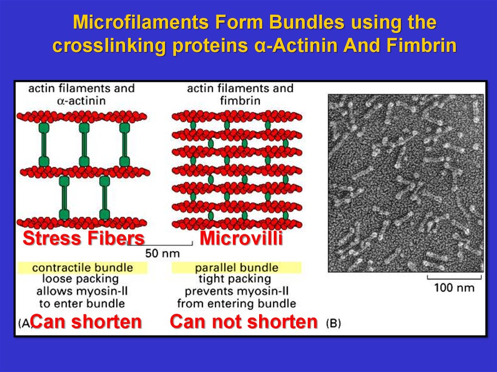
Microfilaments Form Bundles using thecrosslinking proteins α-Actinin And Fimbrin
Stress Fibers
Microvilli
Can shorten
Can not shorten
12.
Microvilli Contain A Microfilament Bundle13.
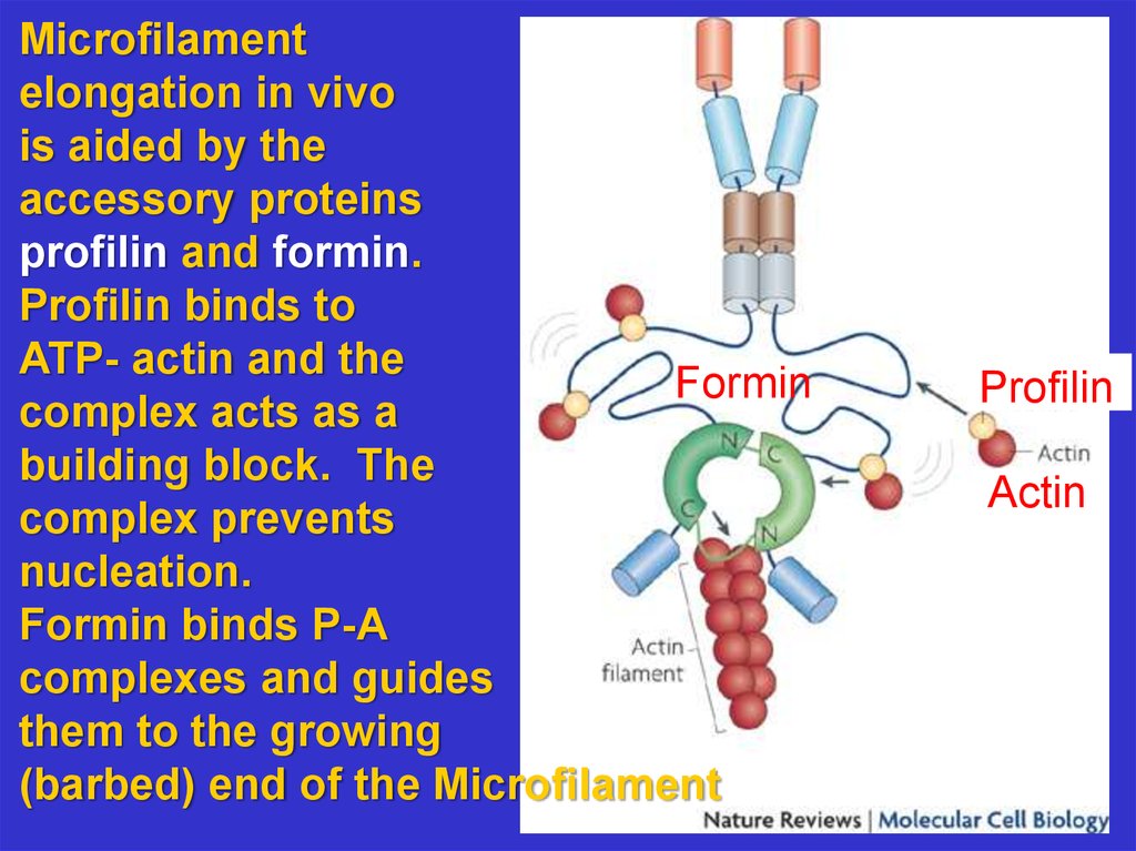
Microfilamentelongation in vivo
is aided by the
accessory proteins
profilin and formin.
Profilin binds to
ATP- actin and the
Formin
complex acts as a
building block. The
complex prevents
nucleation.
Formin binds P-A
complexes and guides
them to the growing
(barbed) end of the Microfilament
Profilin
Actin
14.
Animation of Actin Polymerization using Formin15.
In cells, the ends of microfilaments are cappedwith other proteins to control assembly. ARP 2
and ARP 3 cap the minus end of microfilaments.
CAPPING
PREVENTS
ACTIN
DISSOCIATION
16.
ARPs allow binding of minus ends to otherfilaments. In this way microfilament networks
can be formed that are used as
superstructures for cell shape and motility.
17.
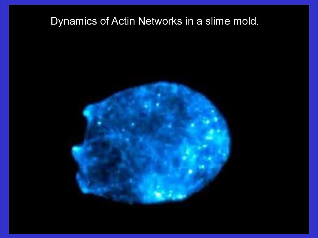
Dynamics of Actin Networks in a slime mold.18.
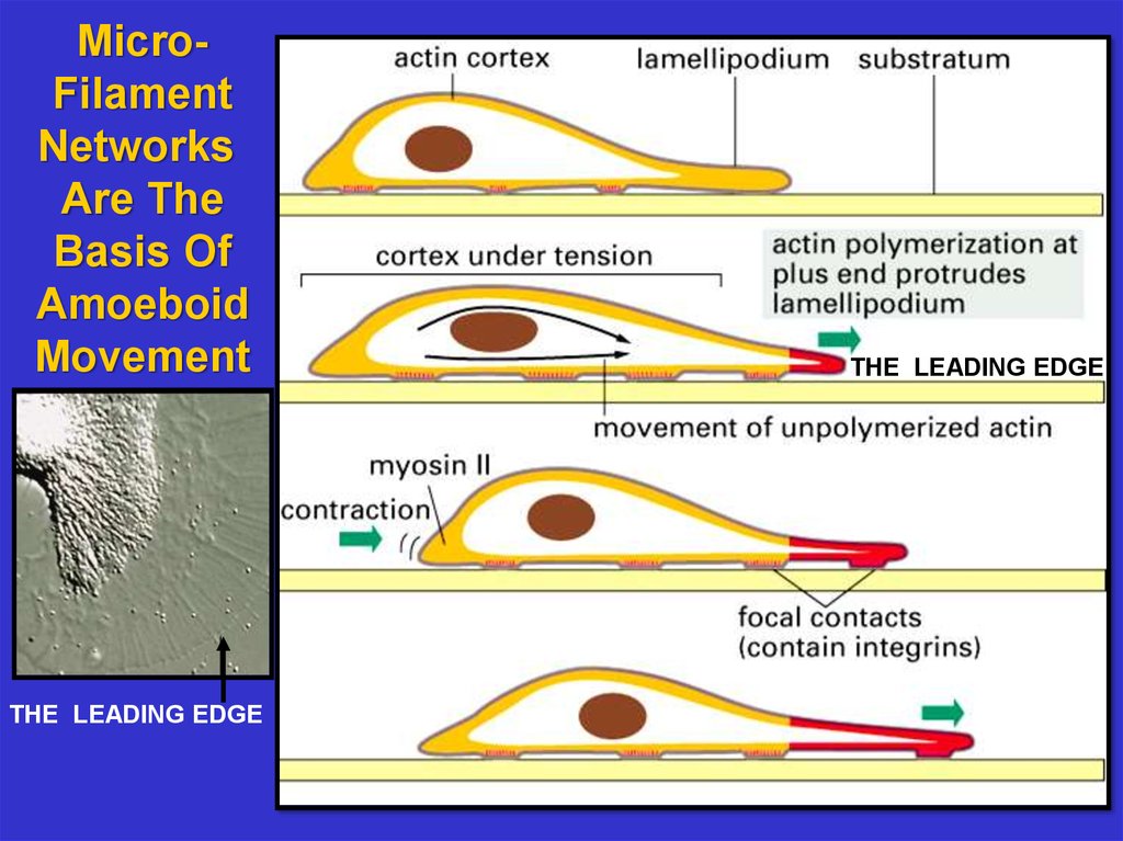
MicroFilamentNetworks
Are The
Basis Of
Amoeboid
Movement
THE LEADING EDGE
THE LEADING EDGE
19.
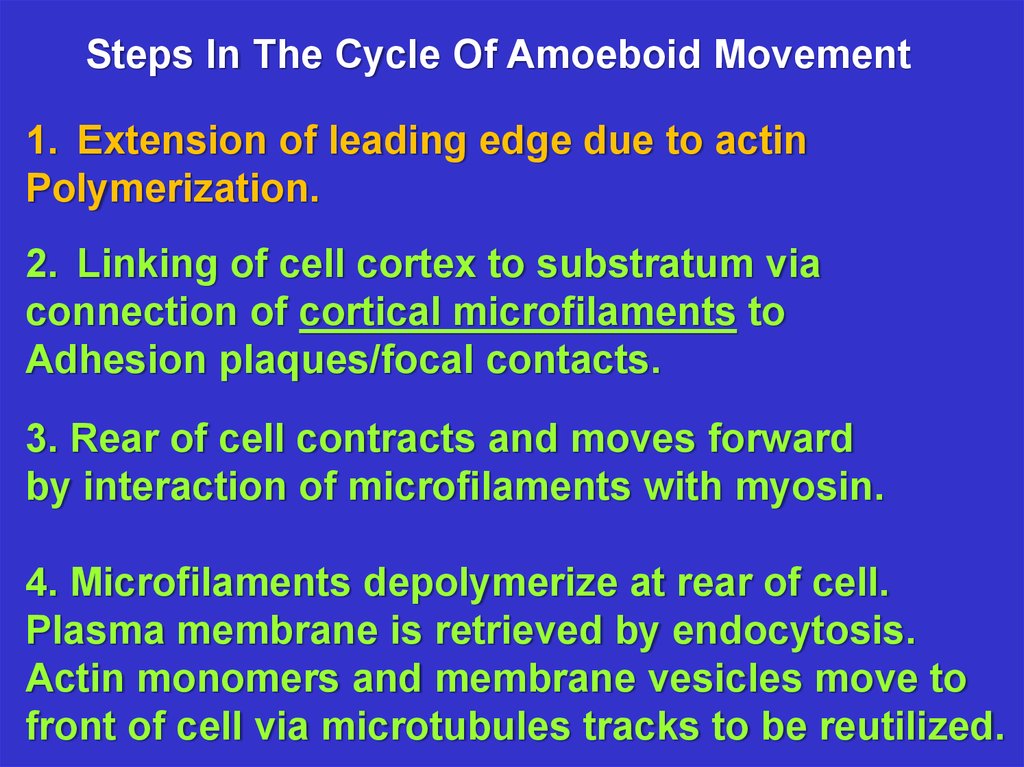
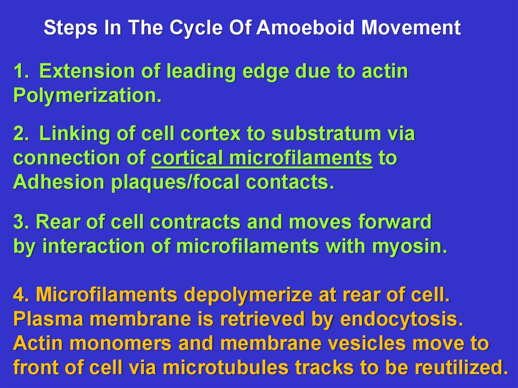
Steps In The Cycle Of Amoeboid Movement1. Extension of leading edge due to actin
Polymerization.
2. Linking of cell cortex to substratum via
connection of cortical microfilaments to
Adhesion plaques/focal contacts.
3. Rear of cell contracts and moves forward
by interaction of microfilaments with myosin.
4. Microfilaments depolymerize at rear of cell.
Plasma membrane is retrieved by endocytosis.
Actin monomers and membrane vesicles move to
front of cell via microtubules tracks to be reutilized.
20.
Microfilament Networks Are Dynamic At TheLeading Edge
21.
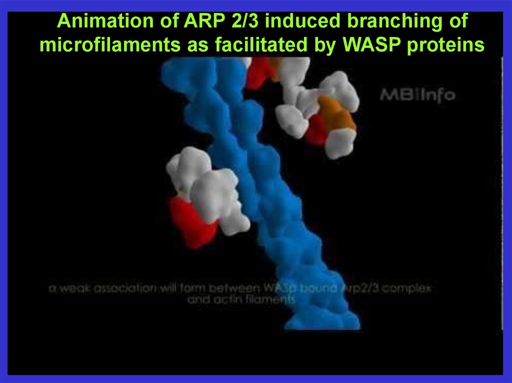
Animation of ARP 2/3 induced branching ofmicrofilaments as facilitated by WASP proteins
22.
Gab1Adaptor
Adaptor
Growth
Factor
Receptor
GEF
Auto-inhibited WASP
Cell Ruffling
Response
Actin
polymerization
and branching
ARP 2/3
Complex
Activated WASP G-Protein
“Switch”
Cell Signaling
Controls
The Actin
Cytoskeleton
Formin
Signal
Rho GTPase
Rac GTPase
Formin
polymerization
filopodia extend
WASP ARP 2/3 branching lamellapodia
23.
MicroFilamentNetworks
Are the
Basis of
Ameoboid
Movement
THE LEADING EDGE
24.
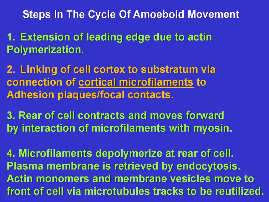
Steps In The Cycle Of Amoeboid Movement1. Extension of leading edge due to actin
Polymerization.
2. Linking of cell cortex to substratum via
connection of cortical microfilaments to
Adhesion plaques/focal contacts.
3. Rear of cell contracts and moves forward
by interaction of microfilaments with myosin.
4. Microfilaments depolymerize at rear of cell.
Plasma membrane is retrieved by endocytosis.
Actin monomers and membrane vesicles move to
front of cell via microtubules tracks to be reutilized.
25.
Focal adhesions use hundreds of transmembrane proteinscalled integrins for linking the actin cytoskeleton inside
the cell to extracellular matrix fibers such as collagen
outsidethe cell. This linkage has a mechanical function.
Focal adhesions are
dynamically controlled
allowing them to bind,
and unbind from
extracellular matrix
fibers in a reversible
manner. This allows
them to serve as
temporary anchors
during cell movement.
26.
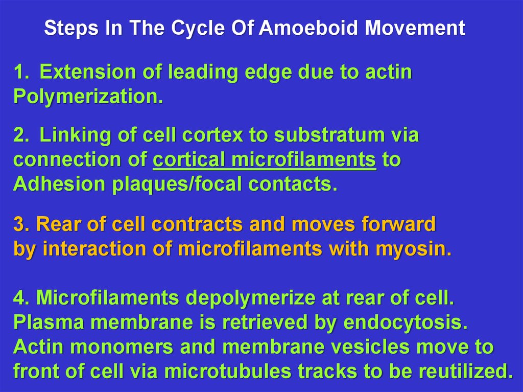
Steps In The Cycle Of Amoeboid Movement1. Extension of leading edge due to actin
Polymerization.
2. Linking of cell cortex to substratum via
connection of cortical microfilaments to
Adhesion plaques/focal contacts.
3. Rear of cell contracts and moves forward
by interaction of microfilaments with myosin.
4. Microfilaments depolymerize at rear of cell.
Plasma membrane is retrieved by endocytosis.
Actin monomers and membrane vesicles move to
front of cell via microtubules tracks to be reutilized.
27.
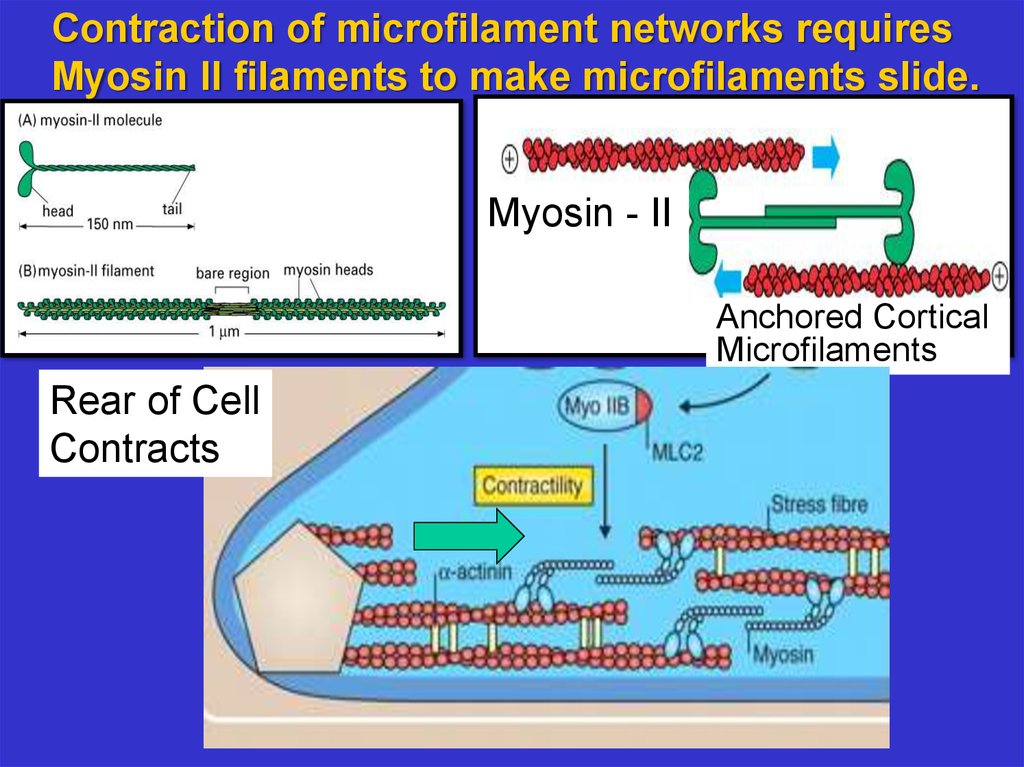
Contraction of microfilament networks requiresMyosin II filaments to make microfilaments slide.
Myosin - II
Anchored Cortical
Microfilaments
Rear of Cell
Contracts
28.
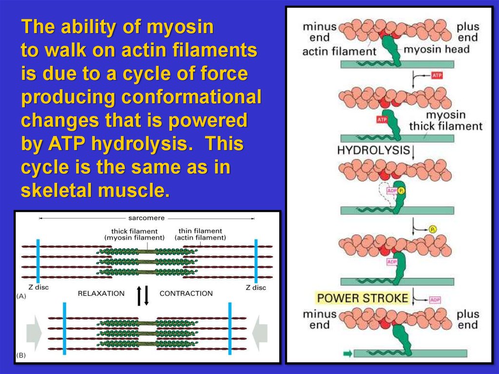
The ability of myosinto walk on actin filaments
is due to a cycle of force
producing conformational
changes that is powered
by ATP hydrolysis. This
cycle is the same as in
skeletal muscle.
29.
Steps In The Cycle Of Amoeboid Movement1. Extension of leading edge due to actin
Polymerization.
2. Linking of cell cortex to substratum via
connection of cortical microfilaments to
Adhesion plaques/focal contacts.
3. Rear of cell contracts and moves forward
by interaction of microfilaments with myosin.
4. Microfilaments depolymerize at rear of cell.
Plasma membrane is retrieved by endocytosis.
Actin monomers and membrane vesicles move to
front of cell via microtubules tracks to be reutilized.
30.
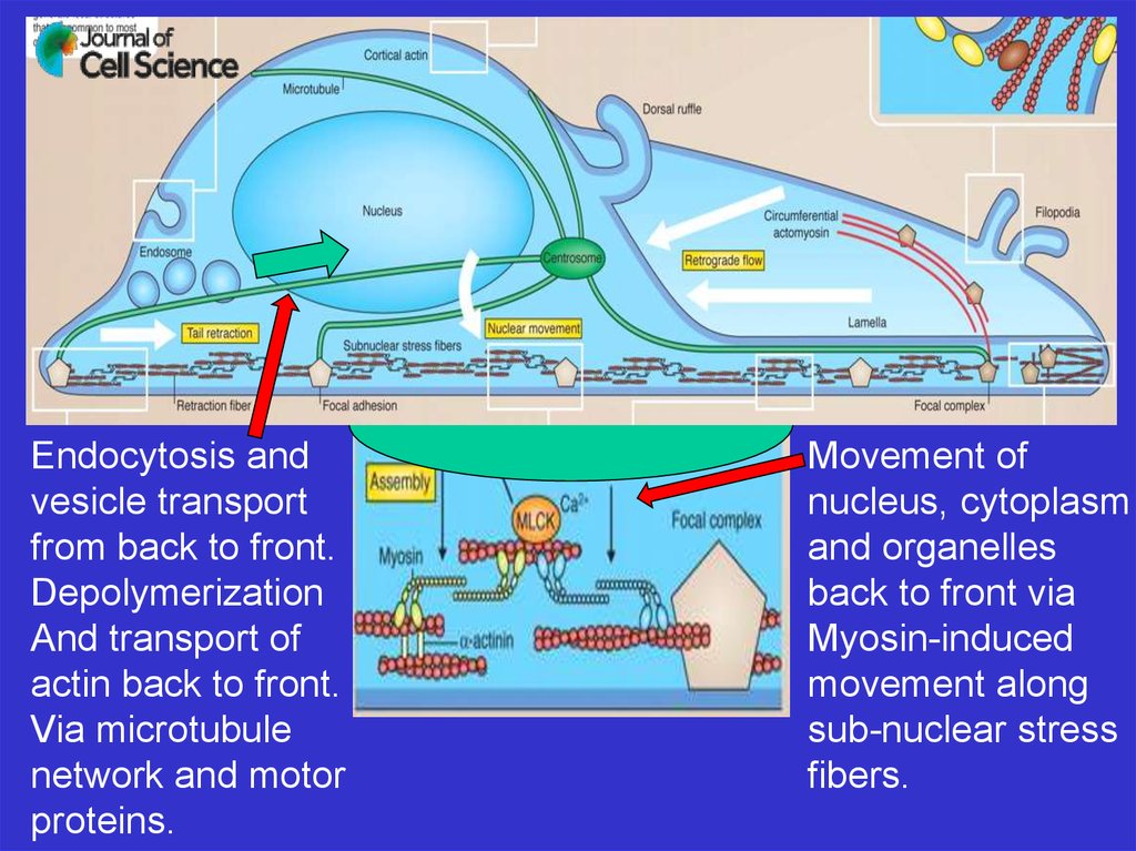
Endocytosis andvesicle transport
from back to front.
Depolymerization
And transport of
actin back to front.
Via microtubule
network and motor
proteins.
Movement of
nucleus, cytoplasm
and organelles
back to front via
Myosin-induced
movement along
sub-nuclear stress
fibers.
31.
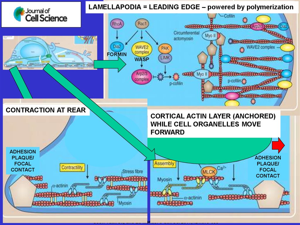
LAMELLAPODIA = LEADING EDGE – powered by polymerizationFORMIN
WASP
CONTRACTION AT REAR
CORTICAL ACTIN LAYER (ANCHORED)
WHILE CELL ORGANELLES MOVE
FORWARD
ADHESION
PLAQUE/
FOCAL
CONTACT
ADHESION
PLAQUE/
FOCAL
CONTACT
Soon-Tuck Sit, and Ed Manser J Cell Sci 2011;124:679-683