


 Биология
Похожие презентации:


The Cytoskeleton: Intermediate Filaments and Microtubules
1.
Lecture 20:The Cytoskeleton:
Intermediate Filaments and
Microtubules
Essential
Cell Biology
Fourth Edition
Chapter 17
2.
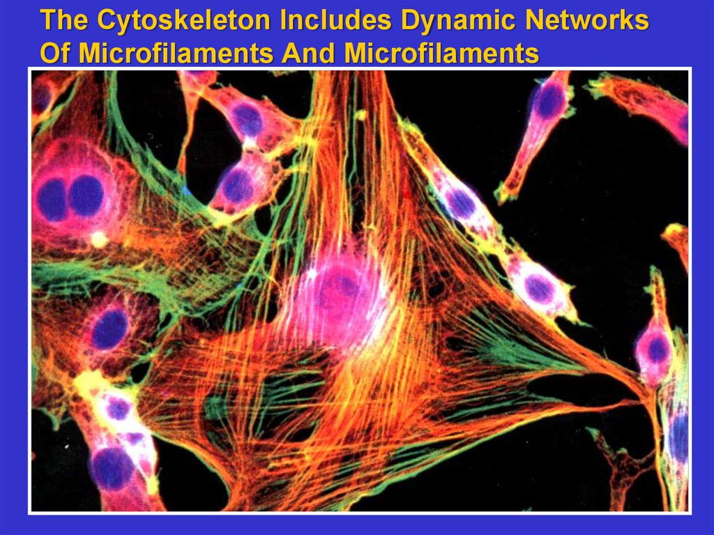
The Cytoskeleton Includes Dynamic NetworksOf Microfilaments And Microfilaments
3.
The cytoskeleton consists of three major typesof filaments plus many filament-associated
proteins including molecular motors
Microfilaments – composed of actin, these
filaments form dynamic networks that form
the basis for cell shape and movement
Microtubules – composed of tubulin, these
tubules act as tracks on which to move
vesicles and organelles. They also form the
basis of cilia and flagella. They are dynamic.
Intermediate filaments – composed of proteins
that associate to form rope-like structures
that are of high mechanical strength. They
position organelles and form a strong, long
lasting cell superstructure.
4.
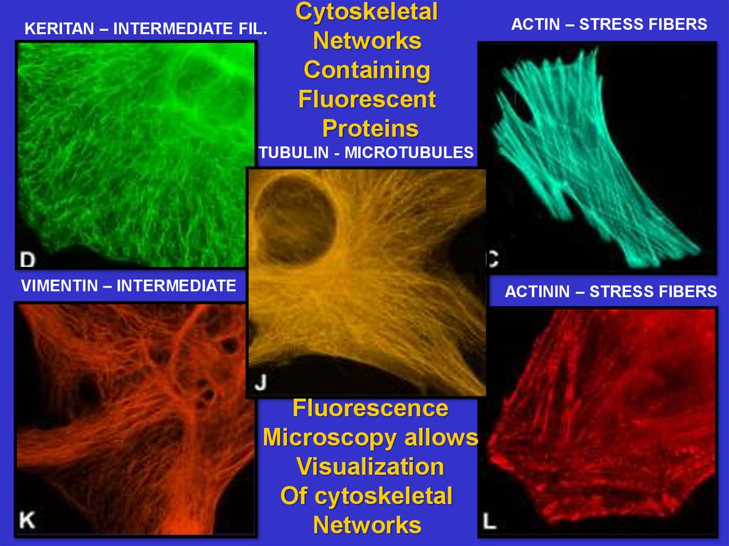
KERITAN – INTERMEDIATE FIL.Cytoskeletal
Networks
Containing
Fluorescent
Proteins
ACTIN – STRESS FIBERS
TUBULIN - MICROTUBULES
VIMENTIN – INTERMEDIATE
ACTININ – STRESS FIBERS
Fluorescence
Microscopy allows
Visualization
Of cytoskeletal
Networks
5.
IntermediateFilaments
are nondynamic and
structural.
They position
the nucleus
and insert into
Desmosomes
to hold
neighboring
cells
together.
6.
IntermediateFilaments
polymerize
to form strong
rope-like fibers.
The basic
structural unit
is a coiled-coil
dimer. These
fibers are
symmetric
7.
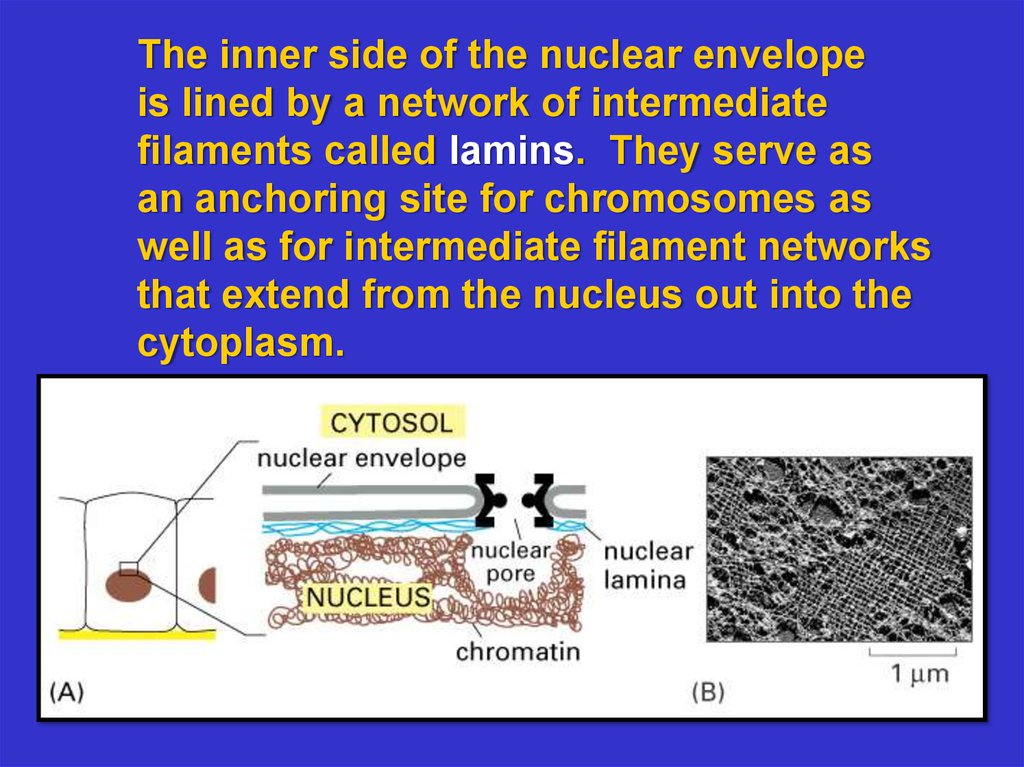
The inner side of the nuclear envelopeis lined by a network of intermediate
filaments called lamins. They serve as
an anchoring site for chromosomes as
well as for intermediate filament networks
that extend from the nucleus out into the
cytoplasm.
8.
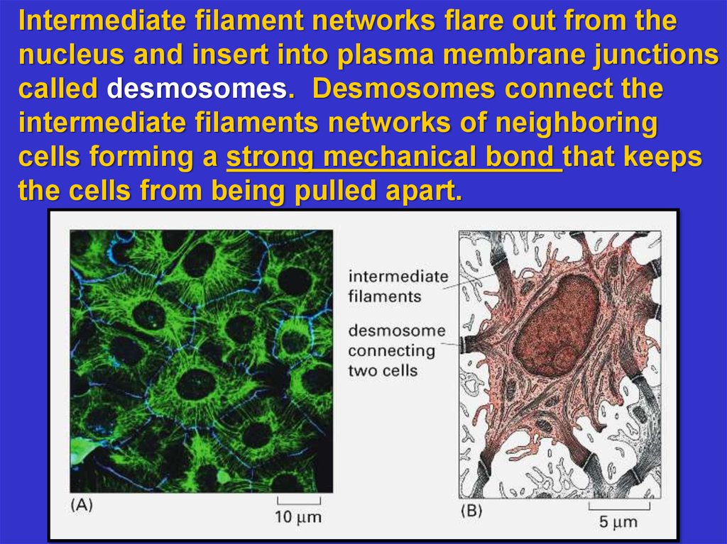
Intermediate filament networks flare out from thenucleus and insert into plasma membrane junctions
called desmosomes. Desmosomes connect the
intermediate filaments networks of neighboring
cells forming a strong mechanical bond that keeps
the cells from being pulled apart.
9.
MicrotubulesMake Up
Dynamic
Networks
10.
Microtubules serve four functions:1. To give shape to the cell.
Example: nerve axons contain numerous microtubules along their length. If disrupted the axon
shrivels.
2. To provide “tracks” on which to move
vesicles carrying cargo.
Example: pigment granules move outward
and inward from cell center using microtubules.
3. To form the mitotic spindle which separates
chromosomes during mitosis and meiosis.
4. To form flagella and cilia – whip like
structures that propel cells.
11.
Microtubules Are Made Of Tubulin Protofilaments12.
Microtubules asseen by Electron
Microscopy
1) thin section
2) freeze dried
And platinum
Shadowed
13.
Microtubules are stabilized by capping at theirPlus and minus ends. Centrosomes and
Microtubule organizing centers (MTOCs) cap the
minus end; special membrane-associated proteins
cap the plus end.
14.
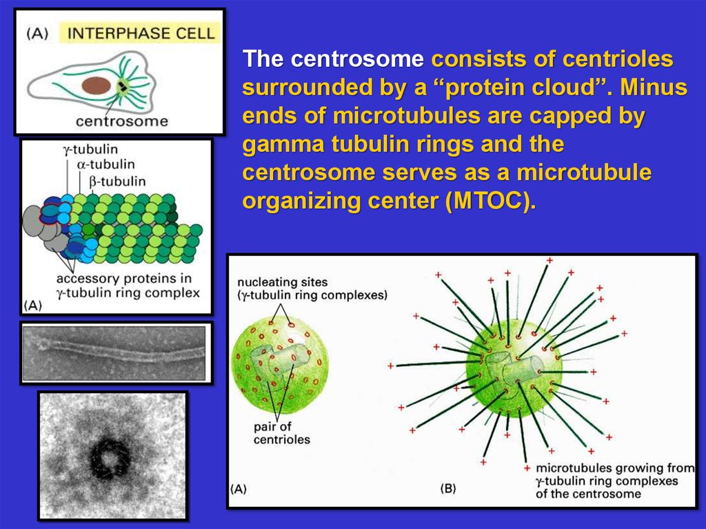
The centrosome consists of centriolessurrounded by a “protein cloud”. Minus
ends of microtubules are capped by
gamma tubulin rings and the
centrosome serves as a microtubule
organizing center (MTOC).
15.
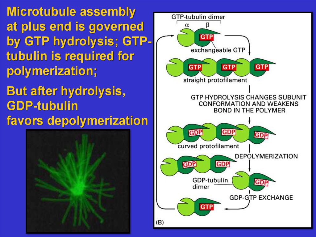
Microtubule assemblyat plus end is governed
by GTP hydrolysis; GTPtubulin is required for
polymerization;
But after hydrolysis,
GDP-tubulin
favors depolymerization
16.
CatastrophicDisassembly can occur
if growth at the plus end
stops or is slow; but
the microtubule starts to
grow at this end again.
17.
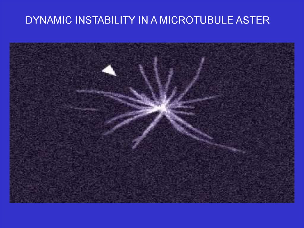
DYNAMIC INSTABILITY IN A MICROTUBULE ASTER18.
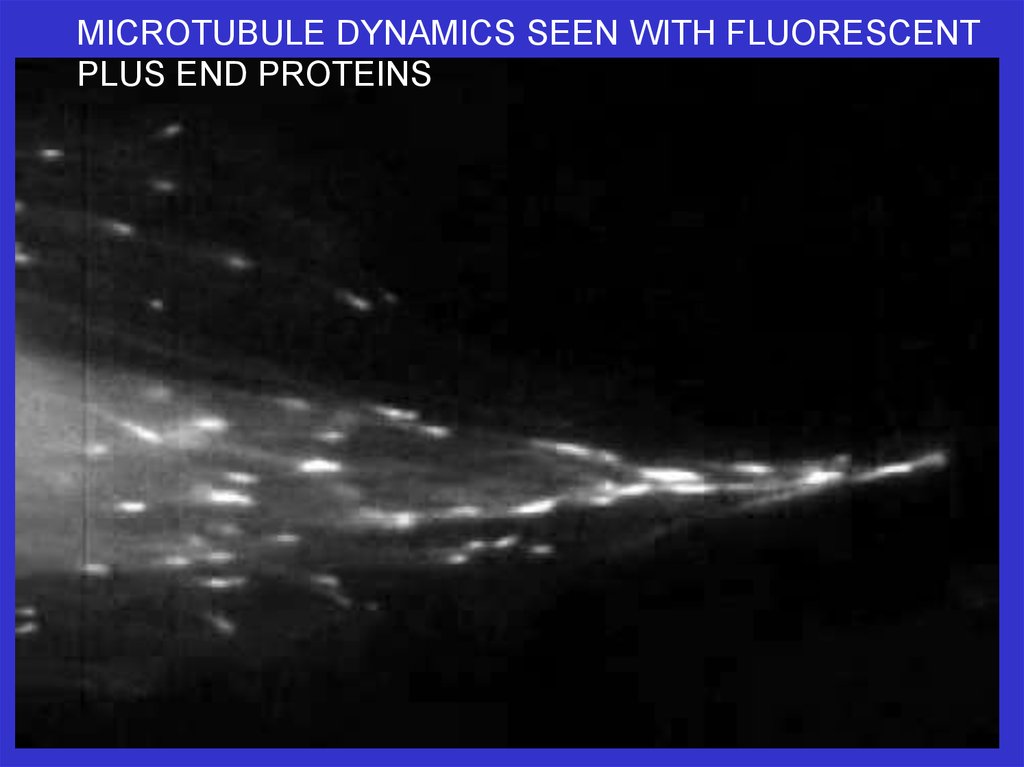
MICROTUBULE DYNAMICS SEEN WITH FLUORESCENTPLUS END PROTEINS
19.
MICROTUBULE DYNAMICS SEEN WITH FLUORESCENTPLUS END PROTEINS
20.
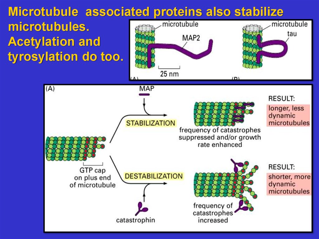
Microtubule associated proteins also stabilizemicrotubules.
Acetylation and
tyrosylation do too.
21.
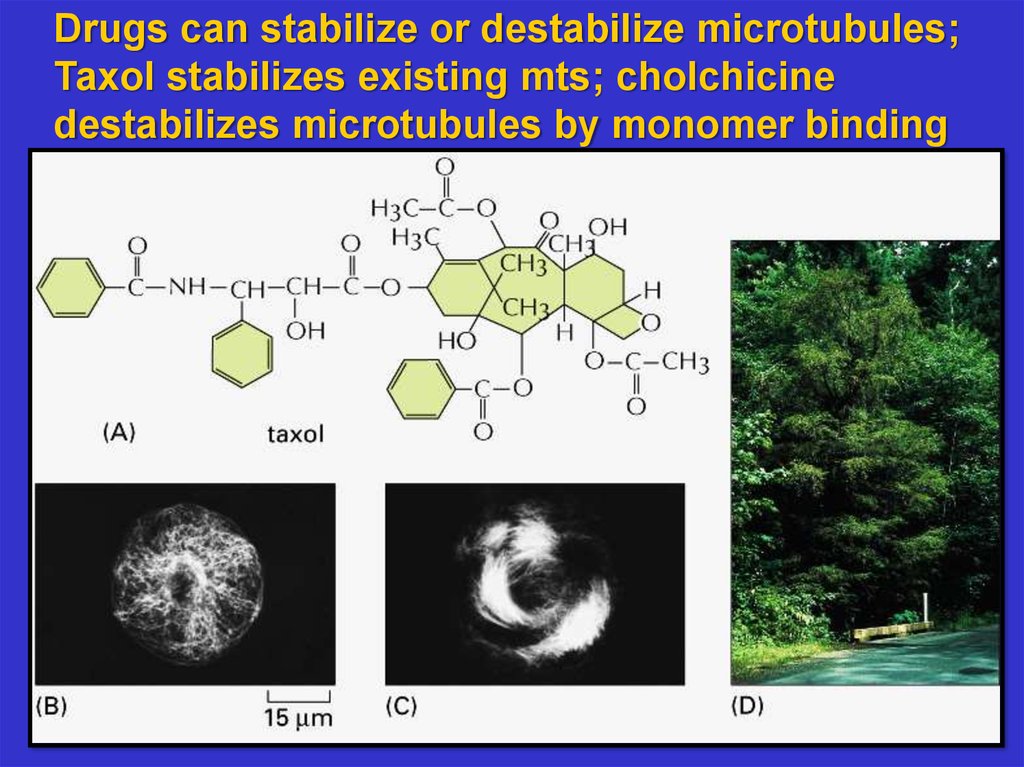
Drugs can stabilize or destabilize microtubules;Taxol stabilizes existing mts; cholchicine
destabilizes microtubules by monomer binding
22.
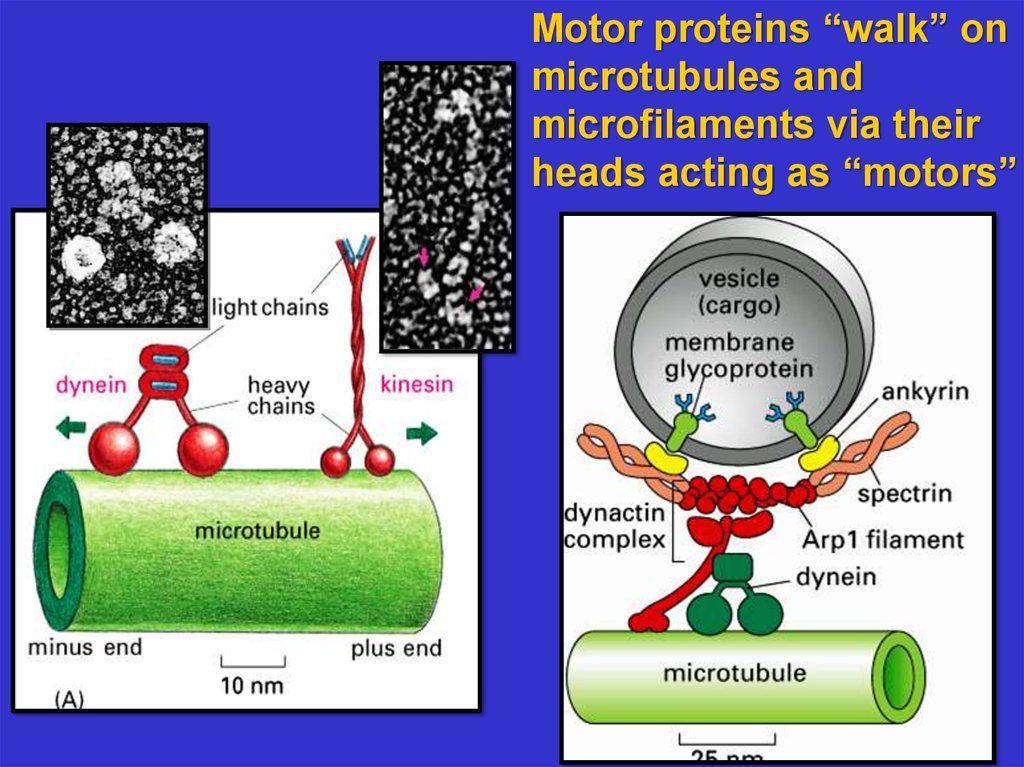
Motor proteins “walk” onmicrotubules and
microfilaments via their
heads acting as “motors”
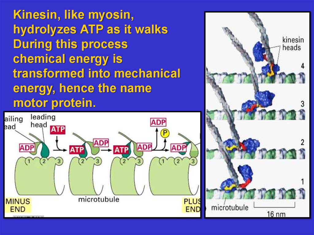
23.
Kinesin, like myosin,hydrolyzes ATP as it walks
During this process
chemical energy is
transformed into mechanical
energy, hence the name
motor protein.
24.
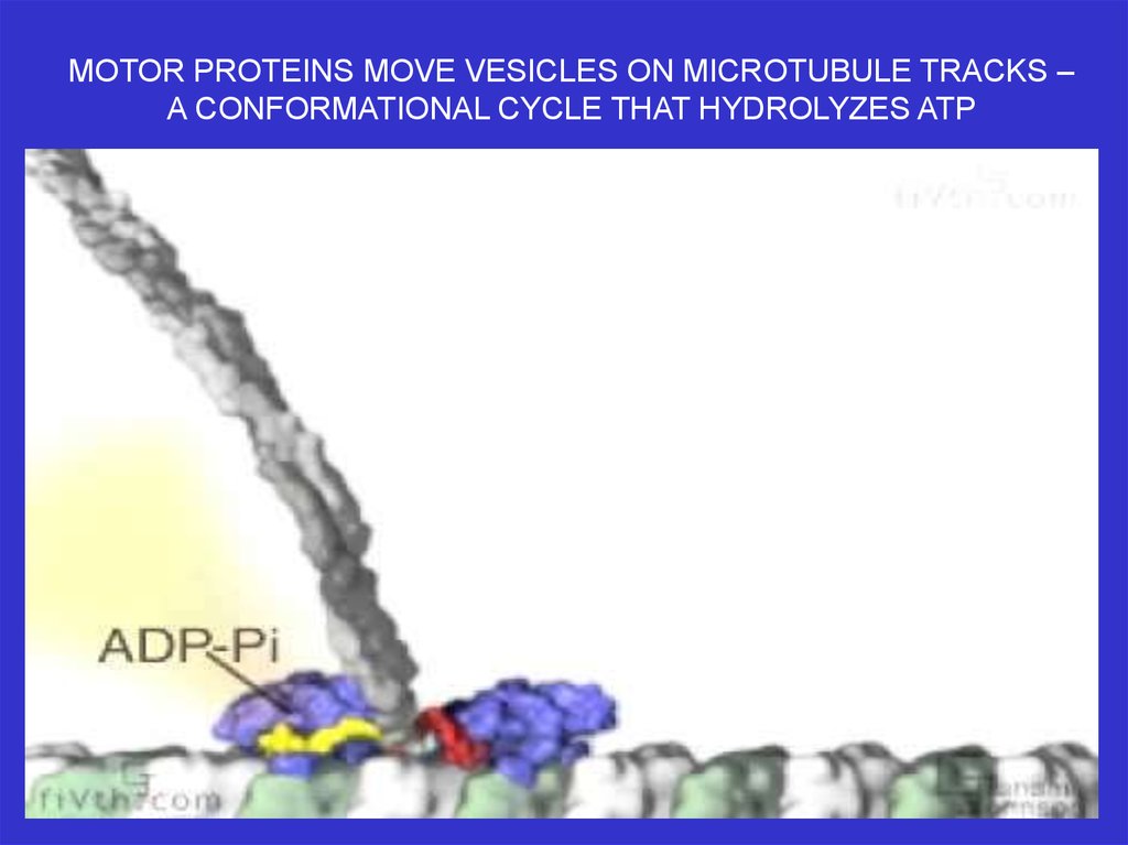
MOTOR PROTEINS MOVE VESICLES ON MICROTUBULE TRACKS –A CONFORMATIONAL CYCLE THAT HYDROLYZES ATP
25.
MOTOR PROTEINS MOVE VESICLES ON MICROTUBULE TRACKS26.
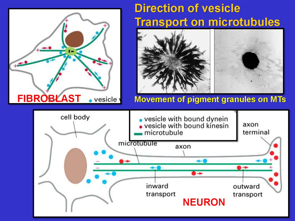
Direction of vesicleTransport on microtubules
FIBROBLAST
Movement of pigment granules on MTs
NEURON
27.
Cilia And Flagella: A Different Form Of Motility28.
The Structure OfFlagella And Cilia
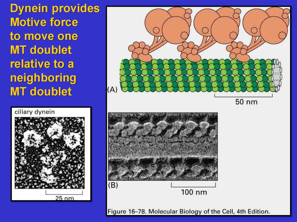
29.
Dynein providesMotive force
to move one
MT doublet
relative to a
neighboring
MT doublet
30.
DyneinMotors
cause
microtubule
sliding in
vitro; these
motors
cause
bending in
an intact
flagellum