


 Медицина
Похожие презентации:


Immunophysiology of reproductive system
1. Immunophysiology of reproductive system
2.
Over 50 years ago, there was the assumption that the placenta is an allograftexpressing paternal proteins and, therefore, under normal immunological
conditions, should be rejected. However, as our knowledge of placental biology
has significantly increased over the last 50 years, we can appreciate that the
placenta is more than a transplanted organ. The placenta is an immune
regulatory organ. The new integrational model takes in consideration the
fetal–placental immune response and the maternal immune system as integrated.
The immunology of pregnancy is the result of the combination of signals and
responses originated from the maternal immune system and the fetal–placental
immune system. The signals originated in the placenta will modulate the way the
maternal immune system will behave in the presence of potential dangerous
signals. The immune system of the mother should not be thought of as
suppressed, but rather modulated and streamlined to focus on pathogen
recognition, communication, trafficking and repair. This suggests that the
mother’s immune system is still able to mount an attack, but only when
absolutely necessary. Such modulated mechanisms allow the mother to
maintain a well-balanced immune system.
3.
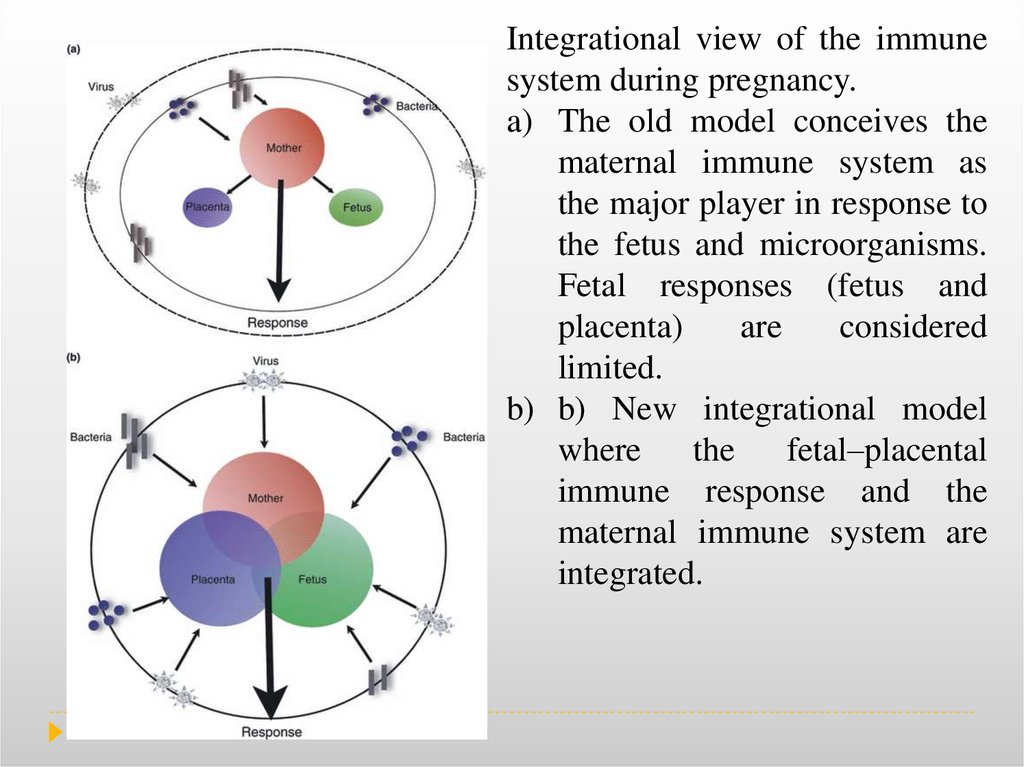
Integrational view of the immunesystem during pregnancy.
a) The old model conceives the
maternal immune system as
the major player in response to
the fetus and microorganisms.
Fetal responses (fetus and
placenta)
are
considered
limited.
b) b) New integrational model
where
the
fetal–placental
immune response and the
maternal immune system are
integrated.
4.
Role of the placenta as a modulator of fetal and maternal responses.Inflammation at the placenta has a bidirectional effect. Activates the maternal
immune system as well as the fetus by creating an inflammatory environment. The
inflammatory response may also influence the development of the fetal immune
system with important consequences during postnatal age.
5.
6.
7.
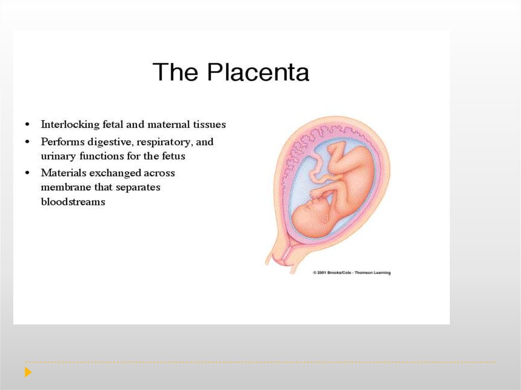
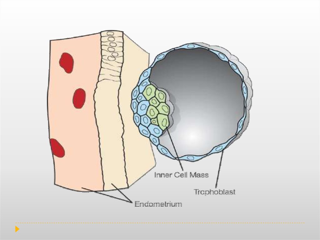
Trophoblasts are specialized cells of the placenta that play an important role in embryoimplantation and interaction with the Decidualised maternal uterus. The core of placental
villi is surrounded by two layers of trophoblast; a single layer of mononuclear
cytotrophoblast that covers the entire surface of the placenta. It is this syncytiotrophoblast
that is in direct contact with the maternal blood that reaches the placental
surface, and thus facilitates the exchange of nutrients, wastes and gases between the
maternal and fetal systems.
In addition, cytotrophoblast can differentiate into another type of trophoblast called the
extravillous trophoblast and penetrate into the decidualised uterus. This process is
essential not only for physically attaching the placenta to the mother, but also for altering
the vasculature in the uterus to allow it to provide an adequate blood supply to the growing
fetus as pregnancy progresses. Some of these trophoblast even replace the endothelial cells
in the uterine spiral arteries as they remodel these vessels into wide bore conduits that are
independent of maternal vasoconstriction. This ensures the fetus receives a steady supply
of blood, and the placenta is not subjected to fluctuations in oxygen that could cause it
damage.
Decidualization is a process that results in significant changes to cells of the endometrium
in preparation for, and during, pregnancy.
8.
During normal pregnancy, the human decidua contains a high number of immune cells, suchas macrophages, natural killer (NK) cells and regulatory T cells (Treg). 70% of decidual
leukocytes are NK cells, 20–25% are macrophages and 1.7% are dendritic cells.
From the adaptive immune system, B cells are absent, but T lymphocytes constitute about
3–10% of the decidual immune cells. During the first trimester, NK cells, dendritic
cells and macrophages infiltrate the decidua and accumulate around the
invading trophoblast cells. Deletion of either macrophages, NK cells or dendritic cells
(DC) has deleterious effects. Elegant studies have shown that in the absence of NK
cells, trophoblast cells are not able to reach the endometrial vascularity leading
to termination of the pregnancy. These studies suggest that uNK cells are
critical for trophoblast invasion in the uterus. Similarly, depletion of DCs
prevented blastocyst implantation and decidual formation. Indeed, this study
suggests that uDC are necessary for decidual formation and may affect the
angiogenic response by inhibiting blood vessel maturation.
These data further support the idea that the fetal–maternal immune
interaction is more complex than the comparison to transplant allograft.
Consequently, the presence of immune cells at the implantation site is not associated with
a response to the ‘foreign’ fetus but to facilitate and protect the pregnancy.
Therefore, the immune system at the implantation site is not suppressed, on the contrary it
is active, functional and is carefully controlled.
9.
10.
Comparison and contrast between a cellular response to a skin allograft and to a semi-allogeneicfetus and its placenta. (A) Transplanted skin cells express allogeneic HLA-A and HLA-B
molecules (blue border). A large fraction of CTLs (cytotoxic T cells) are activated by allogeneic
MHC class I molecules through the direct allorecognition pathway and extravasate into the tissue,
where they destroy the allograft. (B) The invading trophoblast cells do not express HLA-A and
HLA-B molecules, but instead express HLA-C molecules which interact with KIRs on maternal
NK cells in the placental bed of the uterus. Here the interactions between KIRs and fetal HLA-C
regulate vascular remodeling of the uterus, allocation of nutrients to the feto-placental unit and
fetal growth. The potentially destructive CTLs rarely extravasate, and few allogeneic HLA-Crestricted CTLs are found in the uterus.
11.
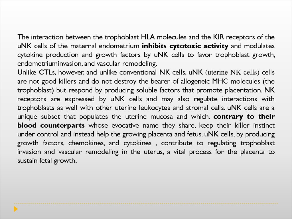
The interaction between the trophoblast HLA molecules and the KIR receptors of theuNK cells of the maternal endometrium inhibits cytotoxic activity and modulates
cytokine production and growth factors by uNK cells to favor trophoblast growth,
endometriuminvasion, and vascular remodeling.
Unlike CTLs, however, and unlike conventional NK cells, uNK (uterine NK cells) cells
are not good killers and do not destroy the bearer of allogeneic MHC molecules (the
trophoblast) but respond by producing soluble factors that promote placentation. NK
receptors are expressed by uNK cells and may also regulate interactions with
trophoblasts as well with other uterine leukocytes and stromal cells. uNK cells are a
unique subset that populates the uterine mucosa and which, contrary to their
blood counterparts whose evocative name they share, keep their killer instinct
under control and instead help the growing placenta and fetus. uNK cells, by producing
growth factors, chemokines, and cytokines , contribute to regulating trophoblast
invasion and vascular remodeling in the uterus, a vital process for the placenta to
sustain fetal growth.
12.
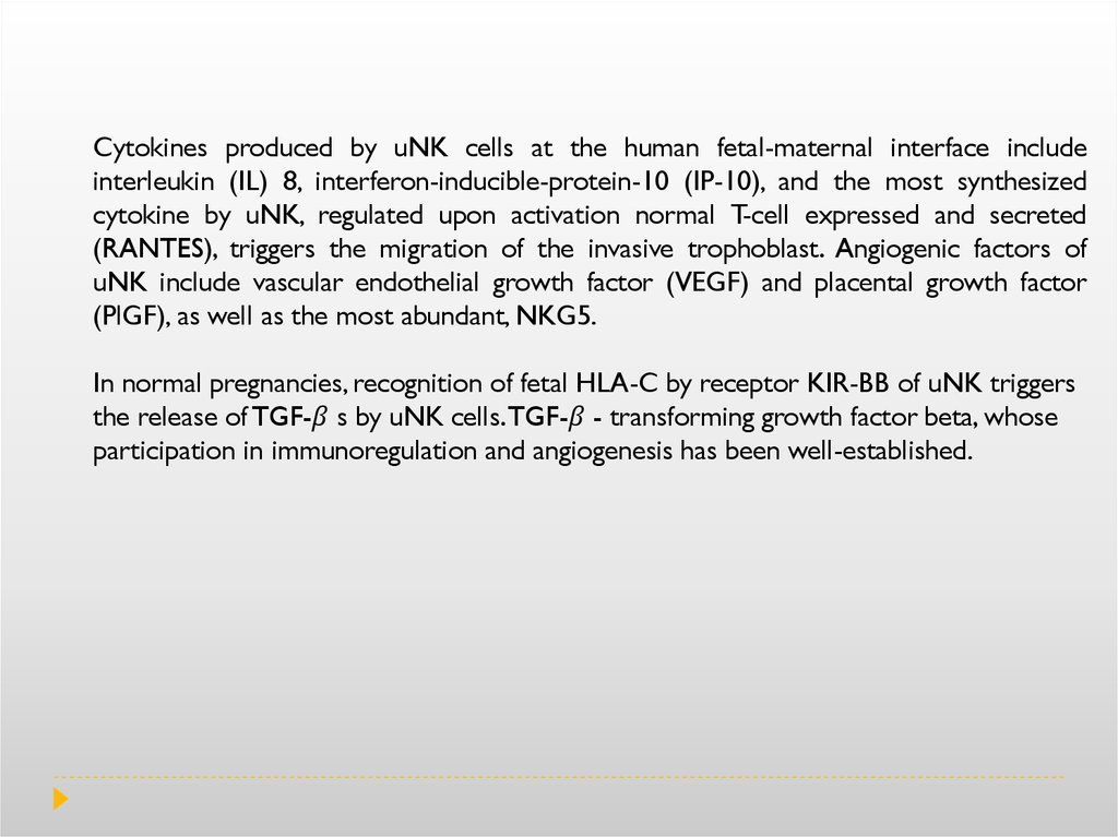
Cytokines produced by uNK cells at the human fetal-maternal interface includeinterleukin (IL) 8, interferon-inducible-protein-10 (IP-10), and the most synthesized
cytokine by uNK, regulated upon activation normal T-cell expressed and secreted
(RANTES), triggers the migration of the invasive trophoblast. Angiogenic factors of
uNK include vascular endothelial growth factor (VEGF) and placental growth factor
(PlGF), as well as the most abundant, NKG5.
In normal pregnancies, recognition of fetal HLA-C by receptor KIR-BB of uNK triggers
the release of TGF-