
")


-1 and IGF-binding protein-3 in Henoch-Schonlein purpura.")


")
")


 Медицина
Похожие презентации:


")


")
")

Schonlein - Hennoch purpura
1. SIW theme: “Schonlein- Hennoch purpura”
JSC “Astana Medical University”Department of Internal Diseases № 1
SIW
THEME:
“SCHONLEIN- HENNOCH PURPURA”
Checked by: Baidurin S.A
Done by: Suleymenova D. 463GM.
Astana 2018y.
2. Henoch-Schönlein Purpura (HSP)
• is a common vasculitis of small vessels withcutaneous and systemic complications.
• It is the most common cause of nonthrombocytopenic
purpura in children.
3. EPIDEMIOLOGY
The etiology is unknown• more frequent in children than adults, with most cases
occurring between 2 and 8 yr of age,
• most frequently in the winter months.
• The overall incidence is estimated to be 9/100,000
population.
• Males are affected twice as frequently as females.
4. PATHOGENESIS
• in specific populations, patients with HSP have a significantly higher frequency ofHLA-DRB1*01 and decreased frequency of the *07 haplotype than controls.
• increased serum concentrations of the cytokines tumor necrosis factor-α (TNF-α) and
interleukin (IL)-6.
• In one study, almost half of the patients had elevated antistreptolysin O (ASO) anti-
bodies, implicating group A streptococcus.
• HSP is an IgA-mediated vasculitis of small vessels.
• Immunofluorescence techniques show deposition of IgA and C3 in the
small vessels of the skin and the renal glomeruli;
5. CTLA-4 +49 A/G genotype and HLA-DRB1 polymorphisms in Turkish patients with Henoch-Schönlein purpura.
• presence of Cytotoxic T lymphocyte-associatedprotein 4( CTLA-4) AG genotype and HLA-DRB1*13
could be a risk factor for developing nephrotic-range
proteinuria in these patients.
.
6.
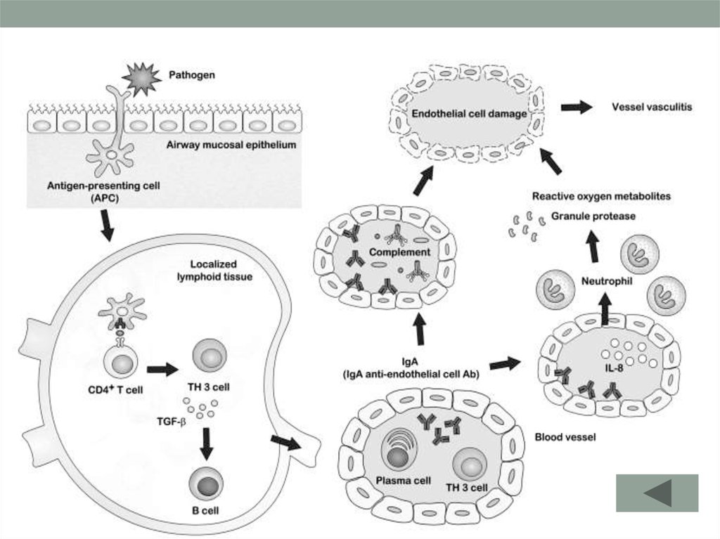
7. The immunobiology of Henoch-Schönlein purpura.
• group A beta-hemolytic streptococcus (GAS) has widely studied and found in20–50% of patients with acute HSP by serological tests or bacterial cultures,
• Bartonella henselae (12 of 18 HSP patient sera were positive )
Parvovirus B19 (only one of 29 HSP patients )
Other HSP-associated pathogens have been reported
• Staphylococcus aureus,
• Helicobacter Pylori,
• Hemophilus parainfluenza,
• Coxsackie virus,
• adenovirus,
• hepatitis A virus,
• hepatitis B virus
8. CLINICAL MANIFESTATIONS
• The disease onset may be acute, or insidious, with sequentialoccurrence of symptoms over a period of weeks or months.
• Low-grade fever and fatigue are present in more than half
of affected children.
• The typical rash and the clinical symptoms of HSP are a
consequence of the usual location of the acute small vessel
damage primarily in the skin, gastrointestinal tract, and
kidneys.
9. CLINICAL MANIFESTATIONS
• Rash (95-100%), especially involving the legs, may not bepresent on initial presentation
Subcutaneous edema (20-50%)
Abdominal pain and vomiting (85%)
Joint pain (60-80%), especially involving the knees and
ankles
Scrotal edema (2-35%)
Bloody stools
10. Rash
beginning as pinkish maculopapules that initially blanch on pressure and progress
to petechiae or purpura,
• characterized clinically as palpable purpura that evolve from red to purple to rusty
brown before they eventually fade
• last from 3-10 days, and may appear at intervals that vary from a few days to as
long as 3-4 mo.
In <10% of children, recurrences of the rash may not end until as late as a yr,
Damage to cutaneous vessels also results in local angioedema, which may
precede the palpable purpura.
• Edema independent of purpura occurs primarily in dependent areas such as below
the waist, over the buttocks (or on the back and posterior scalp in the infant), or in
areas of greater tissue distensibility such as the eyelids, lips, scrotum, or dorsum of
the hands and feet.
11. Rash
12. Arthritis
• present in more than ⅔ of children with HSP,• is usually localized to the knees and ankles and
appears to be concomitant with edema.
• The effusions are serous, not hemorrhagic,
• resolve after a few days without residual deformity or
articular damage.
• They may recur during a subsequent reactive phase of
the disease.
13. Gastrointestinal tract
• intermittent abdominal pain that is often colicky in nature.• There may be peritoneal exudate, enlarged mesenteric lymph
nodes, segmental edema, and hemorrhage into the bowel.
More than half of patients have occult heme-positive stools,
diarrhea (with or without visible blood), or hematemesis.
Intussusception may occur, which may rarely be followed by
complete obstruction or infarction with bowel perforation.
If not resolved by hydrostatic reduction during a contrast study,
surgical intervention is necessary.
14. Renal involvement
• occurs in 25-50% of childrenmay manifest with:
• hematuria,
• proteinuria, or both;
• nephritis or nephrosis;
• acute renal failure.
• Renal involvement at presentation may lead to chronic
hypertension or end-stage renal disease in the future
15. Increased serum levels of insulin-like growth factor (IGF)-1 and IGF-binding protein-3 in Henoch-Schonlein purpura.
Serum IGF-1 levels were significantly higher in HSP with proteinuriathan those without proteinuria and controls (p = 0.001 and p =
0.001, respectively).
Also, IGFBP-3 levels were greater in HSP with proteinuria compared
to those without proteinuria and controls (p = 0.005 and p =
0.0001).
Serum immunoglobulin-A/complement-C3 ratio was higher in HSP
than in the controls (p = 0.0001) but this ratio did not change
according to proteinuria, hematuria or positive SOB.
In conclusion, IGF-1 and IGFBP-3 levels could be new markers for
determination of renal involvement in HSP.
16. What is the difference between IgA nephropathy and Henoch-Schönlein purpura nephritis
Clinical featuresIgAN
HSPN
Extra-renal symptoms
-
+
Age at onset
15<y
15>y
Nephritic/nephrotic syndrome
-/+
+++
Risk of chronic renal failure (CRF)
+
++
Hypersensitivity
-
+
Secondary forms
++
-/+
Endocapillary proliferation
-/+
++
Epithelial crescents
-/+
++
Perivascular glomerular IgA
-/+
++
Subepithelial/subendothelial dense deposits
-/+
++
Fibrin deposits
-/+
++
17. Relationship between initial clinical signs and risk of chronic renal failure in Henoch-Schönlein purpura nephritis
Relationship between initial clinical signs andrisk of chronic renal failure in HenochSchönlein purpura nephritis
18. Complications of Henoch-Schönlein Purpura
HepatosplenomegalyMyocardial infarction
Pulmonary hemorrhage
Pleural effusion
Unnecessary abdominal surgery
Intussusception
Hemorrhage
Shock
Gastrointestinal bleeding
Bowel infarction
Renal failure
Hematuria
Proteinuria
Seizures
Mononeuropathies
Testicular torsion
19. DIAGNOSIS
Diagnostic uncertainty arises when the symptom complex ofedema, rash, arthritis with abdominal complaints, and renal
findings occurs for a prolonged period.
20. DIAGNOSIS
• Affected children often have a moderate thrombocytosis andleukocytosis.
The erythrocyte sedimentation rate (ESR) may be elevated.
Anemia may result from chronic or acute gastrointestinal blood loss.
Immune complexes are often present, and 50% of patients have elevated
concentrations of IgA as well as IgM
usually negative for antinuclear antibodies (ANAs), antibodies to
nuclear cytoplasmic antigens (ANCAs), and rheumatoid factor (even in
the presence of rheumatoid nodules).
Anticardiolipin or antiphospholipid antibodies may be present and
contribute to the intravascular coagulopathy.
Intussusception is usually ileoileal in location;
Renal involvement manifests in red blood cells, white blood cells, casts,
or albumin in the urine and azotemia
21. Definitive diagnosis confirmed by biopsy
• cutaneous site showing leukocytoclastic angiitis.• Renal biopsy may show mesangial deposition of IgA and
occasionally IgM, C3, and fibrin.
22.
H & E stain of skin biopsy showing leukocytoclastic vasculitis with infiltration ofneutrophils.
23. Henoch-Schönlein purpura.
A: Cutaneous purpura;B: Urine sediment red blood
cell cast;
C: Acute glomerular
inflammation and crescent
formation;
D: Details of basal membrane
mesangial proliferation
and IgA deposits
24. Immunofluorescence micrograph of a glomerulus from a patient with HSP nephropathy stained for the presence of IgA.
Immunofluorescence micrograph of a glomerulus from apatient with HSP nephropathy stained for the presence of IgA .
25. Differential Diagnosis of Henoch-Schönlein Purpura
Acute abdomen
Meningococcal meningitis or septicemia
Rheumatoid arthritis
Rheumatic fever
Idiopathic thrombocytopenic purpura
Systemic lupus erythematosus
poly-arteritis nodosa,
Child abuse
Drug reactions
Bacterial endocarditis
Rocky Mountain spotted fever
familial Mediterranean fever
inflammatory bowel disease.
Kawasaki disease.
26. Acute hemorrhagic edema (AHE)
• is an acute cutaneous benign leukocytoclastic vasculitis• seen in children ≤2 yr of age
• AHE presents with fever; tender edema of the face, scrotum, hands, and feet;
and ecchymosis (usually larger than the purpura of HSP) on the face and
extremities
• petechiae may be seen in mucous membranes.
• The patient usually appears well except for the rash.
• The platelet count is normal or elevated;
• the urinalysis is normal.
The younger age, nature of the lesions, absence of other organ involvement,
and biopsy may help distinguish AHE from HSP.
27. Acute hemorrhagic edema (AHE)
28. TREATMENT
Symptomatic treatment• adequate hydration,
• bland diet,
• pain control with acetaminophen is provided for self-
limited complaints of arthritis, edema, fever, and malaise.
• Avoidance of competitive activities and avoidance of
maintaining the lower extremities in a dependent position
may decrease local edema.
• If edema involves the scrotum, elevation of the scrotum
and local cooling, as tolerated, may decrease discomfort.
29. TREATMENT
• Therapy with oral or intravenous corticosteroids (1-2mg/kg/day) is often associated with dramatic improvement of
both gastrointestinal and CNS complications.
• the effects of corticosteroids on renal manifestations are not
clear.
• intussusception may be life-threatening and managed with
cortico-steroids and, when necessary, hydrostatic reduction (by
air or with contrast) or resection of the intussusception.
30. TREATMENT
• is the same as for other forms of acute glomerulonephritis• If anti-cardiolipin or antiphospholipid antibodies are identified
and thrombotic events have occurred, aspirin (81 mg) given
once may decrease the risks associated with a hypercoagulable
state.
• Rheumatoid nodules may respond to alternate-day colchicine
(0.6 mg every other day).
31. Prognosis
• More than 80% of patients have a single isolated episode lastinga few weeks.
• Approximately 10-20% of patients have recurrences.
• Fewer than 5% of patients develop chronic HSP.
• Abdominal pain resolves spontaneously within 72 hours in most
patients