


 sequences of serine / threonine protein phosphatases. The catalytic")


 into eight subfamilies (R1-R8), based on sequence")


 Английский язык
Похожие презентации:


")


Enzyme-l nked receptors
1. Enzyme-l nked receptors
Done by: Naizabayeva D.Alibekova A.
Maulenbay A.
BT 16-02
2. Content
1.Introduction2.Classification
3.Structure
4.Mechanism of action
4.1 Activation and regulation
3. ntroduction
Enzyme-linked receptors (catalytic receptos) are asecond major type of cell-surface receptor. They were
recognized initially through their role in responses to
extracellular signal proteins that promote the growth,
proliferation, differentiation, or survival of cells in
animal tissues.
The responses to them are typically slow (on the
order of hours) and usually require many intracellular
signaling steps that eventually lead to changes in gene
expression. in gene expression.
4. Classification
1. Receptor tyrosine kinases2. Tyrosine-kinase-associated receptors
3. Receptorlike tyrosine phosphatases
4. Receptor serine/threonine kinases
5. Receptor guanylyl cyclases
6. Histidine-kinase-associated receptors
5.
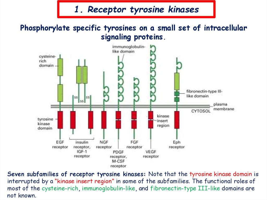
1. Receptor tyrosine kinasesPhosphorylate specific tyrosines on a small set of intracellular
signaling proteins.
Seven subfamilies of receptor tyrosine kinases: Note that the tyrosine kinase domain is
interrupted by a “kinase insert region” in some of the subfamilies. The functional roles of
most of the cysteine-rich, immunoglobulin-like, and fibronectin-type III-like domains are
not known.
6.
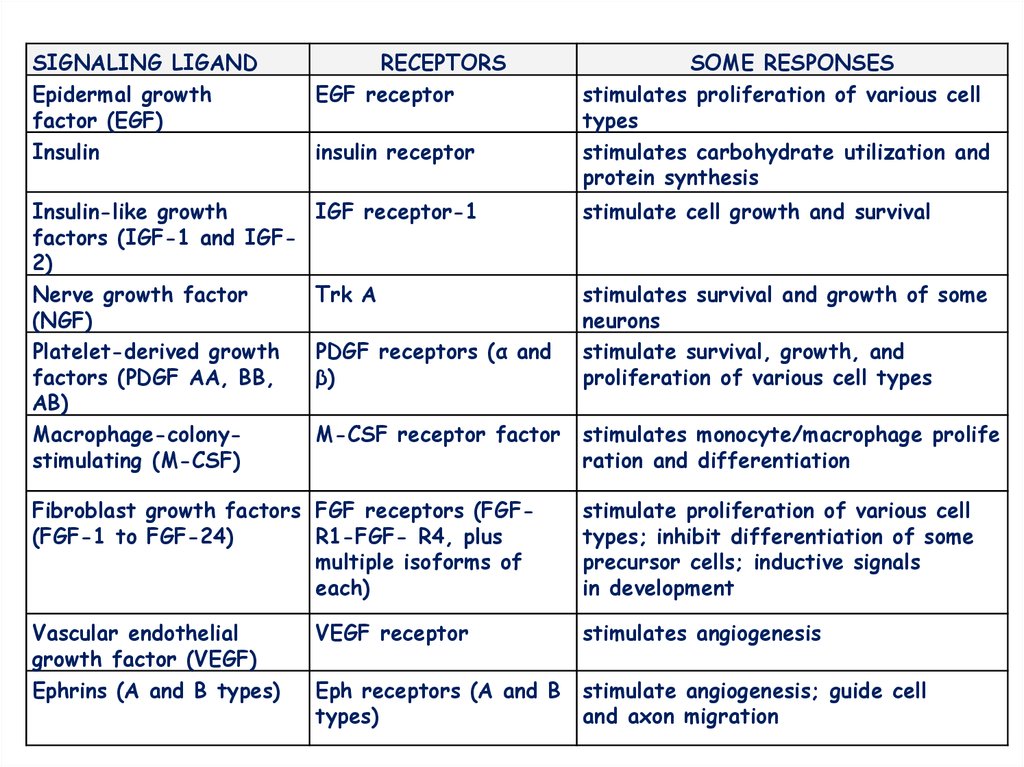
SIGNALING LIGANDEpidermal growth
factor (EGF)
Insulin
RECEPTORS
EGF receptor
Insulin-like growth
factors (IGF-1 and IGF2)
Nerve growth factor
(NGF)
Platelet-derived growth
factors (PDGF AA, BB,
AB)
Macrophage-colonystimulating (M-CSF)
IGF receptor-1
stimulate cell growth and survival
Trk A
stimulates survival and growth of some
neurons
stimulate survival, growth, and
proliferation of various cell types
insulin receptor
PDGF receptors (α and
β)
M-CSF receptor factor
SOME RESPONSES
stimulates proliferation of various cell
types
stimulates carbohydrate utilization and
protein synthesis
stimulates monocyte/macrophage prolife
ration and differentiation
Fibroblast growth factors FGF receptors (FGF(FGF-1 to FGF-24)
R1-FGF- R4, plus
multiple isoforms of
each)
stimulate proliferation of various cell
types; inhibit differentiation of some
precursor cells; inductive signals
in development
Vascular endothelial
growth factor (VEGF)
Ephrins (A and B types)
VEGF receptor
stimulates angiogenesis
Eph receptors (A and B
types)
stimulate angiogenesis; guide cell
and axon migration
7.
StructureLigand (Insulin)
Tyrosine-kinase
receptor
Tyrosine-kinase
domain (enzymatic)
8.
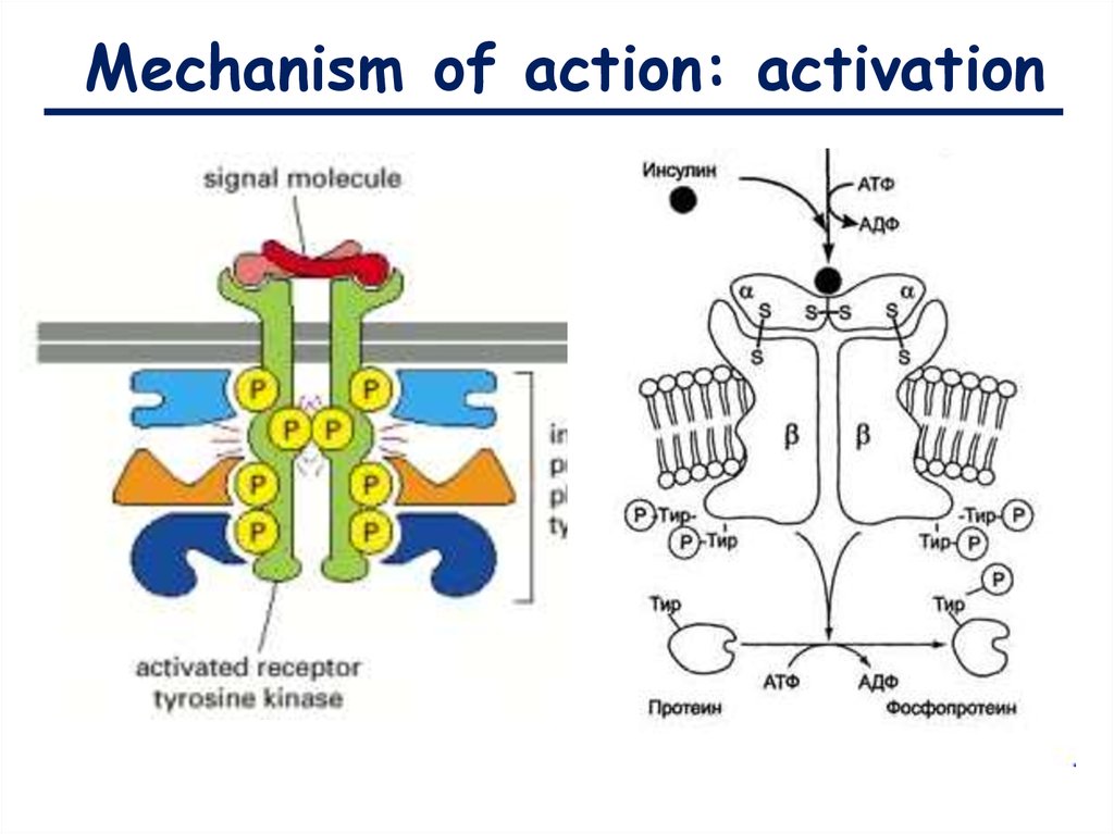
Mechanism of action: activation9.
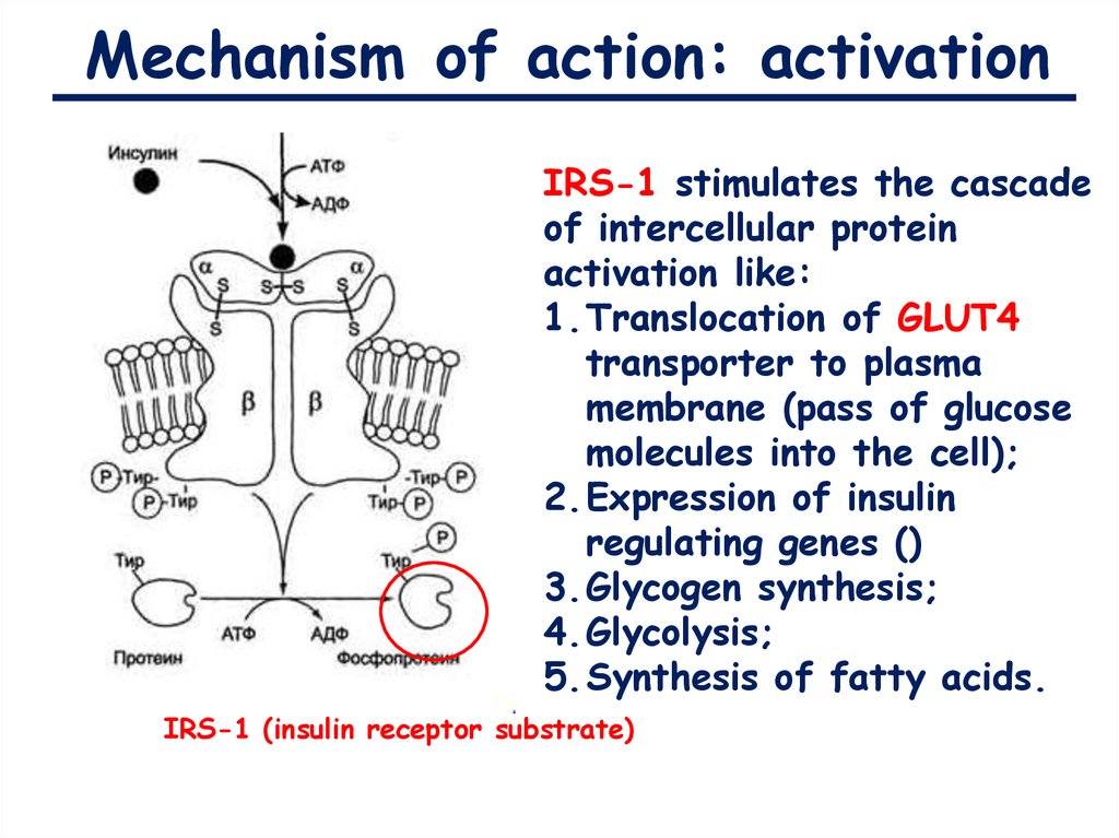
Mechanism of action: activationIRS-1 stimulates the cascade
of intercellular protein
activation like:
1.Translocation of GLUT4
transporter to plasma
membrane (pass of glucose
molecules into the cell);
2.Expression of insulin
regulating genes ()
3.Glycogen synthesis;
4.Glycolysis;
5.Synthesis of fatty acids.
IRS-1 (insulin receptor substrate)
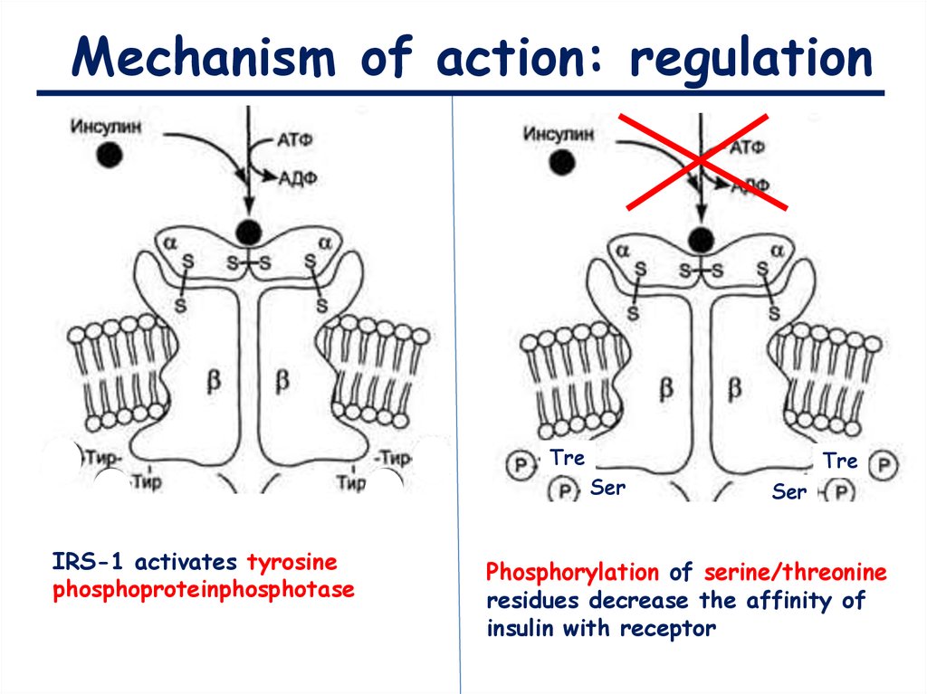
10.
Mechanism of action: regulationTre
Ser
IRS-1 activates tyrosine
phosphoproteinphosphotase
Tre
Ser
Phosphorylation of serine/threonine
residues decrease the affinity of
insulin with receptor
11.
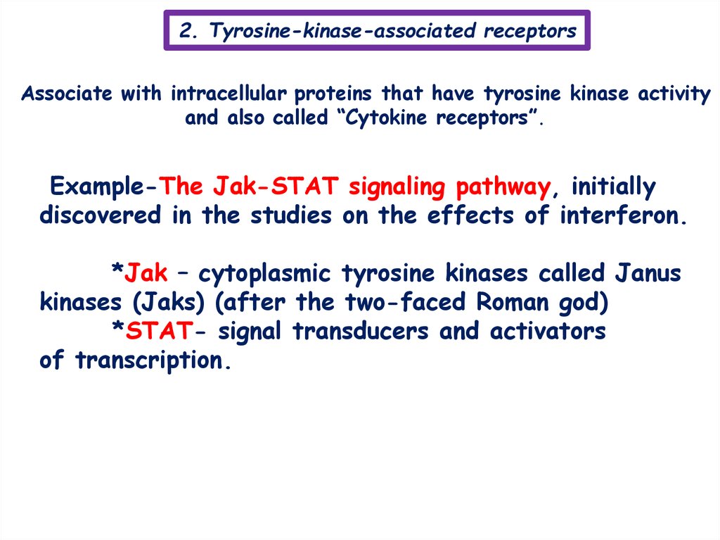
2. Tyrosine-kinase-associated receptorsAssociate with intracellular proteins that have tyrosine kinase activity
and also called “Cytokine receptors”.
Example-The Jak-STAT signaling pathway, initially
discovered in the studies on the effects of interferon.
*Jak – cytoplasmic tyrosine kinases called Janus
kinases (Jaks) (after the two-faced Roman god)
*STAT- signal transducers and activators
of transcription.
12.
2. Tyrosine-kinase-associated receptorsSIGNALING
LIGAND
RECEPTORASSOCIATE
D JAKS
Jak1 and
Jak2
STATS
SOME RESPONSES
ACTIVATED
Tyk2 and
Jak2
Jak2
STAT1
STAT2
STAT5
STAT5
Growth hormone
Jak1 and
Jak2
Jak2
GM-CSF
IL-3
γ-interferon
α-interferon
Erythropoietin
Prolactin
STAT1
activates macrophages;
increases MHC protein expres
sion
increases cell resistance to
viral infection
stimulates production of
erythrocytes
stimulates milk production
Jak2
STAT1
STAT5
STAT5
stimulates growth by inducing
IGF-1 production
stimulates production of
granulocytes and macrophages
Jak2
STAT5
stimulates early blood cell
production
13.
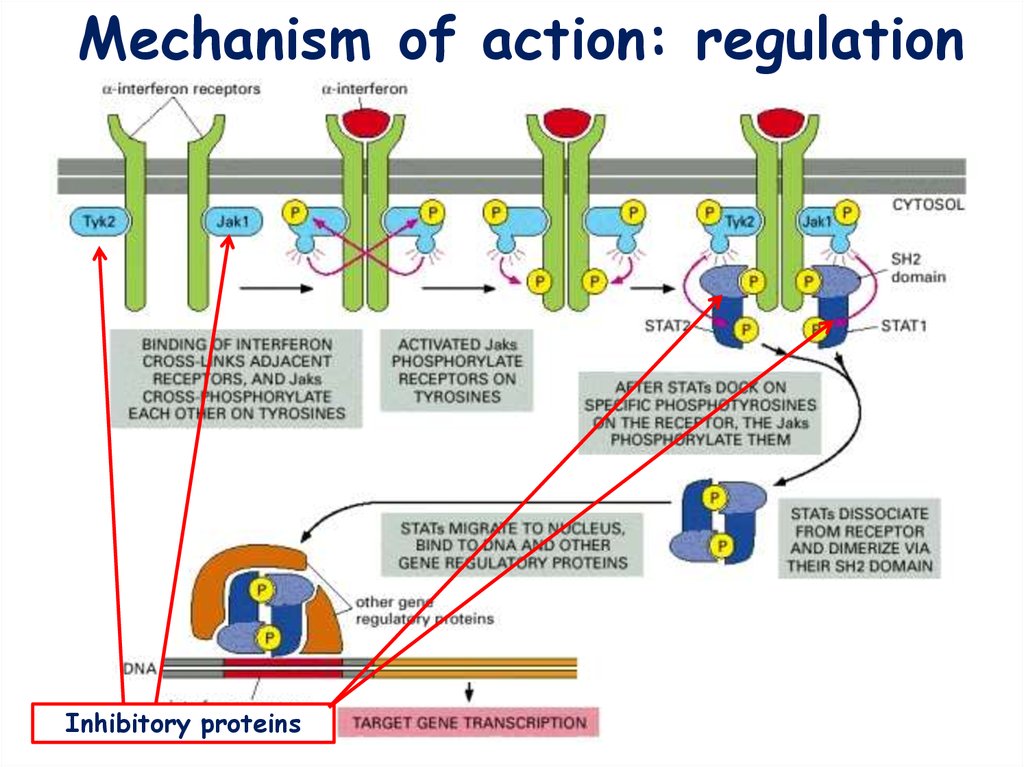
Mechanism of action: activation14.
Mechanism of action: regulationInhibitory proteins
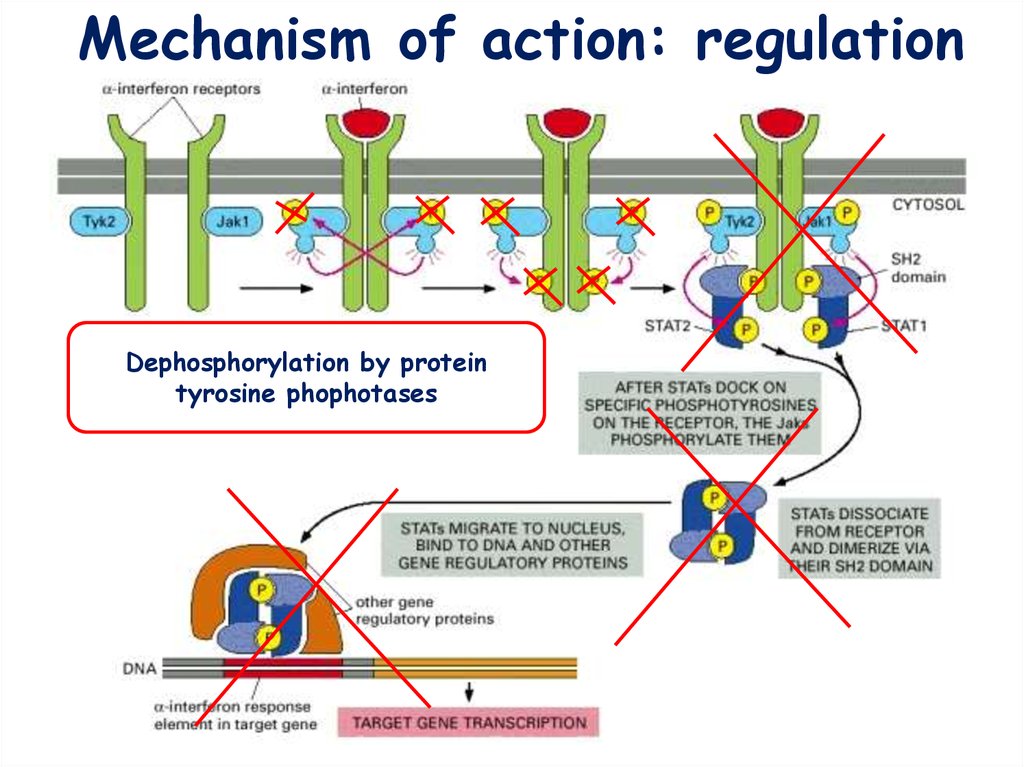
15.
Mechanism of action: regulationDephosphorylation by protein
tyrosine phophotases
16.
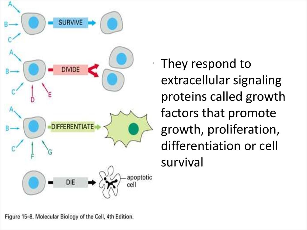
• They respond toextracellular signaling
proteins called growth
factors that promote
growth, proliferation,
differentiation or cell
survival
17.
• also known as a catalytic receptor•transmembrane receptor, where the binding
of an extracellular ligand causes enzymatic
activity on the intracellular side
• •integral membrane protein possessing both
enzymatic catalytic and receptor functions
•Upon ligand binding a conformational
change is transmitted which activates the
enzyme, initiating signaling cascades
18. Physiology and diseases
• involved in growth, proliferation, differentiation,or survival
• Because of this, their ligands are collectively
called growth factors.
• The effects of enzyme-linked receptors typically
are slow requiring the expression of new genes
• Mutations in receptor tyrosine kinases are
responsible for a wide array of diseases, including
cancers, neurodegeneration, achondroplasia and
atherosclerosis.
19. Six classes of enzyme-linked receptors have thus far been identified:
Six classes of enzyme-linked receptorshave thus far been identified:
• 1.Receptor tyrosine kinases phosphorylate specific tyrosines on a small set
of intracellular signaling proteins.
• 2.Tyrosine-kinase-associated receptors associate with intracellular proteins
that have tyrosine kinase activity.
• 3.Receptorlike tyrosine phosphatases remove phosphate groups from
tyrosines of specific intracellular signaling proteins. (They are called
“receptorlike” because the presumptive ligands have not yet been
identified, and so their receptor function has not been directly
demonstrated.)
• 4.Receptor serine/threonine kinases phosphorylate specific serines or
threonines on associated latent gene regulatory proteins.
• 5.Receptor guanylyl cyclases directly catalyze the production of cyclic
GMP in the cytosol.
• 6.Histidine-kinase-associated receptors activate a “two-component”
signaling pathway in which the kinase phosphorylates itself on histidine
and then immediately transfers the phosphate to a second intracellular
signaling protein.
20.
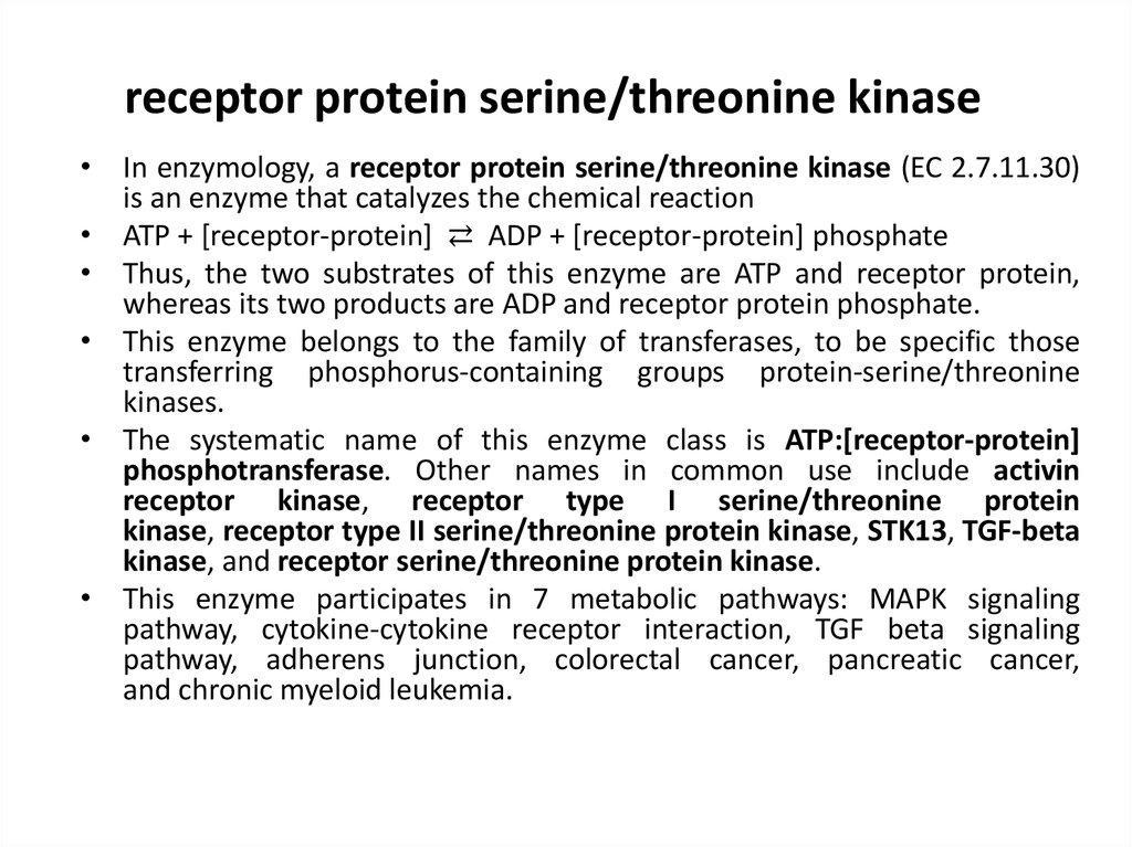
receptor protein serine/threonine kinase• In enzymology, a receptor protein serine/threonine kinase (EC 2.7.11.30)
is an enzyme that catalyzes the chemical reaction
• ATP + [receptor-protein] ⇄ ADP + [receptor-protein] phosphate
• Thus, the two substrates of this enzyme are ATP and receptor protein,
whereas its two products are ADP and receptor protein phosphate.
• This enzyme belongs to the family of transferases, to be specific those
transferring phosphorus-containing groups protein-serine/threonine
kinases.
• The systematic name of this enzyme class is ATP:[receptor-protein]
phosphotransferase. Other names in common use include activin
receptor kinase, receptor type I serine/threonine protein
kinase, receptor type II serine/threonine protein kinase, STK13, TGF-beta
kinase, and receptor serine/threonine protein kinase.
• This enzyme participates in 7 metabolic pathways: MAPK signaling
pathway, cytokine-cytokine receptor interaction, TGF beta signaling
pathway, adherens junction, colorectal cancer, pancreatic cancer,
and chronic myeloid leukemia.
21. Receptor serine/threonine kinases
• phosphorylate specific Serine/ Threonine• There are two types of serine/threonine kinase receptors, both of
which contain an intracellular kinase domain. They are each dimeric
proteins, so an active receptor complex is made up of four
receptors.
1. Type I receptors
• Inactive unless in complex with type II receptors.
• Do not interact with ligand dimers.
• Contain conserved sequences of serine and threonine residues near
to their kinase domains.
2. Type II receptors
• Constitutively active kinase domains (even in the absence of the
bound ligand).
• Able to phosphorylate and activate the type I receptor.
22.
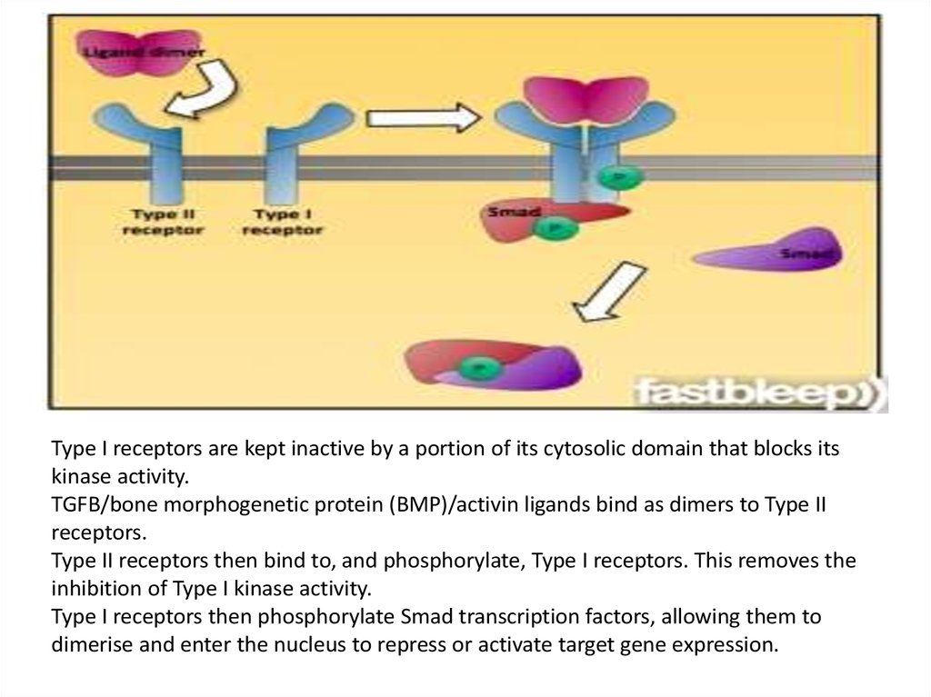
Type I receptors are kept inactive by a portion of its cytosolic domain that blocks itskinase activity.
TGFB/bone morphogenetic protein (BMP)/activin ligands bind as dimers to Type II
receptors.
Type II receptors then bind to, and phosphorylate, Type I receptors. This removes the
inhibition of Type I kinase activity.
Type I receptors then phosphorylate Smad transcription factors, allowing them to
dimerise and enter the nucleus to repress or activate target gene expression.
23.
• Serine/Threonine Kinase receptors play a role in the regulation ofcell proliferation, programmed cell death (apoptosis), cell
differentiation, and embryonic development.
Selectivity
While serine/threonine kinases all phosphorylate serine or threonine
residues in their substrates, they select specific residues to
phosphorylate on the basis of residues that flank the
phosphoacceptor site, which together comprise the consensus
sequence. Since the consensus sequence residues of a target
substrate only make contact with several key amino acids within the
catalytic cleft of the kinase (usually through hydrophobic forces
and ionic bonds), a kinase is usually not specific to a single
substrate, but instead can phosphorylate a whole "substrate family"
which share common recognition sequences. While the catalytic
domain of these kinases is highly conserved, the sequence variation
that is observed in the kinome (the subset of genes in the genome
that encode kinases) provides for recognition of distinct substrates.
Most kinases are inhibited by a pseudosubstrate that binds to the
kinase like a real substrate but lacks the amino acid to be
phosphorylated. When the pseudosubstrate is removed, the kinase
can
perform
its
normal
function.
24.
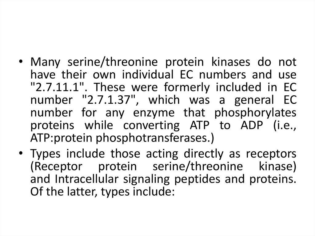
• Many serine/threonine protein kinases do nothave their own individual EC numbers and use
"2.7.11.1". These were formerly included in EC
number "2.7.1.37", which was a general EC
number for any enzyme that phosphorylates
proteins while converting ATP to ADP (i.e.,
ATP:protein phosphotransferases.)
• Types include those acting directly as receptors
(Receptor protein serine/threonine kinase)
and Intracellular signaling peptides and proteins.
Of the latter, types include:
25.
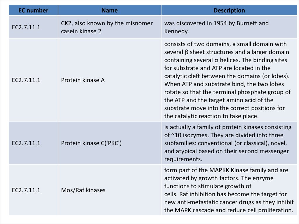
EC numberEC2.7.11.1
EC2.7.11.1
EC2.7.11.1
EC2.7.11.1
Name
Description
CK2, also known by the misnomer
casein kinase 2
was discovered in 1954 by Burnett and
Kennedy.
Protein kinase A
consists of two domains, a small domain with
several β sheet structures and a larger domain
containing several α helices. The binding sites
for substrate and ATP are located in the
catalytic cleft between the domains (or lobes).
When ATP and substrate bind, the two lobes
rotate so that the terminal phosphate group of
the ATP and the target amino acid of the
substrate move into the correct positions for
the catalytic reaction to take place.
Protein kinase C('PKC')
is actually a family of protein kinases consisting
of ~10 isozymes. They are divided into three
subfamilies: conventional (or classical), novel,
and atypical based on their second messenger
requirements.
Mos/Raf kinases
form part of the MAPKK Kinase family and are
activated by growth factors. The enzyme
functions to stimulate growth of
cells. Raf inhibition has become the target for
new anti-metastatic cancer drugs as they inhibit
the MAPK cascade and reduce cell proliferation.
26.
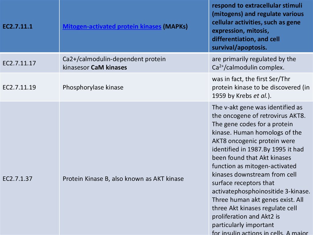
EC2.7.11.1Mitogen-activated protein kinases (MAPKs)
respond to extracellular stimuli
(mitogens) and regulate various
cellular activities, such as gene
expression, mitosis,
differentiation, and cell
survival/apoptosis.
EC2.7.11.17
Ca2+/calmodulin-dependent protein
kinasesor CaM kinases
are primarily regulated by the
Ca2+/calmodulin complex.
Phosphorylase kinase
was in fact, the first Ser/Thr
protein kinase to be discovered (in
1959 by Krebs et al.).
EC2.7.11.19
EC2.7.1.37
Protein Kinase B, also known as AKT kinase
The v-akt gene was identified as
the oncogene of retrovirus AKT8.
The gene codes for a protein
kinase. Human homologs of the
AKT8 oncogenic protein were
identified in 1987.By 1995 it had
been found that Akt kinases
function as mitogen-activated
kinases downstream from cell
surface receptors that
activatephosphoinositide 3-kinase.
Three human akt genes exist. All
three Akt kinases regulate cell
proliferation and Akt2 is
particularly important
for insulin actions in cells. A major
27.
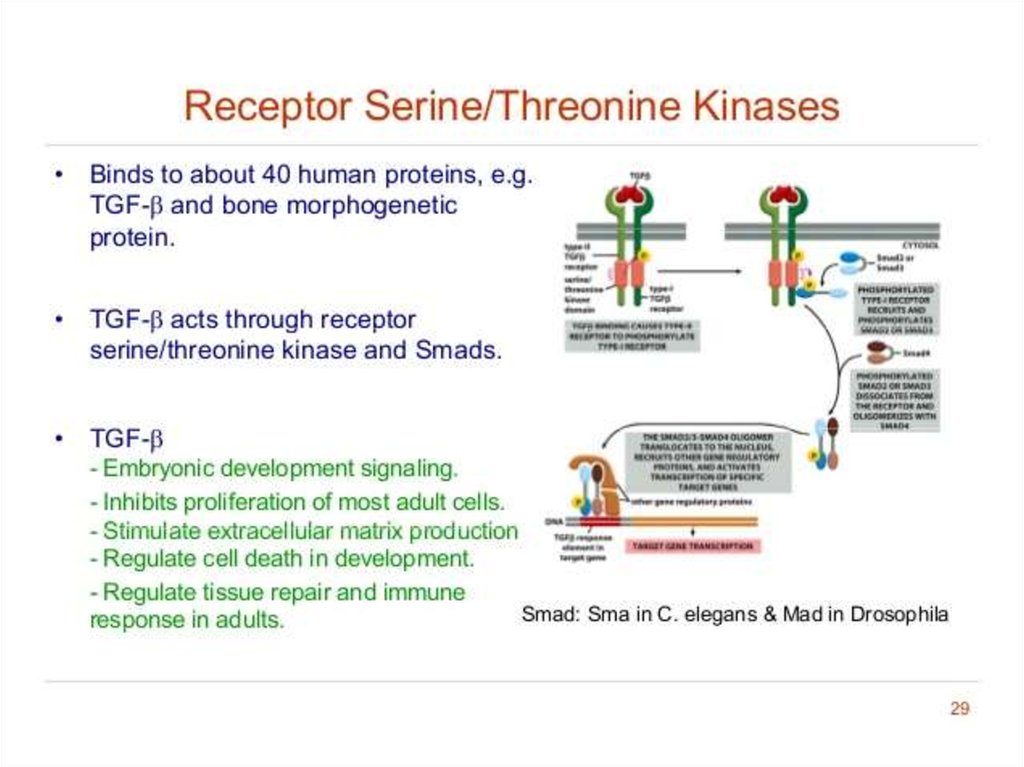
28. Receptor Serine/Threonine Protein Kinase Smad-dependent signaling pathway activated by TGF-β
• Transforming growth factor(TGF-β) consists of a large
number of structurally related,
secreted, dimeric proteins. They
mediate a wide range of
biological functions in animals:
proliferation,
differentiation,
ECM production, cell death,
tissue repair and immune
regulation.
• They act through receptor
serine/threonine kinases type I
and type II
• Smad family members are
directly phosphorylated by the
type I receptor, and moves to
the nucleus to direct gene
transcription
29. The size and location of protein kinases
30. Receptor like tyrosine phosphatases
• Receptor like tyrosine phosphatases remove phosphate groups fromtyrosines of specific intracellular signaling proteins. (They are called
“receptorlike” because the presumptive ligands have not yet been
identified, and so their receptor function has not been directly
demonstrated.)
• Together with tyrosine kinases, PTPs regulate
the phosphorylation state of many important signalling molecules,
such as the MAP kinase family. PTPs are increasingly viewed as
integral components of signal transduction cascades, despite less
study and understanding compared to tyrosine kinases
• PTPs have been implicated in regulation of many cellular processes,
including, but not limited to:
• Cell growth
• Cellular differentiation
• Mitotic cycles
• Oncogenic transformation
• Receptor endocytosis
31.
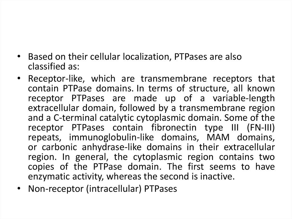
• Based on their cellular localization, PTPases are alsoclassified as:
• Receptor-like, which are transmembrane receptors that
contain PTPase domains. In terms of structure, all known
receptor PTPases are made up of a variable-length
extracellular domain, followed by a transmembrane region
and a C-terminal catalytic cytoplasmic domain. Some of the
receptor PTPases contain fibronectin type III (FN-III)
repeats, immunoglobulin-like domains, MAM domains,
or carbonic anhydrase-like domains in their extracellular
region. In general, the cytoplasmic region contains two
copies of the PTPase domain. The first seems to have
enzymatic activity, whereas the second is inactive.
• Non-receptor (intracellular) PTPases
32. Mechanism of action and highly conserved (signature) sequences of serine / threonine protein phosphatases. The catalytic
subunits have broad specificity33. Specificity of serine/threonine phosphatases is largely determined by association of regulatory subunits that affect
subcellular localisation as wellas substrate specificity
34.
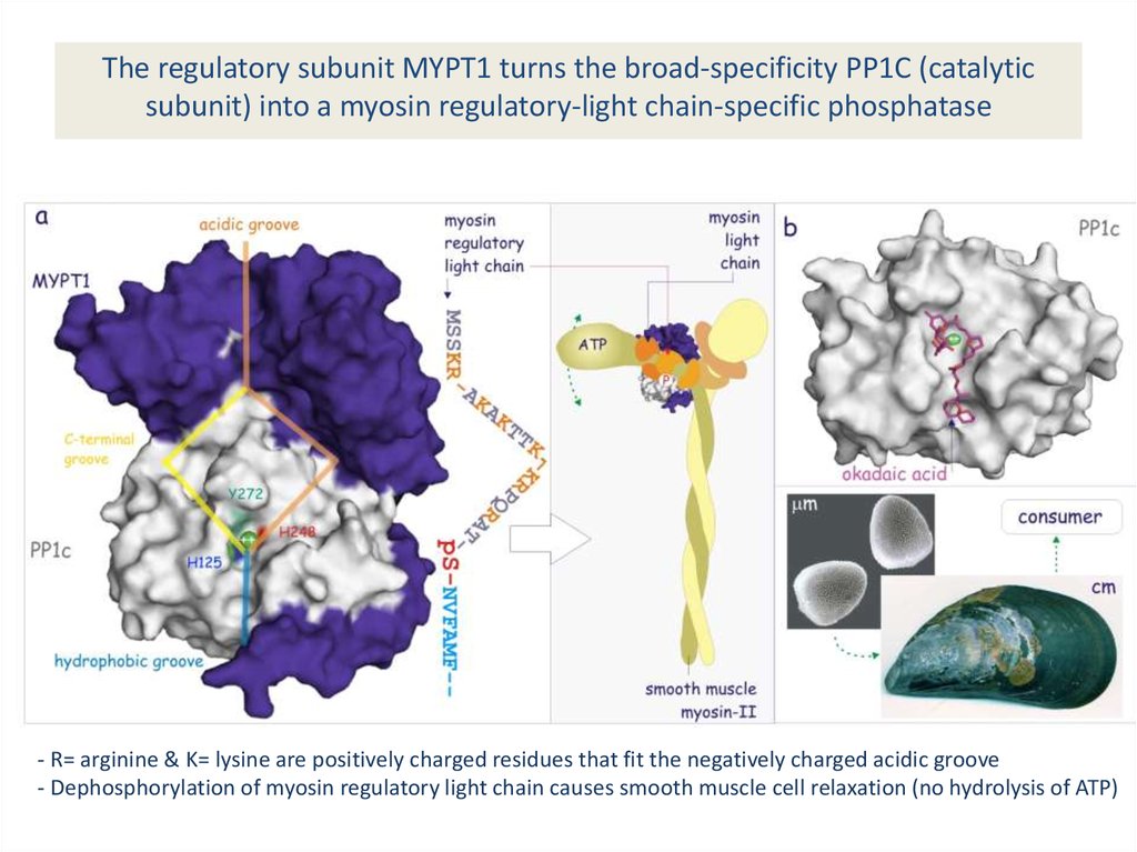
The regulatory subunit MYPT1 turns the broad-specificity PP1C (catalyticsubunit) into a myosin regulatory-light chain-specific phosphatase
- R= arginine & K= lysine are positively charged residues that fit the negatively charged acidic groove
- Dephosphorylation of myosin regulatory light chain causes smooth muscle cell relaxation (no hydrolysis of ATP)
35. Clinical significance
Serine/threonine kinase (STK) expression is altered in many types of cancerSerine/threonine protein kinase p90-kDa ribosomal S6 kinase (RSK) is in involved in
development of some prostate cancers.
Raf inhibition has become the target for new anti-metastatic cancer drugs as they
inhibit the MAPK cascade and reduce cell proliferation.
Act as Cell-Surface Receptors Protein tyrosine phosphatases (PTPs) remove selected
phosphotyrosines on a subset of tyrosine-phosphorylated proteins. Exhibit high
degree of substrate selectivity. These enzyme ensure that the tyrosine
phosphorylations are short-lived and are responsible for regulating the intensity of
the signal. There are about 30 known PTPs and occur as both transmembrane and
cytoplasmic forms.
36.
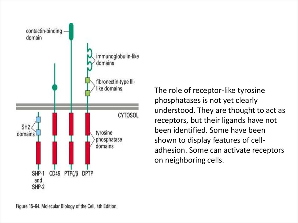
The role of receptor-like tyrosinephosphatases is not yet clearly
understood. They are thought to act as
receptors, but their ligands have not
been identified. Some have been
shown to display features of celladhesion. Some can activate receptors
on neighboring cells.
37. Classification of receptor-like protein tyrosine phosphatases (RPTPs) into eight subfamilies (R1-R8), based on sequence
similarity among PTP catalyticdomains . PTPμ, κ, ρ and PCP-2 are members of the R2B subfamily.
RPTPs have been divided into eight major
subfamilies, based on phylogenetic analysis of the
phosphatase domains. Four of these subfamilies
(R2A, R2B, R3, and R4) play critical roles in CNS
development. 2 RPTPs is an extracellular segment
containing a combination of multiple fibronectin
and immunoglobulin (Ig)-like domains, and a
single transmembrane region. The intracellular
region
contains
a
membrane
proximal
juxtamembrane domain, followed by a
catalytically active tyrosine phosphatase domain
and a second inactive domain. Type 2 RPTPs have
been further subdivided into two distinct classes
(R2A and R2B). Genes in the R2B class are
differentiated from the R2A class by an additional
MAM (Meprin/ A5/PTP mu) domain at the Nterminus, R2B molecules have cell adhesive
properties. Because no invertebrate homologues
of the four R2B molecules have been found to
date, and no ESTs indicative of R2Bs have been
isolated from invertebrates, the function(s) of
these phosphatases is likely to be highly specific to
vertebrate species.
38. Receptor guanylyl cyclases
39. Receptor guanylyl cyclases
• Single-pass transmembrane proteins with anextracellular binding site for a signal molecule
and an intracellular guanylyl cyclase catalytic
domain. The binding of the signal molecule
activates the cyclase domain to produce cyclic
GMP, which in turn binds to and activates a
cyclic GMP-dependent protein kinase (PKG),
which phosphorylates specific proteins on
serine or threonine.
40. Catalytic domain of human soluble guanylate cyclase 1
EC 4.6.1.2, also known as guanyl cyclase,guanylate cyclase, or GC) is a lyase enzyme.
Guanylyl cyclase is often part of the G
protein signaling cascade that is activated by
low intracellular calcium levels and inhibited
by high intracellular calcium levels.
41. Guanylyl cyclase catalyzes the reaction …
3',5'-cyclic guanosinemonophosphate (cGMP)
of guanosine triphosphate (GTP)
42. Some of the protein kinases
43. Histidine-kinase-associated receptors
44. Histidine-kinase-associated receptors
• Activate a “two-component” signalingpathway in which the kinase phosphorylates
itself on histidine and then immediately
transfers the phosphate to a second
intracellular signaling protein.
45. Protein histidine kinase
Crystallographicstructure of
ATP:protein-Lhistidine Nphosphotransferase
46.
• Multifunctional, typically transmembrane,proteins of the transferase class of enzymes
that play a role insignal transduction across
the cellular membrane.
47.
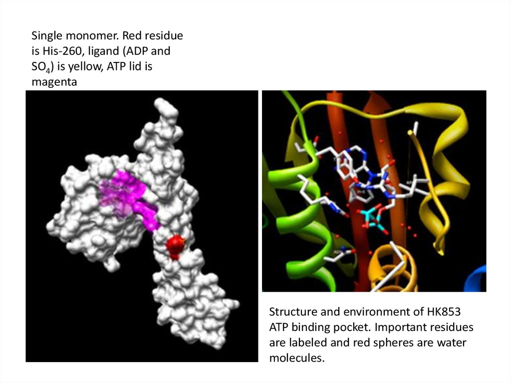
Single monomer. Red residueis His-260, ligand (ADP and
SO4) is yellow, ATP lid is
magenta
Structure and environment of HK853
ATP binding pocket. Important residues
are labeled and red spheres are water
molecules.
48. The bacterial flagellar motor
49. Positions of the flagella on E. coli during swimming
(A) Flagella rotate counterclockwise,they are drawn together into a single
bundle, which acts as a propeller to
produce smooth swimming
(B) Flagella rotate clockwise, they fly apart
and produce tumbling
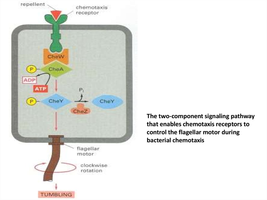
50.
The two-component signaling pathwaythat enables chemotaxis receptors to
control the flagellar motor during
bacterial chemotaxis
51. Conclusion
(1) receptor tyrosine kinases
(2) tyrosine-kinase-associated receptors
(3) receptor serine/threonine kinases
(4) transmembrane guanylyl cyclases
(5) histidine-kinase-associated receptors.
52.
• Some transmembrane tyrosine phosphatases,which remove phosphate from
phosphotyrosine side chains of specific
proteins, are thought to function as receptors,
although for the most part their ligands are
unknown. The first two classes of receptors
are by far the most numerous.
53.
• Tyrosine-kinase-associated receptors depend on variouscytoplasmic tyrosine kinases for their action. These kinases include
members of the Src family, which associate with many kinds of
receptors, and the focal adhesion kinase (FAK), which associates
with integrins at focal adhesions. The cytoplasmic tyrosine kinases
then phosphorylate a variety of signaling proteins to relay the signal
onward. The largest family of receptors in this class is the cytokine
receptors family. When stimulated by ligand binding, these
receptors activate Jak cytoplasmic tyrosine kinases, which
phosphorylate STATs. The STATs then dimerize, migrate to the
nucleus, and activate the transcription of specific genes. Receptor
serine/threonine kinases, which are activated by signaling proteins
of the TGF-β superfamily, act similarly: they directly phosphorylate
and activate Smads, which then oligomerize with another Smad,
migrate to the nucleus, and activate gene transcription.
54.
• Bacterial chemotaxis is mediated by histidinekinase-associated chemotaxis receptors. Whenactivated by the binding of a repellent, the
receptors stimulate their associated protein
kinase to phosphorylate itself on histidine and
then transfer that phosphate to a messenger
protein, which relays the signal to the flagellar
motor to alter the bacterium's swimming
behavior. Attractants have the opposite effect on
this kinase and therefore on swimming.
55.
Thanksfor
attention