


 Медицина
Похожие презентации:


 – Digestion and Organs of digestive system")


Esophagus stomach
1. Esophagus & stomach
Esophagus & stomach2. Alimentary Canal
Same basic construction end toend
Four distinct layers:
Mucosa
Epithelium
lamina propria,
MUSCULARIS MUCOSAE
Submucosa
dense irregular connective
tissue
Muscularis
two layers of smooth muscle
Serosa /adventitia
serosa is a mesothelium and
underlying connective tissue
adventitia is found where the
wall of the gut is attached to a
structure
?
3. Mucosa
Mucosa has three functions:Barrier
separates the lumen (which is in contact with the
environment) from the body interior
Secretory
Absorptive
3
4. Mucosa: Epithelium
Epithelium secretes:Digestive enzymes
into lumen
onto apical plasma membrane
Hormones
Mucous
Antibodies
which it receives from connective tissue (diffuse
lymphatic tissue)
Epithelium absorbs products of digestion
transport to vascular system
absorption occurs in small and large intestine
4
5. Mucosa: Lamina Propria
Areolar (loose) connective tissue underepithelium
Contains:
- glands
- vessels to receive absorbed substances
- fenestrated type
- numerous lymphatic capillaries; receive lipids and
some proteins
- components of the immune system
Diffuse lymphatic tissue
Lymphatic nodules
Gut-associate lymphatic tissue (GALT)
and Peyer’s patches
5
6. Mucosa: Lamina propria cont.
67. Mucosa: Muscularis Mucosae
Layer of smoothmuscles that forms
boundary between
mucosa and
submucosa
EP
M
LP
MM
Consists of 2 layers:
Inner circular
Outer longitudinal
CT
SM
Can produce movement
of mucosa independent
of movement of entire
gut wall
7
8. Submucosa
Consists ofmoderately dense
irregular connective
tissue
larger blood vessels
send branches to
mucosa, to muscularis
externa and serosa
LV
BV
lymphatics
LV
9. Submucosa cont.
Submucosa also containsnerve plexuses
AKA Submucosal
(Meissner’s) plexus
Nerve networks contain cell
bodies (ganglion cells) of
postganglionic neurons
Represent the third (enteric)
division of autonomic nervous
system
Innervate the smooth muscle of the
alimentary canal
9
10. Submucosa cont.
Neurons of the enteric division show the samepathologic changes that can occur in neurons of
the brain
e.g., Lewy bodies associated with Parkinson’s disease
Amyloid plaques and neurofibrillary tangles
associated with Alzheimer’s disease
These finding may lead to development of
routine rectal biopsies for early diagnosis of
these conditions as it is not possible to biopsy
the brain
10
11. Submucosa cont.
Glands occur in submucosaof esophagus and initial
part of duodenum
Presence of these glands
aids in identification of
particular regions of gut
MM
EG
11
12. Muscularis Externa
Also called the MUSCULARISUsually consists of two
concentric thick layers of
smooth muscle
Inner layer forms tight spiral
seen as a CIRCULAR LAYER
outer loose spiral described as a
LONGITUDINAL layer
ME
OL
G
IC
SM
M
13. Muscularis cont.
Located between the 2muscle layers is a thin
connective tissue layer
contains the MYENTERIC
PLEXUS (AUERBACH’S
PLEXUS)
Part of the enteric
division of the
autonomic NS
Also contains blood
vessels and lymphatic
vessels
14.
1415. Serosa & Adventitia
Serosa & AdventitiaSerosa is a membrane containing simple
squamous epithelium
the MESOTHELIUM
and a small amount of underlying connective
tissue
equivalent to visceral peritoneum of gross anatomy
Continuous with the MESENTERY which
holds the digestive tract in place
Contains large blood and lymphatic vessels
travel to and from mesentery to gut
15
16. Serosa & Adventitia cont.
Serosa & Adventitia cont.Large amounts of fat can accumulate in
serosa
Where gut has no serosa (esophagus,
duodenum, ascending colon, and
descending colon)
it is attached by loose connective tissue
ADVENTITIA
Adventitia blends with connective tissue of
surrounding structures
16
17. Esophagus
Mucosa:Epithelium non-keratinized
stratified squamous
A
Lamina propria has diffuse
lymphatic tissue
muscularis externa
and lymphatic nodules
Muscularis mucosae
EG
composed of longitudinally
organized smooth muscle
D
M
EP
EG
MM
SM
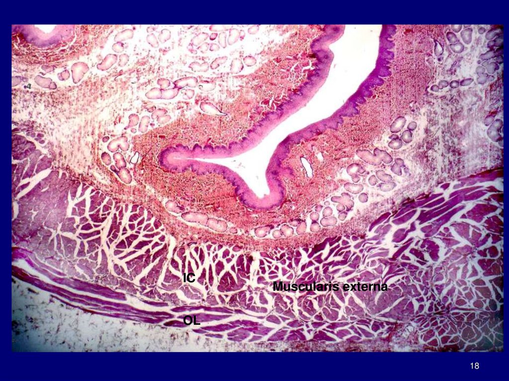
18.
ICMuscularis externa
OL
18
19. Esophagus cont.
Submucosa along withmuscularis mucosae
forms longitudinal folds
create very irregular luminal
profile when seen in XS
Muscularis externa
upper 1/3 is
striated muscle
Middle third
striated and smooth
muscle interwoven
Lower third
Smooth muscle
Continuous with the rest of
the gut
19
20.
StMSM
StM
21. Esophagus cont.
Esophagus has adventitia until it entersabdominal cavity
where it is covered by SEROSA
21
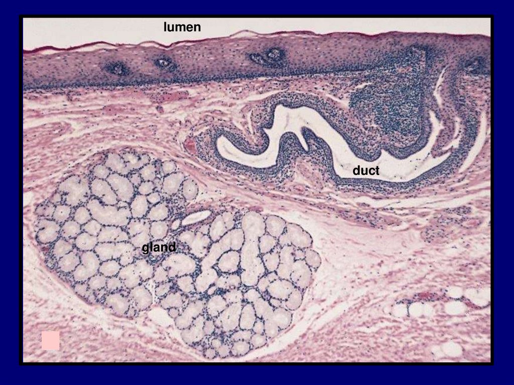
22. Esophagus: Glands
Two types:ESOPHAGEAL GLANDS
PROPER
ESOPHAGEAL CARDIAC
GLANDS
ESOPHAGEAL GLANDS
PROPER
Occur in submucosa
Scattered through out the
length of esophagus;
most in upper one half
Small compound
tubuloalveolar glands
Produce acidic mucous
22
23.
lumenduct
gland
24. Esophagus: Glands cont.
ESOPHAGEAL CARDIACGLANDS
occur in lamina propria of mucosa
Similar to cardiac glands of
stomach
Present in terminal parts of
esophagus
Esoph. Cardiac glands produce
neutral mucous
protect against regurgitated
material
Cs
Se
Es
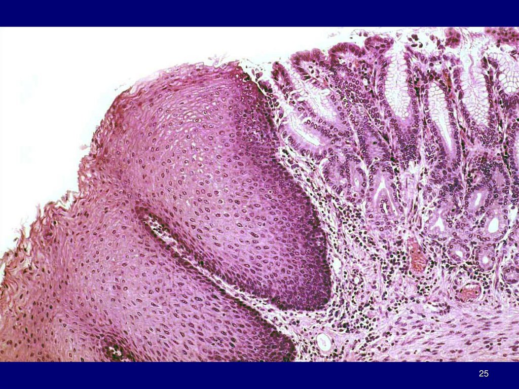
25.
2526. Stomach
Expanded part of alimentary canalSame organization as remaining gut
Mucosa
submucosa
muscularis
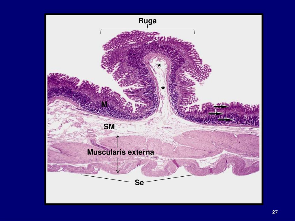
serosa
Inner surface has longitudinal folds
called RUGAE
Poorly developed in upper stomach
more elaborate in lower part
Disappear when stomach is
distended
Accommodate expansion
26
27.
RugaM
SM
Muscularis externa
Se
27
28. Stomach cont.
Numerous openings seen inmucosal surface
GASTRIC PITS or FOVEOLAE
Easily seen in SEM
Gastric glands empty into
bottom of gastric pits
b
28
29. Stomach cont.
Histologically dividedinto 3 regions:
Cardia
pylorus
Fundus
29
30. Stomach cont.
CARDIA (cardiac region)part near esophagus
contains cardiac glands
PYLORUS (pyloric region)
part proximal to pyloric
sphincter
contains pyloric glands
FUNDUS (fundic region)
called the body
largest part
between cardia and pylorus
Contains fundic glands
Called gastric glands
30
31. Stomach: Gastric Secretion
2 liters of fluid/dayGastric secretions include:
Pepsinogen
inactive precursor of proteolytic enzyme PEPSIN
HCl- (0.16N HCl)
acid pH promotes hydrolysis of food
converts pepsinogen to pepsin
Intrinsic factor
glycoprotein used for absorption of vitamin B
Hormone gastrin and others
produced by ENTEROENDOCRINE cells in gastric
epithelium
31
32. Stomach: Absorption
Stomach lining absorbssome water
salts
lipid-soluble drugs
certain drugs
Asprin enters by damaging surface epithelium
Alcohol
32
33. Stomach: Gastric Mucosa
Simple columnarepithelium
Called SURFACE
MUCOUS CELLS
Cells have a large apical
deposit (cap) of mucin
granules
Mucous forms a thick gel-like
coat on surface
33
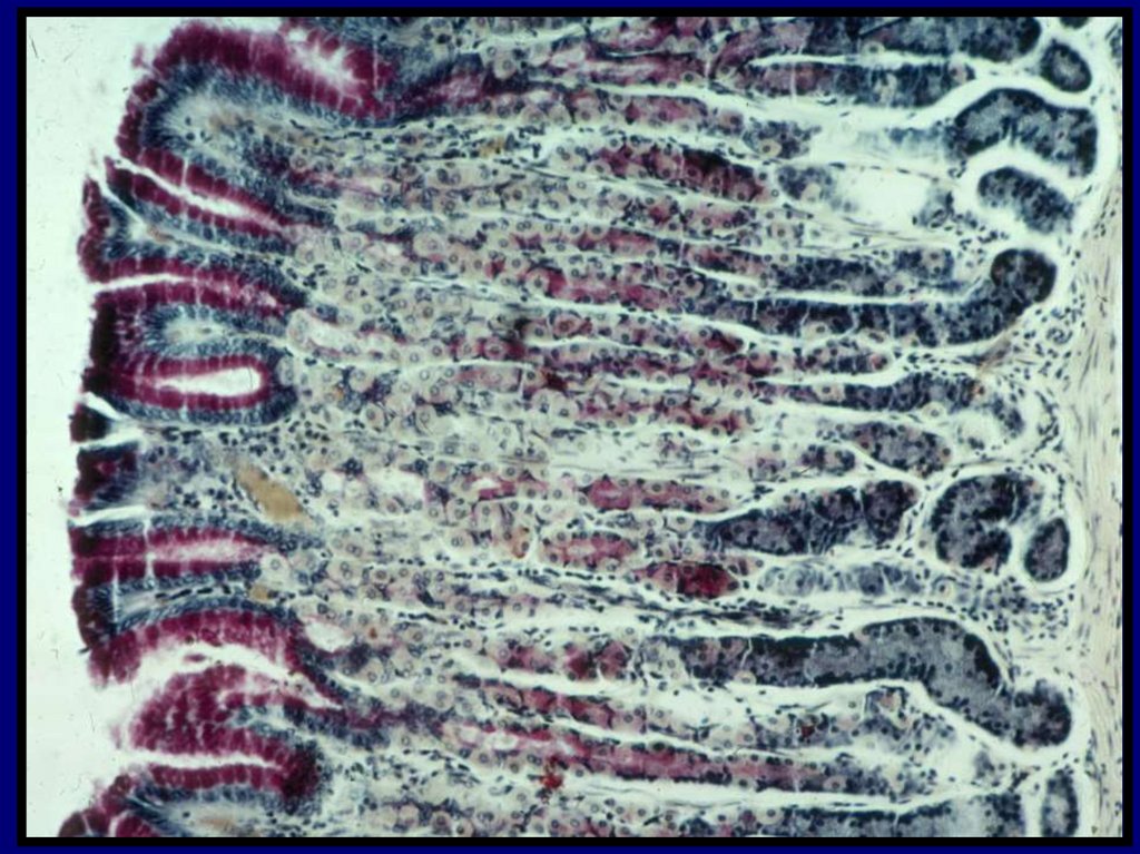
34. Fundic Glands
Fundic glands; also called gastricglands
Produce digestive juice of stomach
Present throughout gastric mucosa
except where cardiac and pyloric
glands occur
Simple branched tubular glands
Extend from bottom of GASTRIC
PITS to muscularis mucosae
34
35. Fundic Glands cont.
Several glands open intoone gastric pit
Each gland has
long NECK SEGMENT
BASE or FUNDIC SEGMENT
Gland may divide into 2 or 3
branches
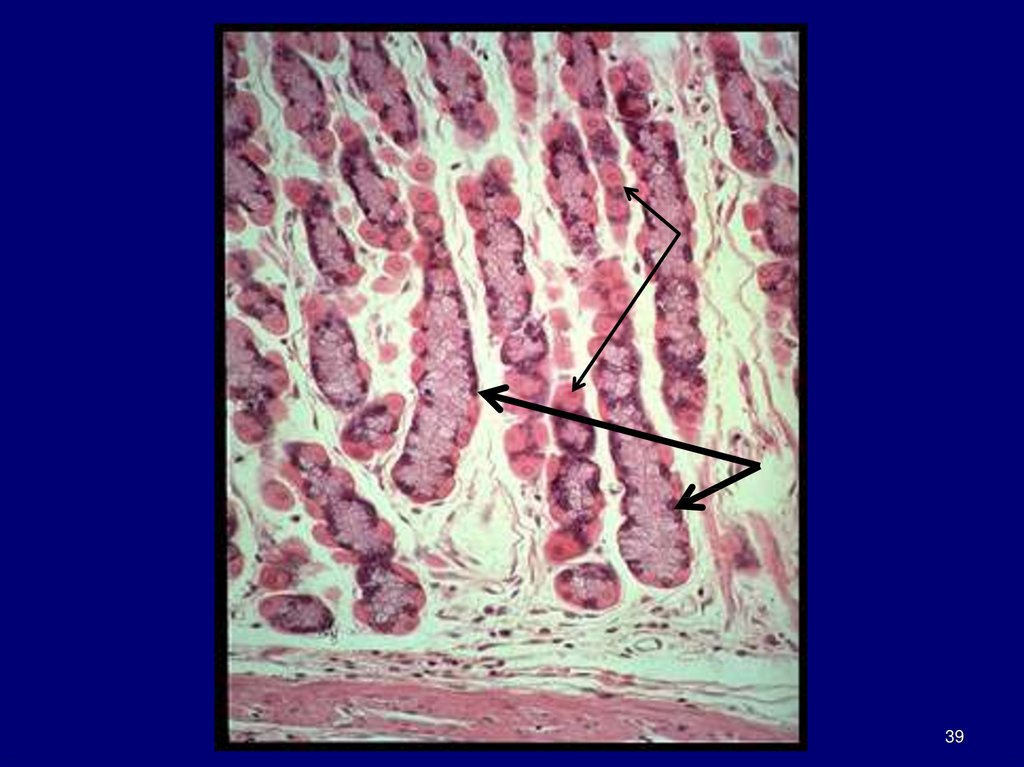
36. Fundic Glands cont.
Composed of 4functional cell types:
MUCOUS NECK CELLS
CHIEF CELLS
PARIETAL CELLS (Oxyntic
cells)
ENTEROENDOCRINE
CELLS
And
UNDIFFERENTIATED
CELLS
located in upper neck
region
give rise to mature cells
listed above
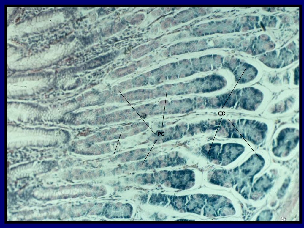
37.
CCPC
L
37
38.
3839.
3940. Fundic Glands: Mucous Neck Cells
Located in neck regionShorter than surface
mucous cell
Nucleus tends to be
spherical rather than
elongate
as in surface cells
Secretes soluble
mucous
compared to viscous
surface mucous
40
41. Fundic Glands: Chief Cells
Typical proteinsecreting cellsOccupy the basal
portion of the gland
Cells easily identified
by intense basophilia
Basal rER and apical
granules
Secrete:
pepsin as inactive
pepsinogen
a weak lipase
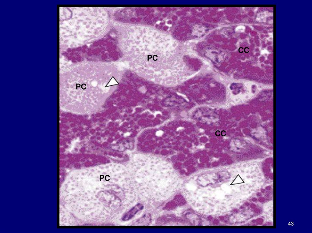
42. Fundic Glands: Parietal Cells
Called OXYNTIC CELLSSecrete HCl
and intrinsic factor
Most numerous in upper
and middle region of the
gland
Large cells
Appear round to triangular
PC
with apex directed toward
lumen of gland
42
43.
CCPC
PC
CC
PC
43
44. Fundic Glands: Parietal Cell cont.
Nucleus is sphericalCytoplasm intensely
eosinophilic
easily recognized by
size and staining
Numerous
mitochondria (eosinophilia)
Provide energy for ion
trafficking
44
45. Fundic Glands: Parietal Cell cont.
EM shows extensiveINTRACELLULAR
CANALICULAR
SYSTEM
communicates with
lumen of fundic gland
Numerous surface
microvilli project from
canaliculi
Secretion of HCl across
membranes of canaliculi and
microvillous extensions
45
46. Fundic Glands: Parietal Cell cont.
TUBULOVESICULARMEMBRANE SYSTEM
located in cytoplasm adjacent
to canalicular system
represent reserve membrane
for insertion into surface
for increased secretory surface
area
In humans INTRINSIC
FACTOR secreted by parietal
cells
absorption of vitamin B
Failure of IF secrection causes
PERNICIOUS anemia
46
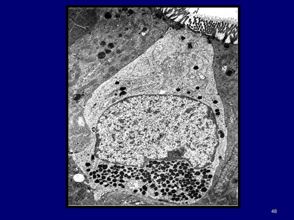
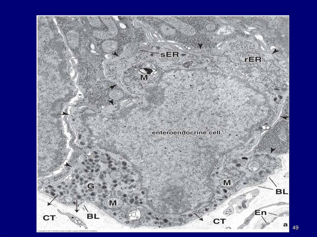
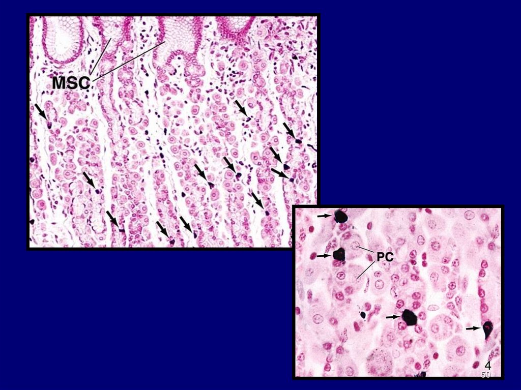
47. Fundic Glands: Enteroendocrines
Open and closed typesOpen are chemoreceptors
Located at any level of gland
sit on basal lamina
EM shows small, membrane-limited
granules
Cells hard to identify
Also called:
Chromaffin cells
argentaffin cells
argyrophil cells
enterochromaffin cells
48.
4849.
4950.
5051. Cardiac Glands
Limited to narrow region ofstomach
near esophageal orifice
Secretion, along with that of
esophageal cardiac glands
contributes to gastric juice
Tubular glands
Tortuous
Composed of mainly mucous
secreting cells
Some enteroendocrine cells
interspersed
51
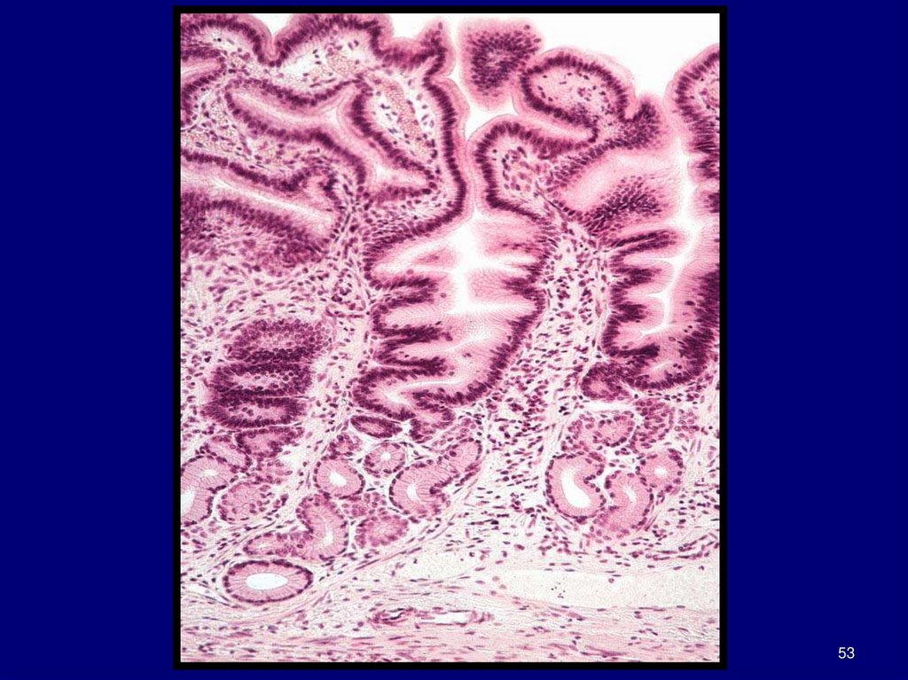
52. Pyloric Glands
Located between fundusand pylorus
Branched tubular glands
Coiled
Cells
Similar to surface mucous
cells
Enteroendocrine cells
interspersed
Glands empty into deep
gastric pits
that occupy ½ the thickness of
the mucosa
52
53.
5354. Stomach: Epithelial Cell Renewal
Mitotic activity found amongimmature cells between the bottom
of pit and neck of gland
New cells migrating upwards along
wall of pit to luminal surface
differentiate
into surface mucous cells
Renewed every 3-5 days
Other new cells migrating downwards
differentiate
Into gastric gland cells (e.g., parietal, chief,
or enteroendocrine cells)
Parietal renewed approx. each ½ year
Chief and entroendocrine each 2-3 months
i.e., migration of the new cells is
bidirectional in the stomach
54