


 Английский язык
Похожие презентации:


Dentin
1. Dentin
Calcified tissueConsists mainly of
harder than bone-higher content of
Calcium salts (70%)
Type I collagen
GAGs
hydroxyapatite crystals
Dentin matrix secreted by
ODONTOBLASTS
Form an epithelial layer over the inner
surface of the dentin
Bear the same relation to dentine as
osteoblasts do to bone
2. Dentin cont.
Odontoblast iselongate
well developed rER
large Golgi
Apical surface in contact
with the forming dentin
Apical junctional
complexes between
odontoblasts separate the
dentin from the pulp
3.
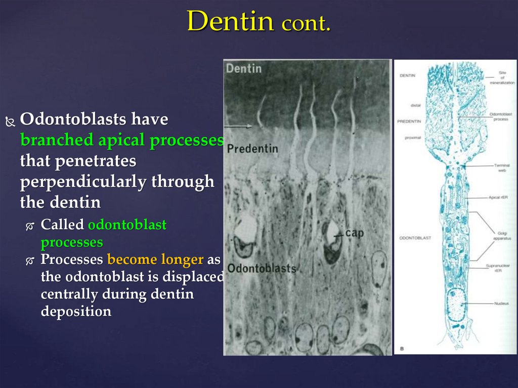
Dentin cont.Odontoblasts have
branched apical processes
that penetrates
perpendicularly through
the dentin
Called odontoblast
processes
Processes become longer as
the odontoblast is displaced
centrally during dentin
deposition
4. Dentin cont.
Processes contained in canals called DENTINAL TUBULESOdontoblast processes are 3-4 um dia near cell body; thinner near
enamel or cementum
A
B
5. Fundic Glands: Parietal Cells
Called OXYNTIC CELLSSecrete HCl
and intrinsic factor
Most numerous in upper and
middle region of the gland
Large cells
Appear round to triangular
with apex directed toward
lumen of gland
PC
6. Fundic Glands: Parietal Cell cont.
Nucleus is sphericalCytoplasm intensely
eosinophilic
easily recognized by
size and staining
Numerous mitochondria
(eosinophilia)
Provide energy for ion
trafficking
7. Small Intestine: Paneth Cell
Found in bases ofintestinal glands
May be seen in colon as
well
Large apical secretory
granules
very eosinophilic
refractile
Granules permit
identification of these cells
8. Small Intestine: Paneth Cell cont.
Granules containLYSOZYME
LYSOZYME digests cell
walls of certain bacteria
α-DEFENSINS
Paneth cells probably
Regulate normal bacterial
flora of small intestine
9. Small Intestine: Submucosa
Consists ofdense connective tissue
aggregates of adipose cells
Conspicuous feature of
duodenum is
submucosal glands
(BRUNNER’S GLANDS)
10. Small Intestine: Submucosa cont.
Cells of Brunner’sglands have
characteristics of both
mucous and serous
secretions
pH of secretions is
8.1-9.3
protects proximal
small intestine
neutralizes acid from
stomach
creates optimal pH for
enzymes
11. Features of Small Intestine Mucosa: Lamina Propria cont.
Lamina propria alsocontains
lymphatic nodules
important part of GALT
Nodules are especially large
in ileum
called PEYER’S PATCHES
Muscularis mucosae
2 thin layers of smooth
muscle
11
inner circular
outer longitudinal
12.
1213. Large Intestine
Composed of :Cecum
Ascending colon
Transverse colon
Descending colon
Sigmoid colon
Rectum
Anal canal
teniae coli
Contain 4 histologic layers of GI tract; exceptions are
Mucosa is smooth (no villi)
Outer muscle layer
13
has 3 equally spaced bands(teania coli)
14. Large Intestine: Rectum & Anal Canal
Large Intestine: Rectum &Anal Canal
Rectum is dilated distal portion of GIT
Upper part is distinguished
TRANSVERSE RECTAL FOLDS
Mucosa similar to distal colon
Anal canal is most distal part of the GIT
Upper part of anal canal has
longitudinal folds
Called ANAL COLUMNS
Depressions between anal columns called ANAL SINUSES