


 Медицина
Похожие презентации:


Хронические гепатиты, циррозы печени
1.
Хронические гепатиты, циррозыпечени
2.
Хронические гепатиты- группа заболеваний печени,
вызываемых различными
причинами, характеризующиеся
различной степенью
выраженности печеночноклеточного некроза и воспаления
и протекающие без улучшения по
меньшей мере 6 месяцев.
3.
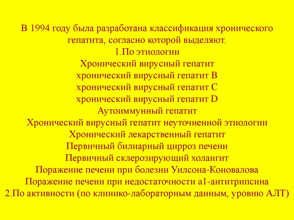
В 1994 году была разработана классификация хроническогогепатита, согласно которой выделяют.
1.По этиологии
Хронический вирусный гепатит
хронический вирусный гепатит B
хронический вирусный гепатит С
хронический вирусный гепатит D
Аутоиммунный гепатит
Хронический вирусный гепатит неуточненной этиологии
Хронический лекарственный гепатит
Первичный билиарный цирроз печени
Первичный склерозирующий холангит
Поражение печени при болезни Уилсона-Коновалова
Поражение печени при недостаточности a1-антитрипсина
2.По активности (по клинико-лабораторным данным, уровню АЛТ)
4.
3.По морфологии (указывают характергистологических изменений, подтверждающих
этиологию и степень активности воспалительного
процесса)
Примечание: отсутствие алкогольного гепатита в
классификации хронических гепатитов обусловлено
тем, что некоторые ученые относят хронический
алкогольный гепатит к алкогольной болезни печени и
не рассматривают его среди других видов
хронических гепатитов
5.
ЭтиологияПервое место среди этиологических факторов хронического гепатита занимают
три вируса В, С, D. При заражении вирусным гепатитом В хронизация процесса
наступает у 5-10% больных, чаще после латентных и легких форм заболевания,
что связано с их редкой диагностикой и отсутствием адекватного лечения. Вирус
гепатита С вызывает хроническое воспаление в 85% случаев и обычно на фоне
алкоголизма. Хронический гепатит D развивается у 90% больных с острым
гепатитом D. Аутоиммунный гепатит расценивают как наследственно
обусловленный иммунными нарушениями. В качестве запускающего фактора
обсуждаются вирусы гепатита А, В, С, D, E, G. Лекарственный гепатит чаще
всего вызывают антибиотики (тетрациклин, левомицетин, гентамицин,
эритромицин, олеандомицин, рифампицин) и другие антибактериальные средства
(нитроксалин, сульфасалазин), препараты для лечения сердечно - сосудистых
заболеваний (пропранолол), непрямые антикоагулянты, салуретики,
психотропные препараты, НВПС, блокаторы Н2-рецепторов гистамина,
иодсодержащие контрастные вещества. Особенно тяжелые формы гепатитов,
протекающих с некрозами печеночной параенхимы, могут вызывать метилдопа,
изониазид, парацетомол, метотрексат, фторотан и некоторые другие препараты.
Алкогольная болезнь печени возникает при ежедневном употреблении более 40
мл чистого этанола на протяжении 6-8 лет. При инфицировании вирусами В и/или
С формирование заболевания происходит в более короткие сроки.
6.
Факторы рискаК ним относятся: инъекционная наркомания, переливание крови и ее
компонентов, стоматологические манипуляции, оперативные вмешательства,
контакт с больным вирусным гепатитом, профессиональный фактор, тату аж,
беспорядочные сексуальные контакты, аборты.
Патогенез
Хронический вирусный гепатит
Его развитие зависит от реакций клеточного иммунитета. Инфицированние
вирусом не всегда сопровождается развитием заболевания. Возможны четыре
типа взаимодействия вируса и иммуной системы:
1.при нормальной иммуной реактивности организма происходит пролиферация
иммунокомпетентных клеток, которые узнают белки вируса, внедрившиеся в
мембрану пораженного гепатоцита. Взаимодействие иммунных клеток с АГ
вызывает цитолиз гепатоцита и гибель содержащегося в нем вируса.
2.при гиперергическом иммуном ответе происходит острый массивный некроз
гепатоцитов и развивается молнеиносная форма острого гепатита.
3.при гипоэргическом иммунном ответе Т-лимфоциты разрушают
инфицированные клетки, но не могут предотвратить распространение вируса
среди здоровых гепатоцитов. Это ведет к развитию хронического гепатита.
7.
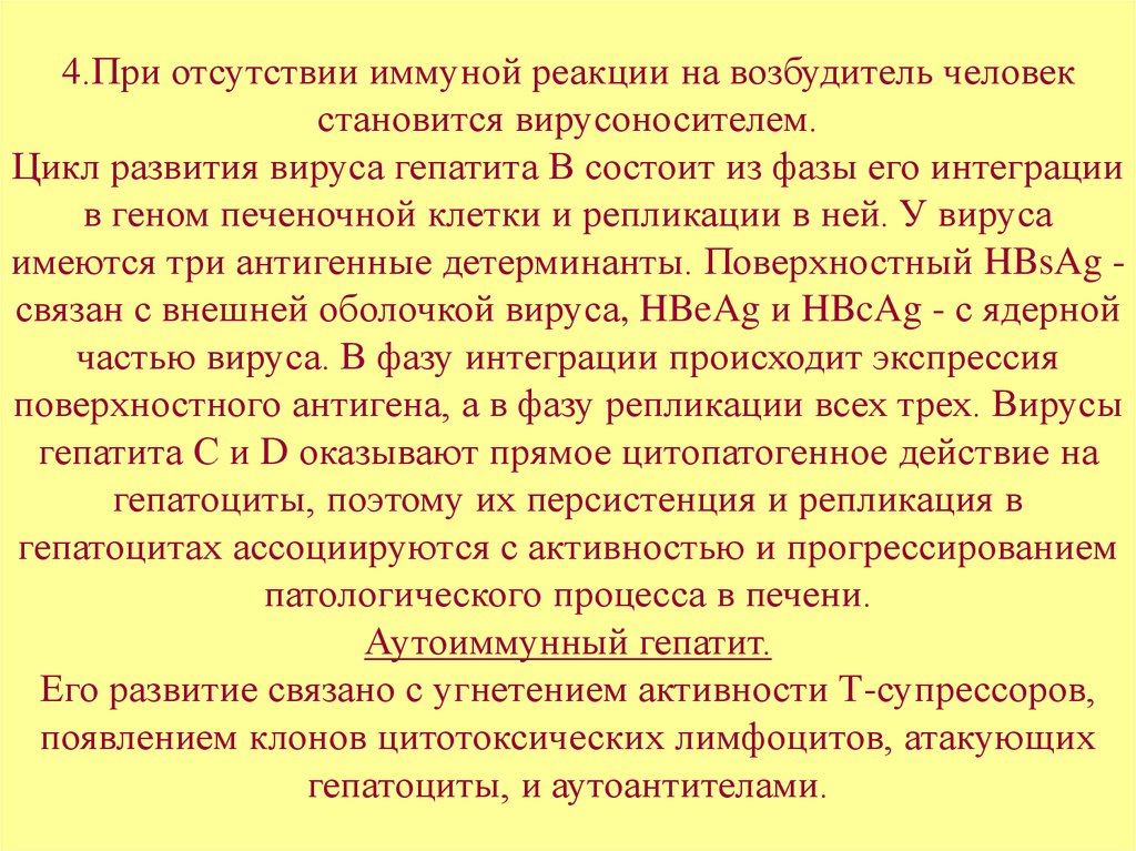
4.При отсутствии иммуной реакции на возбудитель человекстановится вирусоносителем.
Цикл развития вируса гепатита В состоит из фазы его интеграции
в геном печеночной клетки и репликации в ней. У вируса
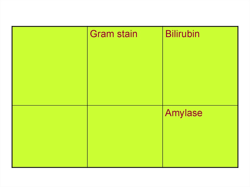
имеются три антигенные детерминанты. Поверхностный HBsAg связан с внешней оболочкой вируса, HBeAg и HBcAg - с ядерной
частью вируса. В фазу интеграции происходит экспрессия
поверхностного антигена, а в фазу репликации всех трех. Вирусы
гепатита С и D оказывают прямое цитопатогенное действие на
гепатоциты, поэтому их персистенция и репликация в
гепатоцитах ассоциируются с активностью и прогрессированием
патологического процесса в печени.
Аутоиммунный гепатит.
Его развитие связано с угнетением активности Т-супрессоров,
появлением клонов цитотоксических лимфоцитов, атакующих
гепатоциты, и аутоантителами.
8.
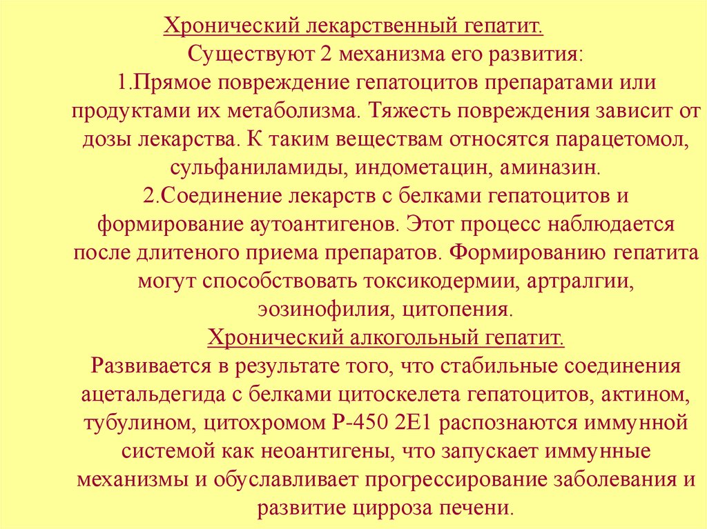
Хронический лекарственный гепатит.Существуют 2 механизма его развития:
1.Прямое повреждение гепатоцитов препаратами или
продуктами их метаболизма. Тяжесть повреждения зависит от
дозы лекарства. К таким веществам относятся парацетомол,
сульфаниламиды, индометацин, аминазин.
2.Соединение лекарств с белками гепатоцитов и
формирование аутоантигенов. Этот процесс наблюдается
после длитеного приема препаратов. Формированию гепатита
могут способствовать токсикодермии, артралгии,
эозинофилия, цитопения.
Хронический алкогольный гепатит.
Развивается в результате того, что стабильные соединения
ацетальдегида с белками цитоскелета гепатоцитов, актином,
тубулином, цитохромом Р-450 2Е1 распознаются иммунной
системой как неоантигены, что запускает иммунные
механизмы и обуславливает прогрессирование заболевания и
развитие цирроза печени.
9.
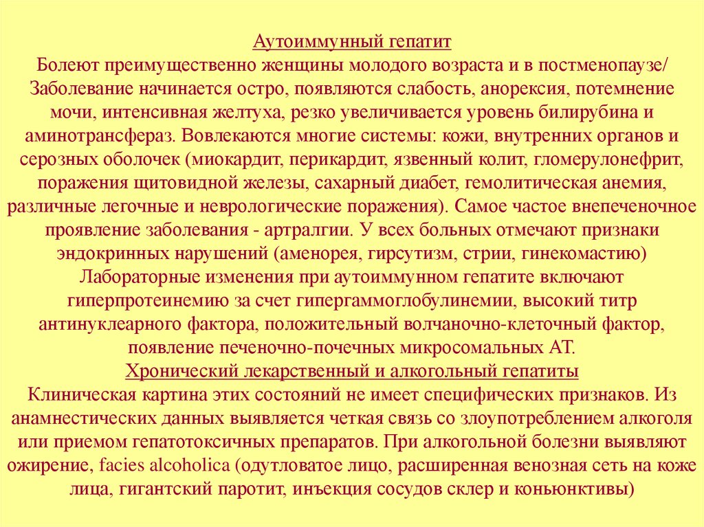
Аутоиммунный гепатитБолеют преимущественно женщины молодого возраста и в постменопаузе/
Заболевание начинается остро, появляются слабость, анорексия, потемнение
мочи, интенсивная желтуха, резко увеличивается уровень билирубина и
аминотрансфераз. Вовлекаются многие системы: кожи, внутренних органов и
серозных оболочек (миокардит, перикардит, язвенный колит, гломерулонефрит,
поражения щитовидной железы, сахарный диабет, гемолитическая анемия,
различные легочные и неврологические поражения). Самое частое внепеченочное
проявление заболевания - артралгии. У всех больных отмечают признаки
эндокринных нарушений (аменорея, гирсутизм, стрии, гинекомастию)
Лабораторные изменения при аутоиммунном гепатите включают
гиперпротеинемию за счет гипергаммоглобулинемии, высокий титр
антинуклеарного фактора, положительный волчаночно-клеточный фактор,
появление печеночно-почечных микросомальных АТ.
Хронический лекарственный и алкогольный гепатиты
Клиническая картина этих состояний не имеет специфических признаков. Из
анамнестических данных выявляется четкая связь со злоупотреблением алкоголя
или приемом гепатотоксичных препаратов. При алкогольной болезни выявляют
ожирение, facies alcoholica (одутловатое лицо, расширенная венозная сеть на коже
лица, гигантский паротит, инъекция сосудов склер и коньюнктивы)
10.
ДиагностикаДиагноз ставят на основании данных анамнеза (связь с
употреблением алкоголя, с приемом гепатотоксичных препаратов,
факторы инфицирования вирусами гепатита) При исследовании
периферической крови наблюдается ускорение СОЭ, тенденци к
лейко- и тромбоцитопении. Отмечают повышение АЛТ,
билирубина, диспротеинемию, гипергаммоглобулинемию, ?глутамилтранс-пептидазы. В случае хронического вирусного
гепатита имеет значение выявление тканевых и сывороточных
маркеров вируса гепатита В, С, D. При аутоиммунном гепатите
считается важным отсутствие маркеров вирусного гепатита,
выявление антинуклеарных, антимитохондриальных аутоантител,
наличие в крови ЦИК, преимущественное увеличение в крови Ig G.
Дифференциальная диагностика Гепатиты дифференцируют между
собой, первичным билиарным циррозом, болезнью УилсонаКоновалова, первичным склерозирующим холангитом.
11.
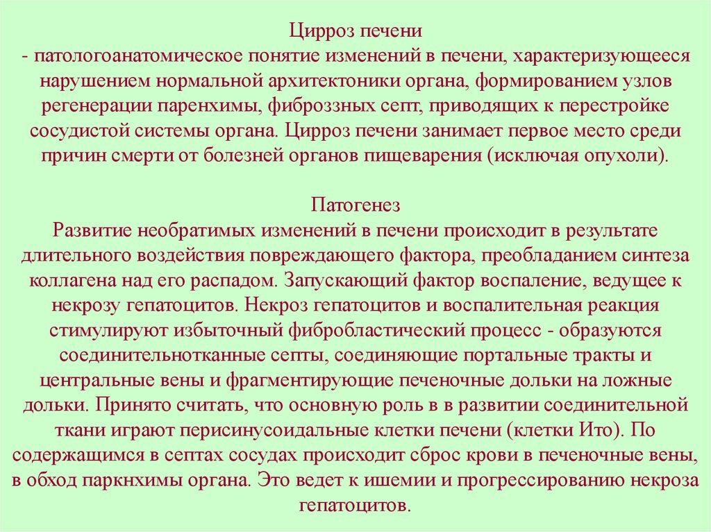
Цирроз печени- патологоанатомическое понятие изменений в печени, характеризующееся
нарушением нормальной архитектоники органа, формированием узлов
регенерации паренхимы, фиброззных септ, приводящих к перестройке
сосудистой системы органа. Цирроз печени занимает первое место среди
причин смерти от болезней органов пищеварения (исключая опухоли).
Патогенез
Развитие необратимых изменений в печени происходит в результате
длительного воздействия повреждающего фактора, преобладанием синтеза
коллагена над его распадом. Запускающий фактор воспаление, ведущее к
некрозу гепатоцитов. Некроз гепатоцитов и воспалительная реакция
стимулируют избыточный фибробластический процесс - образуются
соединительнотканные септы, соединяющие портальные тракты и
центральные вены и фрагментирующие печеночные дольки на ложные
дольки. Принято считать, что основную роль в в развитии соединительной
ткани играют перисинусоидальные клетки печени (клетки Ито). По
содержащимся в септах сосудах происходит сброс крови в печеночные вены,
в обход паркнхимы органа. Это ведет к ишемии и прогрессированию некроза
гепатоцитов.
12.
Продукты распада гепатоцитовстимулируют регенераторные
процессы. Результатом этого
является формирование узлов
регенерации, сдавливающих
сосуды и усугубляющее
нарушение кровообращения.
13.
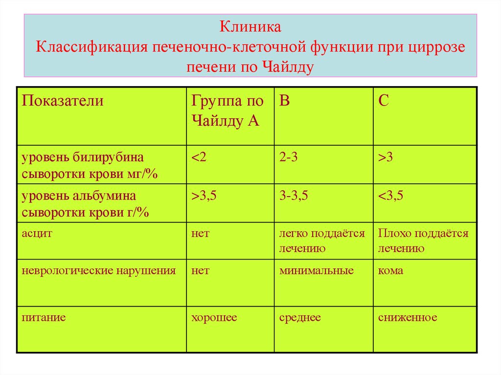
КлиникаКлассификация печеночно-клеточной функции при циррозе
печени по Чайлду
Показатели
Группа по В
Чайлду A
С
уровень билирубина
сыворотки крови мг/%
<2
2-3
>3
уровень альбумина
сыворотки крови г/%
>3,5
3-3,5
<3,5
асцит
нет
легко поддаётся
лечению
Плохо поддаётся
лечению
неврологические нарушения
нет
минимальные
кома
питание
хорошее
среднее
сниженное
14.
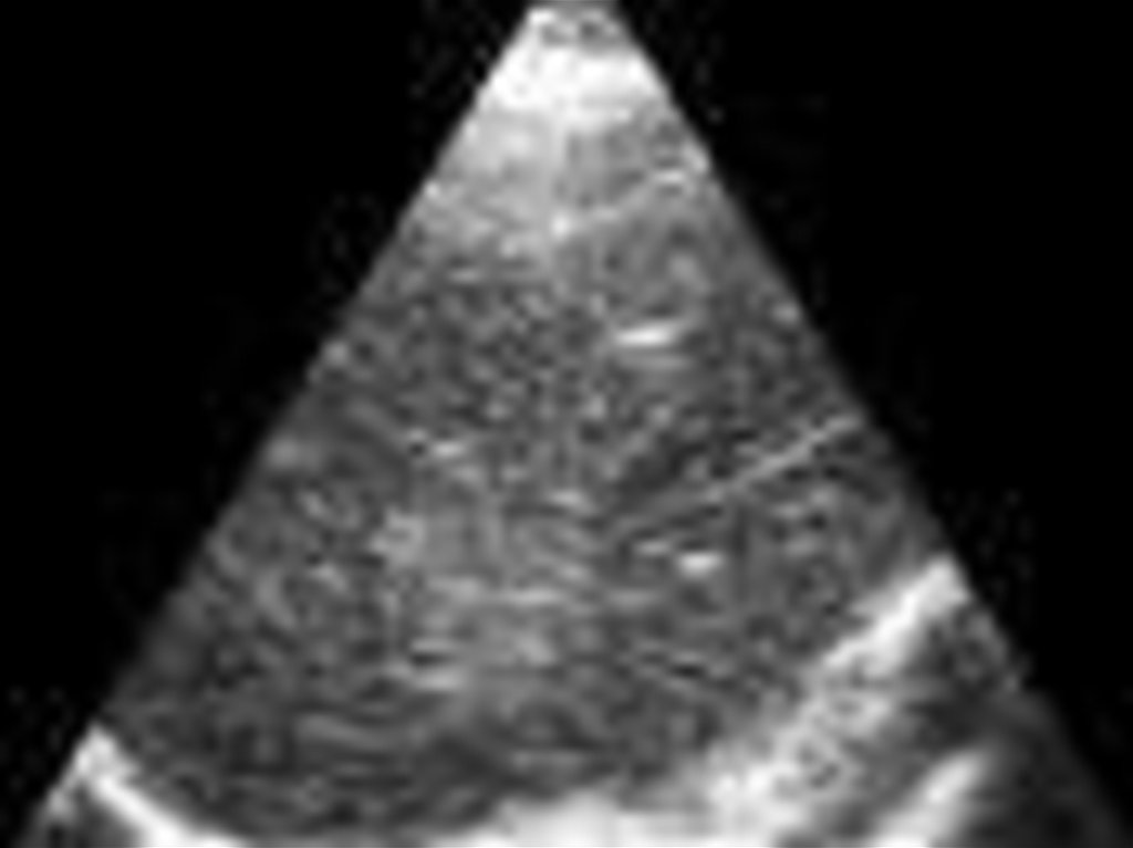
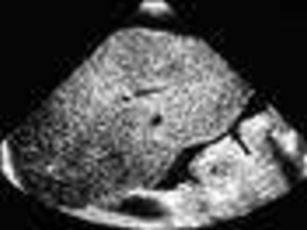
УЗИ печени позволяет обнаружить изменение размеровпечени и селезенки, выявить акустическую
неодносродность печеночной паренхимы и признаки
портальной гипертензии. КТ - более информативный
метод. Ангиографическое исследование - целиакография и
спеленопортография позволяют выявить наличие и
характер портальной гипертензии. С этой же целью
проводят эндоскопическое и рентгенографическое
исследование пищевода и желудка (выявление
расширенных вен пищевода, реже кардиального отдела
желудка). Пункционная биопсия печени позволяет
провести гистологическое исследование биоптата и
выявить стадию процесса.
15.
Редкие формы цирроза печени.Первичный билиарный цирроз.
Распространенность 25-30 человек на 1 млн. Заболевают в основном
женщины в период менопаузы.
Этиология и патогенез. На первой стадии формируется негнойный
деструктивный холангит мелких желчных протоков, этиология которого
неизвестна. Важная роль в патогенезе отводится образованию антител к
внутренней мембране митохондрий.
Клиническая картина. Первым и основным проявлением заболевания
является синдром холестаза, позже присоединяется желтуха. Потом
присоединяются оссалгии, похудание, слабость. Объективно выявляется
иктеричность кожи, следы расчесов, ксантелазмы на коже век.
Диагностика. При биохимическом исследовании выявляется
гипербилирубинемия, повышение активности щелочной фосфотазы, ?глютамилтрансферазы (индикаторы холестаза). Активность трансфераз
повышается в 2-10 раз. В сыворотке крови можно обнаружить
антимитохондриальные АТ. Для исключения внепеченочного холестаза
проводят УЗИ печени (изучение головки поджелудочной железы, общего
желчного протока) и ФГДС (большой дуоденальный сосочек).
16.
Вторичный билиарный циррозРаспространенность неизвестна.
Этиология и патогенез. В основе заболевания лежит
внепеченочный холестаз в результате ЖКБ, врожденных
дефектов желчевыводящих путей, послеоперационных
стриктур).
Клиническая картина. Беспокоят в основном боли в правом
подреберье. Обьективно можно выявить увеличение печени и
желчного пузыря.
Диагностика. Проводится УЗИ и эндоскопическая
ретроградная панкреатохолангиография, позволяющая выявить
причину и уровень обструкции.
Осложнения. Асцит, печеночная энцефалопатия, кровотечение
из расширенных вен пищевода, реже кардиального отдела
желудка, развитие печеночно-клеточной карциномы при
вирусном и алкогольном циррозе.
17.
Этапная диагностика диффузных заболеваний печениПоликлиника
Клиническое обследование больного
Клинический анализ крови (включая подсчет количества
тромбоцитов и ретикулоцитов).
Группа крови, резус-фактор.
Общий анализ мочи.
Исследование кала на скрытую кровь.
Биохимический анализ крови: общий белок, альбумин, общий и
прямой билирубин, глюкоза, креатинин, АлАт, АсАт, ГГТ, ЩФ.
HВsAg, анти-HCV.
Анти-ВИЧ, RW.
УЗИ брюшной полости.
ЭГДС (при необходимости).
18.
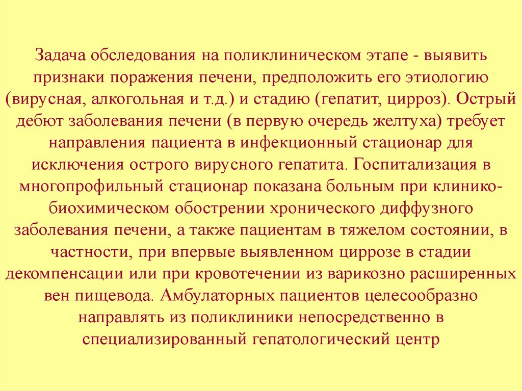
Задача обследования на поликлиническом этапе - выявитьпризнаки поражения печени, предположить его этиологию
(вирусная, алкогольная и т.д.) и стадию (гепатит, цирроз). Острый
дебют заболевания печени (в первую очередь желтуха) требует
направления пациента в инфекционный стационар для
исключения острого вирусного гепатита. Госпитализация в
многопрофильный стационар показана больным при клиникобиохимическом обострении хронического диффузного
заболевания печени, а также пациентам в тяжелом состоянии, в
частности, при впервые выявленном циррозе в стадии
декомпенсации или при кровотечении из варикозно расширенных
вен пищевода. Амбулаторных пациентов целесообразно
направлять из поликлиники непосредственно в
специализированный гепатологический центр
19.
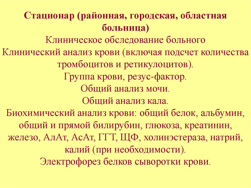
Стационар (районная, городская, областнаябольница)
Клиническое обследование больного
Клинический анализ крови (включая подсчет количества
тромбоцитов и ретикулоцитов).
Группа крови, резус-фактор.
Общий анализ мочи.
Общий анализ кала.
Биохимический анализ крови: общий белок, альбумин,
общий и прямой билирубин, глюкоза, креатинин,
железо, АлАт, АсАт, ГГТ, ЩФ, холинэстераза, натрий,
калий (при необходимости).
Электрофорез белков сыворотки крови.
20.
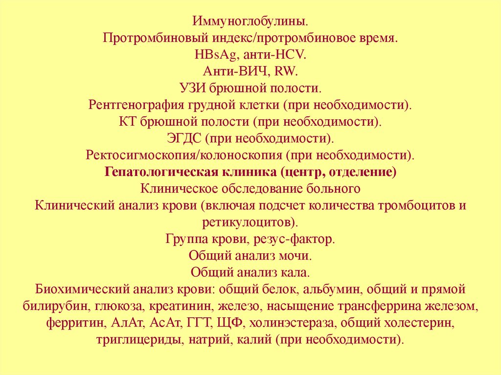
Иммуноглобулины.Протромбиновый индекс/протромбиновое время.
HВsAg, анти-HCV.
Анти-ВИЧ, RW.
УЗИ брюшной полости.
Рентгенография грудной клетки (при необходимости).
КТ брюшной полости (при необходимости).
ЭГДС (при необходимости).
Ректосигмоскопия/колоноскопия (при необходимости).
Гепатологическая клиника (центр, отделение)
Клиническое обследование больного
Клинический анализ крови (включая подсчет количества тромбоцитов и
ретикулоцитов).
Группа крови, резус-фактор.
Общий анализ мочи.
Общий анализ кала.
Биохимический анализ крови: общий белок, альбумин, общий и прямой
билирубин, глюкоза, креатинин, железо, насыщение трансферрина железом,
ферритин, АлАт, АсАт, ГГТ, ЩФ, холинэстераза, общий холестерин,
триглицериды, натрий, калий (при необходимости).
21.
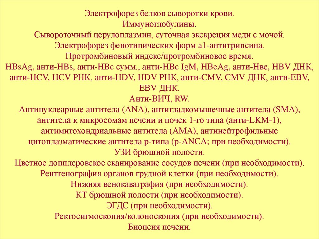
Электрофорез белков сыворотки крови.Иммуноглобулины.
Сывороточный церулоплазмин, суточная экскреция меди с мочой.
Электрофорез фенотипических форм a1-антитрипсина.
Протромбиновый индекс/протромбиновое время.
HВsAg, анти-HBs, анти-HBc сумм., анти-HBc IgM, HВeAg, анти-Нве, HBV ДНК,
анти-HCV, HCV РНК, анти-HDV, HDV РНК, анти-CMV, CMV ДНК, анти-ЕВV,
ЕВV ДНК.
Анти-ВИЧ, RW.
Антинуклеарные антитела (ANA), антигладкомышечные антитела (SMA),
антитела к микросомам печени и почек 1-го типа (анти-LKM-1),
антимитохондриальные антитела (АМА), антинейтрофильные
цитоплазматические антитела р-типа (p-ANCA; при необходимости).
УЗИ брюшной полости.
Цветное допплеровское сканирование сосудов печени (при необходимости).
Рентгенография органов грудной клетки (при необходимости).
Нижняя венокаваграфия (при необходимости).
КТ брюшной полости (при необходимости).
ЭГДС (при необходимости).
Ректосигмоскопия/колоноскопия (при необходимости).
Биопсия печени.
22.
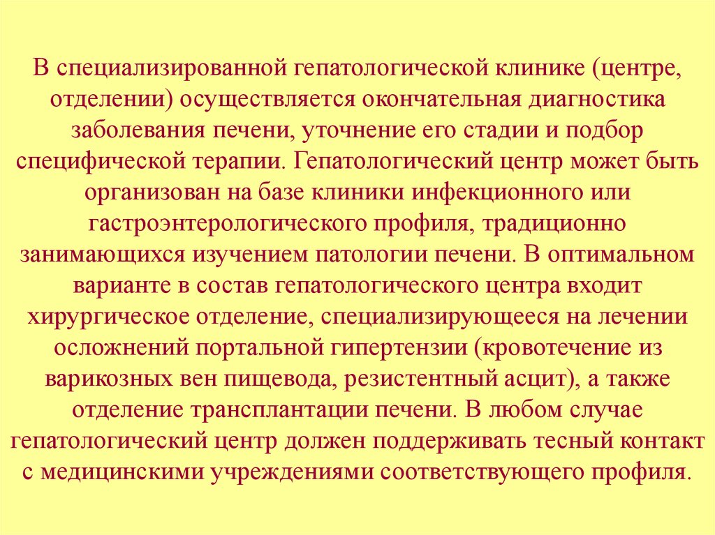
В специализированной гепатологической клинике (центре,отделении) осуществляется окончательная диагностика
заболевания печени, уточнение его стадии и подбор
специфической терапии. Гепатологический центр может быть
организован на базе клиники инфекционного или
гастроэнтерологического профиля, традиционно
занимающихся изучением патологии печени. В оптимальном
варианте в состав гепатологического центра входит
хирургическое отделение, специализирующееся на лечении
осложнений портальной гипертензии (кровотечение из
варикозных вен пищевода, резистентный асцит), а также
отделение трансплантации печени. В любом случае
гепатологический центр должен поддерживать тесный контакт
с медицинскими учреждениями соответствующего профиля.
23.
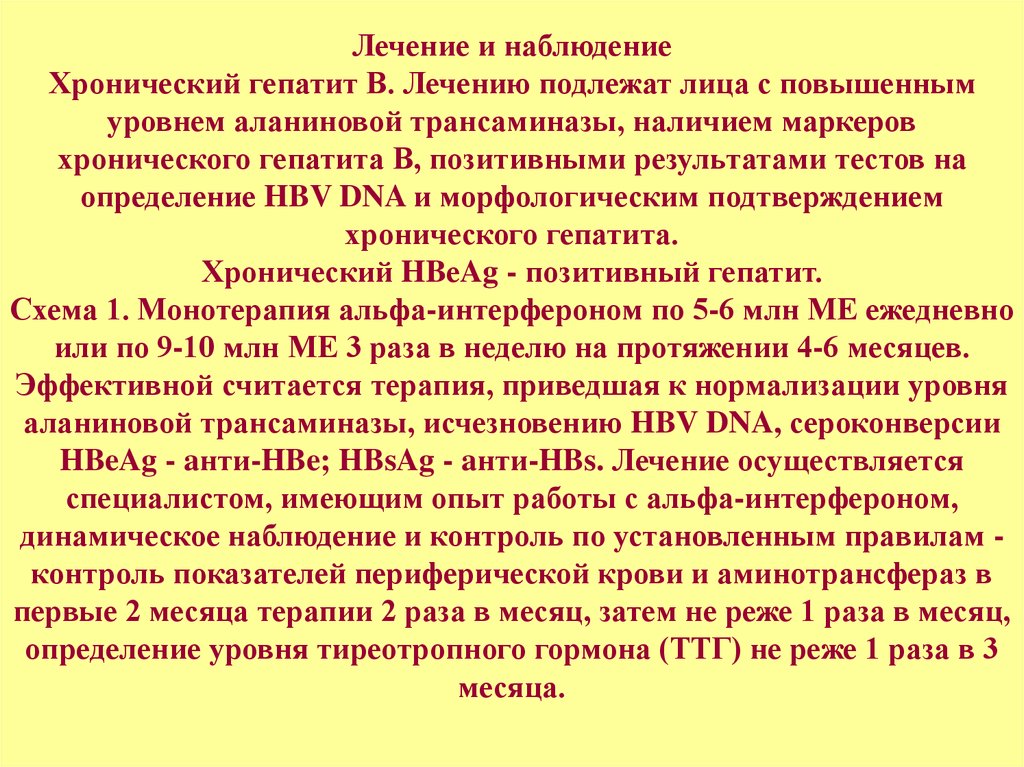
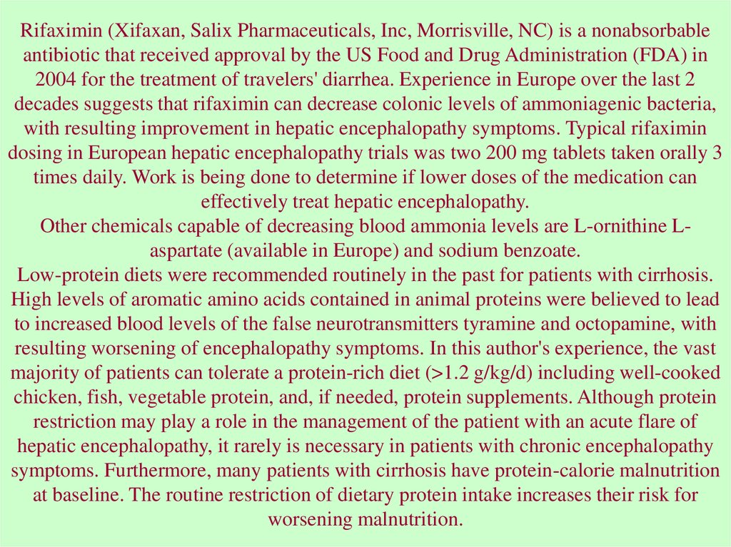
Лечение и наблюдениеХронический гепатит В. Лечению подлежат лица с повышенным
уровнем аланиновой трансаминазы, наличием маркеров
хронического гепатита В, позитивными результатами тестов на
определение HBV DNA и морфологическим подтверждением
хронического гепатита.
Хронический HBеAg - позитивный гепатит.
Схема 1. Монотерапия альфа-интерфероном по 5-6 млн МЕ ежедневно
или по 9-10 млн МЕ 3 раза в неделю на протяжении 4-6 месяцев.
Эффективной считается терапия, приведшая к нормализации уровня
аланиновой трансаминазы, исчезновению HBV DNA, сероконверсии
HBeAg - aнти-HBe; HBsAg - aнти-HBs. Лечение осуществляется
специалистом, имеющим опыт работы с альфа-интерфероном,
динамическое наблюдение и контроль по установленным правилам контроль показателей периферической крови и аминотрансфераз в
первые 2 месяца терапии 2 раза в месяц, затем не реже 1 раза в месяц,
определение уровня тиреотропного гормона (ТТГ) не реже 1 раза в 3
месяца.
24.
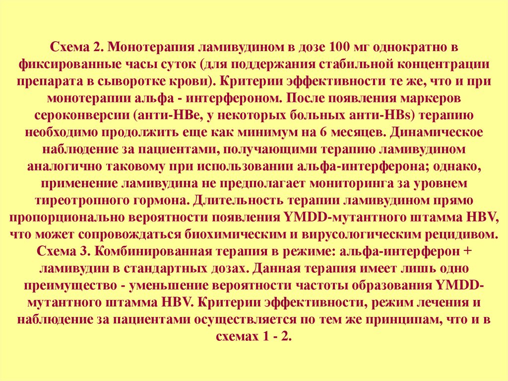
Схема 2. Монотерапия ламивудином в дозе 100 мг однократно вфиксированные часы суток (для поддержания стабильной концентрации
препарата в сыворотке крови). Критерии эффективности те же, что и при
монотерапии альфа - интерфероном. После появления маркеров
сероконверсии (aнти-HВe, у некоторых больных aнти-HBs) терапию
необходимо продолжить еще как минимум на 6 месяцев. Динамическое
наблюдение за пациентами, получающими терапию ламивудином
аналогично таковому при использовании альфа-интерферона; однако,
применение ламивудина не предполагает мониторинга за уровнем
тиреотропного гормона. Длительность терапии ламивудином прямо
пропорционально вероятности появления YMDD-мутантного штамма HBV,
что может сопровождаться биохимическим и вирусологическим рецидивом.
Схема 3. Комбинированная терапия в режиме: альфа-интерферон +
ламивудин в стандартных дозах. Данная терапия имеет лишь одно
преимущество - уменьшение вероятности частоты образования YMDDмутантного штамма HBV. Критерии эффективности, режим лечения и
наблюдение за пациентами осуществляется по тем же принципам, что и в
схемах 1 - 2.
25.
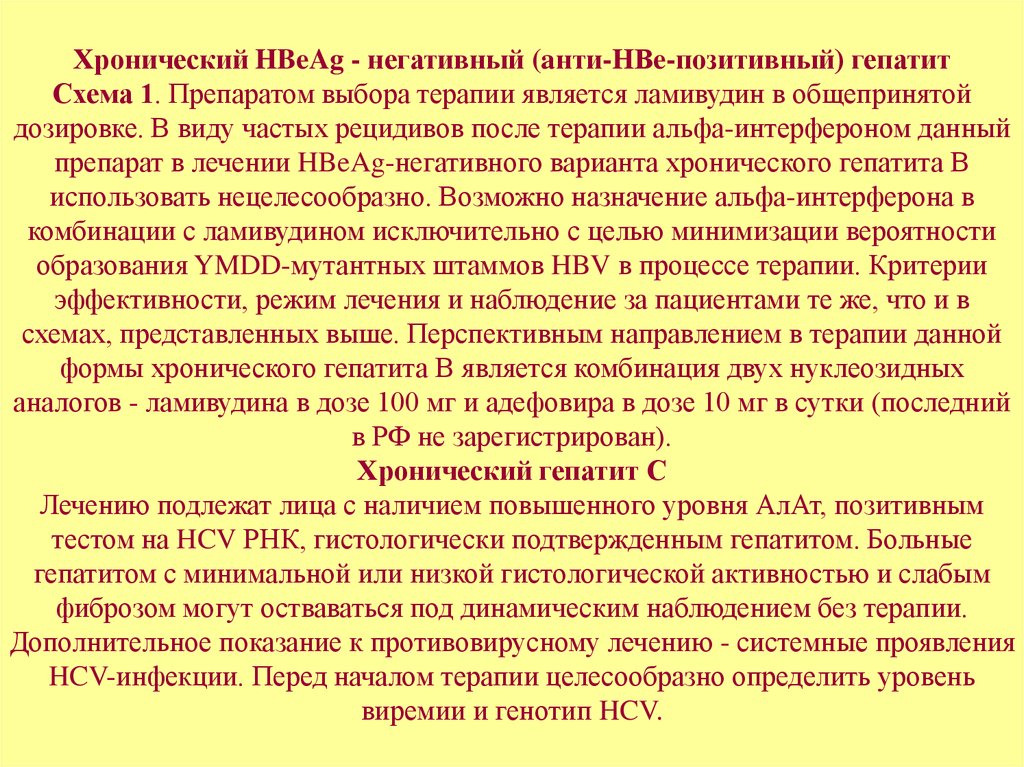
Хронический HBeAg - негативный (aнти-HВe-позитивный) гепатитСхема 1. Препаратом выбора терапии является ламивудин в общепринятой
дозировке. В виду частых рецидивов после терапии альфа-интерфероном данный
препарат в лечении HBeAg-негативного варианта хронического гепатита В
использовать нецелесообразно. Возможно назначение альфа-интерферона в
комбинации с ламивудином исключительно с целью минимизации вероятности
образования YMDD-мутантных штаммов HBV в процессе терапии. Критерии
эффективности, режим лечения и наблюдение за пациентами те же, что и в
схемах, представленных выше. Перспективным направлением в терапии данной
формы хронического гепатита В является комбинация двух нуклеозидных
аналогов - ламивудина в дозе 100 мг и адефовира в дозе 10 мг в сутки (последний
в РФ не зарегистрирован).
Хронический гепатит С
Лечению подлежат лица с наличием повышенного уровня АлАт, позитивным
тестом на HCV РНК, гистологически подтвержденным гепатитом. Больные
гепатитом с минимальной или низкой гистологической активностью и слабым
фиброзом могут остваваться под динамическим наблюдением без терапии.
Дополнительное показание к противовирусному лечению - системные проявления
HCV-инфекции. Перед началом терапии целесообразно определить уровень
виремии и генотип HCV.
26.
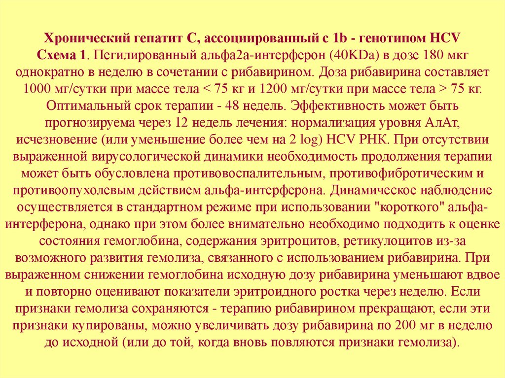
Хронический гепатит С, ассоциированный с 1b - генотипом HCVCхема 1. Пегилированный альфа2а-интерферон (40KDa) в дозе 180 мкг
однократно в неделю в сочетании с рибавирином. Доза рибавирина составляет
1000 мг/сутки при массе тела < 75 кг и 1200 мг/сутки при массе тела > 75 кг.
Оптимальный срок терапии - 48 недель. Эффективность может быть
прогнозируема через 12 недель лечения: нормализация уровня АлАт,
исчезновение (или уменьшение более чем на 2 log) HCV РНК. При отсутствии
выраженной вирусологической динамики необходимость продолжения терапии
может быть обусловлена противовоспалительным, противофибротическим и
противоопухолевым действием альфа-интерферона. Динамическое наблюдение
осуществляется в стандартном режиме при использовании "короткого" альфаинтерферона, однако при этом более внимательно необходимо подходить к оценке
состояния гемоглобина, содержания эритроцитов, ретикулоцитов из-за
возможного развития гемолиза, связанного с использованием рибавирина. При
выраженном снижении гемоглобина исходную дозу рибавирина уменьшают вдвое
и повторно оценивают показатели эритроидного ростка через неделю. Если
признаки гемолиза сохраняются - терапию рибавирином прекращают, если эти
признаки купированы, можно увеличивать дозу рибавирина по 200 мг в неделю
до исходной (или до той, когда вновь повляются признаки гемолиза).
27.
Обязательным условием применения рибавирина являетсястрогое соблюдение мер контрацепции на весь период
терапии и еще в течение 6 месяцев после ее окончания
(эмбрио- и спермотоксигенное действие). Использование
подобного лечения для пациентов с хроническим
гепатитом С должно осуществляться под наблюдением
специалистов, имевших опыт работы с препаратами.
28.
Схема 2. Комбинированное использование пегилированного альфа 2bинтерферона (12KDa) в дозе 1,5 мкг на 1 килограмм веса пациента один раз внеделю в сочетании с рибавирином; доза последнего аналогична
представленной в предыдущей схеме. Критерии эффективности, особенности
динамического наблюдения, сроки лечения - см. предыдущую схему.
Хронический гепатит С, ассоциированный с не 1b - генотипом HCV
Сочетание пегилированных альфа-интерферонов в дозировках,
представленных ранее, с рибавирином (800 мг). Общая продолжительность
лечения может быть ограничена сроком 24 недели. Критерии эффективности,
особенности динамического наблюдения аналогичны тому, что указано выше.
При отсутствии возможности применения пегилированных интерферонов
возможна замена их альфа-интерферонами "короткого" действия (дозировка 3 млн. МЕ 3 раза в неделю). При этом вероятность достижения стойкого
вирусологического ответа снижается приблизительно в 2 раза. При наличии
противопоказаний к применению рибавирина (тяжелая анемия, патология
сердечно-сосудистой системы и др.) альфа-интерфероны могут назначаться с
целью профилактики прогрессирования фиброза и развития
гепатоцеллюлярной карциномы.
29.
Хронический гепатит DЛечению подлежат пациенты с повышенным уровнем АлАт, наличием маркеров
HBV/HDV, позитивными результатами на определение HDV РНК,
гистологически подтвержденным гепатитом.
Схема терапии. Использование альфа-интерферона в дозе 9 млн. MЕ через день
на протяжении как минимум 48 недель. В настоящее время прогностических
критериев эффективности не определено. Иногда позитивные результаты лечения
(нормализация уровня АлАт, реже исчезновение или значительное снижение
титра HDV РНК) может происходить только к концу лечения. После отмены
терапии часто возникает рецидив. Для определения дальнейшей тактики лечения
в этом случае целесообразно выполнение повторной пункционной биопсии с
целью установления факта позитивных результатов предшествующей терапии
(уменьшение воспаления и фиброза). Если позитивное влияние
предшествующего лечения установлено, ставится вопрос о неопределенно
длительном использовании малых доз альфа-интерферона (1 млн. MЕ ежедневно
на протяжении ряда лет). Особенности динамического наблюдения за пациентами
аналогичны таковым при использовании альфа - интерферона. Дополнительное
назначение ламивудина оправдано лишь при наличии маркеров репликации HBV,
однако назначение последнего по данным имеющихся исследований никак не
влияет на кинетику HDV и гистологические проявления гепатита.
30.
При хроническом ВГ в стадии субкомпенсированного цирроза (класс Впо Чайлд-Пью) помимо возможных вышеперечисленных лечебных
мероприятий назначаются калийсберегающие мочегонные средства
(верошпирон 100 мг/сут, триампур и др.), лактулоза (дюфалак внутрь по
30-50 мл 3 р/сут), орнитин-аспартат внутривенно 20 г/сут или внутрь по 1
пакетику гранулята 2-3 р/сут. В стадии декомпенсированного цирроза
(класс С по Чайлд-Пью) к терапии добавляют парентеральное введение
концентрированных (10-20%) растворов альбумина, плазмы,
калийсодержащих растворов на фоне ограничения поступления в
организм хлорида натрия с пищей и инфузионными растворами,
антибактериальные препараты. При отсутствии эффекта назначается
фуросемид (лазикс) по 40-80 мг/сут, при этом кратность введения зависит
от показателей диуреза. Обязательно строгое соблюдение суточного
баланса жидкости. При наличии стойкого асцита и нарастании
хронической печеночной недостаточности, не поддающихся адекватному
медикаментозному лечению, угрозе развития желудочно-кишечного
кровотечения показана консультация хирурга-гепатолога/трансплантолога.
31.
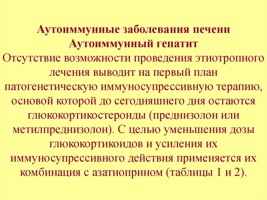
Аутоиммунные заболевания печениАутоиммунный гепатит
Отсутствие возможности проведения этиотропного
лечения выводит на первый план
патогенетическую иммуносупрессивную терапию,
основой которой до сегодняшнего дня остаются
глюкокортикостероиды (преднизолон или
метилпреднизолон). С целью уменьшения дозы
глюкокортикоидов и усиления их
иммуносупрессивного действия применяется их
комбинация с азатиоприном (таблицы 1 и 2).
32.
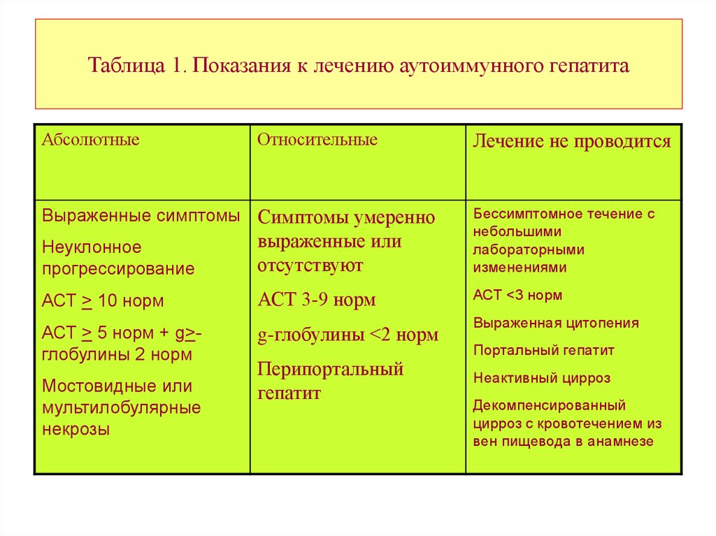
Таблица 1. Показания к лечению аутоиммунного гепатитаАбсолютные
Относительные
Лечение не проводится
Неуклонное
прогрессирование
выраженные или
отсутствуют
Бессимптомное течение с
небольшими
лабораторными
изменениями
АСТ > 10 норм
АСТ 3-9 норм
АСТ <3 норм
АСТ > 5 норм + g>глобулины 2 норм
g-глобулины <2 норм
Выраженные симптомы Симптомы умеренно
Мостовидные или
мультилобулярные
некрозы
Перипортальный
гепатит
Выраженная цитопения
Портальный гепатит
Неактивный цирроз
Декомпенсированный
цирроз с кровотечением из
вен пищевода в анамнезе
33.
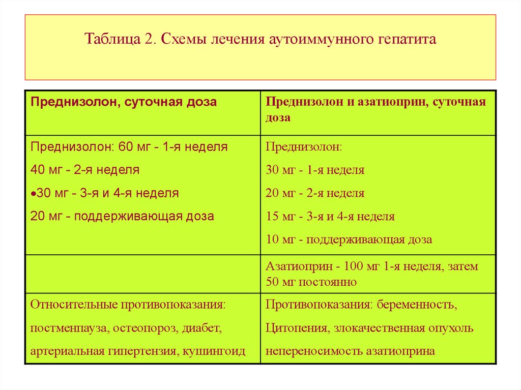
Таблица 2. Схемы лечения аутоиммунного гепатитаПреднизолон, суточная доза
Преднизолон и азатиоприн, суточная
доза
Преднизолон: 60 мг - 1-я неделя
Преднизолон:
40 мг - 2-я неделя
30 мг - 1-я неделя
30 мг - 3-я и 4-я неделя
20 мг - 2-я неделя
20 мг - поддерживающая доза
15 мг - 3-я и 4-я неделя
10 мг - поддерживающая доза
Азатиоприн - 100 мг 1-я неделя, затем
50 мг постоянно
Относительные противопоказания:
Противопоказания: беременность,
постменпауза, остеопороз, диабет,
Цитопения, злокачественная опухоль
артериальная гипертензия, кушингоид
непереносимость азатиоприна
34.
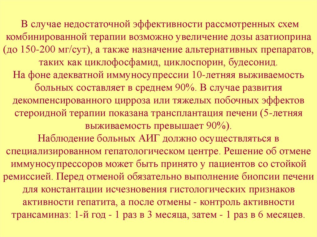
В случае недостаточной эффективности рассмотренных схемкомбинированной терапии возможно увеличение дозы азатиоприна
(до 150-200 мг/сут), а также назначение альтернативных препаратов,
таких как циклофосфамид, циклоспорин, будесонид.
На фоне адекватной иммуносупрессии 10-летняя выживаемость
больных составляет в среднем 90%. В случае развития
декомпенсированного цирроза или тяжелых побочных эффектов
стероидной терапии показана трансплантация печени (5-летняя
выживаемость превышает 90%).
Наблюдение больных АИГ должно осуществляться в
специализированном гепатологическом центре. Решение об отмене
иммуносупрессоров может быть принято у пациентов со стойкой
ремиссией. Перед отменой обязательно выполнение биопсии печени
для константации исчезновения гистологических признаков
активности гепатита, а после отмены - контроль активности
трансаминаз: 1-й год - 1 раз в 3 месяца, затем - 1 раз в 6 месяцев.
35.
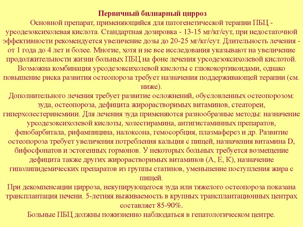
Первичный билиарный циррозОсновной препарат, применяющийся для патогенетической терапии ПБЦ урсодезоксихолевая кислота. Стандартная дозировка - 13-15 мг/кг/сут, при недостаточной
эффективности рекомендуется увеличение дозы до 20-25 мг/кг/сут. Длительность лечения от 1 года до 4 лет и более. Многие, хотя и не все исследования указывают на увеличение
продолжительности жизни больных ПБЦ на фоне лечения урсодезоксихолевой кислотой.
Возможна комбинация урсодезоксихолевой кислоты с глюкокортикоидами, однако
повышение риска развития остеопороза требует назначения поддерживающей терапии (см.
ниже).
Дополнительного лечения требует развитие осложнений, обусловленных остеопорозом:
зуда, остеопороза, дефицита жирорастворимых витаминов, стеатореи,
гиперхолестеринемиии. Для лечения зуда применяются разнообразные методы: назначение
урсодезоксихолевой кислоты, холестирамина, антигистаминных препаратов,
фенобарбитала, рифампицина, налоксона, гемосорбция, плазмаферез и др. Развитие
остеопороза требует увеличения потребления кальция с пищей, назначения витамина D,
бифосфонатов и эстогенных гормонов. У некоторых больных требуется возмещение
дефицита также других жирорастворимых витаминов (А, Е, К), назначение
гиполипидемических препаратов из группы статинов, уменьшение поступления жира с
пищей.
При декомпенсации цирроза, некупирующегося зуда или тяжелого остеопороза показана
трансплантация печени. 5-летняя выживаемость в крупных трансплантационных центрах
составляет 85-90%.
Больные ПБЦ должны пожизненно наблюдаться в гепатологическом центре.
36.
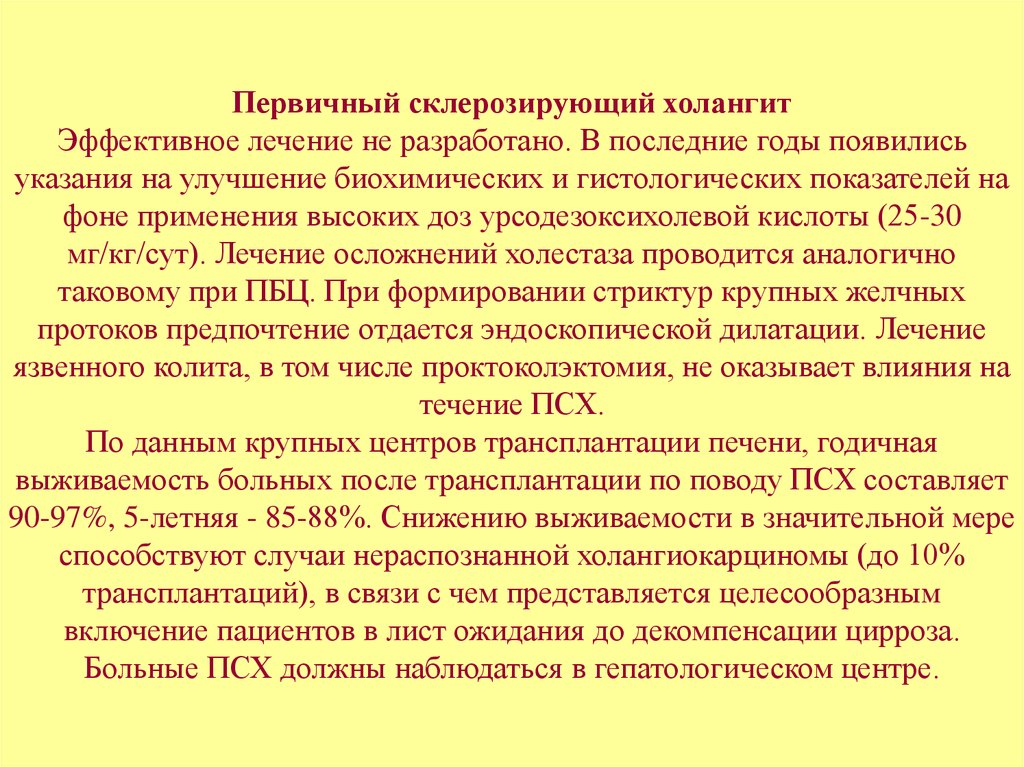
Первичный склерозирующий холангитЭффективное лечение не разработано. В последние годы появились
указания на улучшение биохимических и гистологических показателей на
фоне применения высоких доз урсодезоксихолевой кислоты (25-30
мг/кг/сут). Лечение осложнений холестаза проводится аналогично
таковому при ПБЦ. При формировании стриктур крупных желчных
протоков предпочтение отдается эндоскопической дилатации. Лечение
язвенного колита, в том числе проктоколэктомия, не оказывает влияния на
течение ПСХ.
По данным крупных центров трансплантации печени, годичная
выживаемость больных после трансплантации по поводу ПСХ составляет
90-97%, 5-летняя - 85-88%. Снижению выживаемости в значительной мере
способствуют случаи нераспознанной холангиокарциномы (до 10%
трансплантаций), в связи с чем представляется целесообразным
включение пациентов в лист ожидания до декомпенсации цирроза.
Больные ПСХ должны наблюдаться в гепатологическом центре.
37.
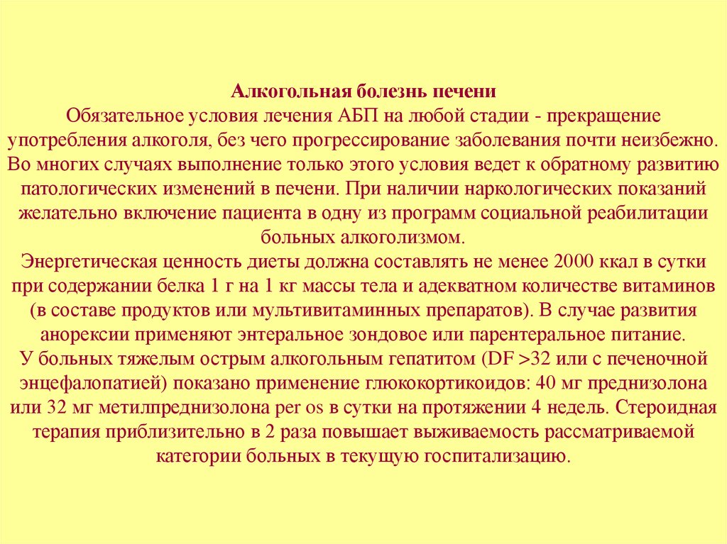
Алкогольная болезнь печениОбязательное условия лечения АБП на любой стадии - прекращение
употребления алкоголя, без чего прогрессирование заболевания почти неизбежно.
Во многих случаях выполнение только этого условия ведет к обратному развитию
патологических изменений в печени. При наличии наркологических показаний
желательно включение пациента в одну из программ социальной реабилитации
больных алкоголизмом.
Энергетическая ценность диеты должна составлять не менее 2000 ккал в сутки
при содержании белка 1 г на 1 кг массы тела и адекватном количестве витаминов
(в составе продуктов или мультивитаминных препаратов). В случае развития
анорексии применяют энтеральное зондовое или парентеральное питание.
У больных тяжелым острым алкогольным гепатитом (DF >32 или с печеночной
энцефалопатией) показано применение глюкокортикоидов: 40 мг преднизолона
или 32 мг метилпреднизолона per os в сутки на протяжении 4 недель. Стероидная
терапия приблизительно в 2 раза повышает выживаемость рассматриваемой
категории больных в текущую госпитализацию.
38.
Антицитокиновые препараты (инфликсимаб) для лечения больныхтяжелым алкогольным гепатитом, резистентным к стандартной
терапии, в настоящее время проходят клинические испытания.
Пациентам на стадии хронического гепатита, компенсированного и
субкомпенсированного цирроза целесообразно назначение
препаратов метаболического действия, таких как эссенциальные
фосфолипиды, адеметионин, силимарин, а также
урсодезоксихолевой кислоты.
На стадии декомпенсированного цирроза (класс С по Чайлд-Пью)
показано включение больного в лист ожидания трансплантации
печени. В большинстве трансплантационных центров в качестве
обязательного условия предусмотрена 6-месячная абстиненция.
39.
Генетически детерминированные заболевания печениБолезнь Вильсона-Коновалова
Установление диагноза болезни Вильсона - Коновалова является
показанием к немедленному началу лечения на любой стадии заболевания.
Схема терапии. Препарат выбора - D-пеницилламин. Начальная доза
обычно составляет 500 мг в сутки с постепенным повышением 1500-2000
мг в сутки и более при условии хорошей переносимости... Доза делится на
три приема, препарат принимают до еды. Уровень свободной меди на фоне
лечения не должен превышать 20мкг/дл, суточная экскреция меди с мочой
не должна превышать 0,5 мг/сутки. В дальнейшем, через 3 - 5 месяцев от
начала лечения поддерживающая доза препарата обычно составляет 20 мг
на килограмм веса пациента в сутки. Поскольку D-пеницилламин является
специфическим антагонистом пиридоксина, больному ежедневно
назначают 25 мг витамина В6. Побочные эффекты терапии:
геморрагический дерматит, панцитопения, миопатия. Терапия
пожизненная.
40.
Идиопатический гемохроматозЛечение гомозиготных больных проводится до развития осложнений, связанных с
перенасыщением железом различных органов.
Схема лечения. Еженедельные кровопускания в объеме не менее 500 мл до
нормализации ферритина в сыворотке крови. После чего кровопускания
проводятся в индивидуальном режиме, обычно это 4 - 6 кровопусканий в год в
объеме 500 мл каждое. Для контроля за эффективностью терапии обычно
используют не уровень сывороточного железа, а содержание ферритина
(приемлемый уровень 30 - 45 мкг/мл).
Использование хелатообразующих препаратов (деферроксамин) в настоящее
время признано нецелесообразным, так как даже при однократном кровопускании
выводится железа приблизительно в 10 раз больше, чем при использовании
данного препарата в сутки в стандартной дозировке. Сочетание дефероксамина и
кровопусканий также не рекомендуется. Применение этого лекарства показано
лишь тогда, когда вторичная тяжелая анемия или гипопротеинемия (на далеко
зашедших стадиях цирроза печени) делают невозможным проведения
кровопусканий и ретрансфузии плазмы.
Дефицит альфа1-антитрипсина
Специфической терапии печеночных проявлений данной формы заболевания не
существует. Лечение обычно симптоматическое, при необходимости решается
вопрос о проведении трансплантации печени.
41.
Портальная гипертензияПортальная гипертензия (ПГ)- симптомокомплекс, связанный с
затруднением тока крови в портальной системе.
Проявления ПГ
варикозное расширение вен (ВРВ) пищевода и желудка,
ректальных сплетений
асцит
спленомегалия
Классификация ПГ
1.Внепеченочная (врожденные дефекты развития воротной
вены, компрессия воротной вены в результате патологического
процесса в гепатопанкреатодуоденальной области,
сегментарный тромбоз селезеночной вены, тромбоз системы
воротной вены при заболеваниях крови)
42.
43.
44.
45.
2.Внутрипеченочная (цирроз, первичные и вторичные очаговыепоражения печени, паразитарные заболевания печени)
3.Надпеченочная ( Болезнь Киари, синдром Бадда-Киари, врожденные
пороки развития нижней полой вены).
Смешанная
Варикозное расширение вен пищевода и желудка
Классификация степени расширения вен пищевода (по А.К.
Ерамишанцеву):
1ст- диаметр вен < 3мм
2ст- от 3 до 5мм
3ст- > 5мм
Dagradi выделяет и 4ст- диаметром более1,0 см.
По Sherlock:
F1- при надавливании эндоскопом размер вен уменьшается,
F2- при надавливании эндоскопом размер вен не уменьшается ,
F3- при надавливании эндоскопом вены сливаются по окружности
пищевода .
Наиболее грозным осложнением ВРВ является кровотечение,
сопровождающееся высокой летальностью.
46.
Цели лечения:Снижение давления в системе воротной вены.
Профилактика кровотечения из ВРВ.
Устранение причины портальной гипертензии.
Методы лечения синдрома портальной гипертензии
Консервативные
Эндоскопические
Эндоваскулярные
Оперативные (паллиативные, радикальные)
Консервативная терапия больных с кровотечением из ВРВ
Баллонная тампонада зондами Сенгстейкена - Блейкмора, Линтона - Нахласса.
Методика постановки зонда Сенгстейкена - Блейкмора.
Премедикация - 1мл 2% раствора промедола.
Зонд смазывают вазелином, после чего проводят через носовой ход в желудок.
Желудочный баллон с интервалом 3-5 мин постепенно раздувают до 100-120 см 3. Затем
зонд подтягивают до упора, фиксируют и опорожняют содержимое желудка . После этого
надувают пищеводный баллон. Воздух также вводят порциями по30-40см3 затем по 10-15
см3 с интервалом 3-5мин. Общий объем вводимого воздуха в пищеводный баллон не
должен превышать 100-150см3 . Каждый час следует проверять натяжение зонда и
давление в пищеводном баллоне. Через 4-6ч пищеводный баллон распускают и при
отсутствии кровотечения манжетку оставляют спущенной. Желудочную манжетку
распускают спустя 1,5-2 часа .
47.
У больных с циррозом печени класса А и В по Чайлд-Пью зонддолжен находиться в желудке до 12 часов для контроля. При
рецидиве кровотечения зонд обтуратор должен быть введен вновь, а
больному предложена операция. У больных с циррозом класса С по
Чайлд-Пью тампонада зондом-обтуратором в сочетании с
медикаментозной терапией является единственной надеждой на
гемостаз. Операция в 90-95% непереносима.
При безуспешности консервативных мероприятий, рецидиве
кровотечения после распускания манжеты или удаления зондаобтуратора больных циррозом (классы А и В) и больных с
внепеченочной портальной гипертензией следует оперировать.
48.
Медикаментозная терапия (проводится одновременно с постановкойзонда Сенгстейкена - Блейкмора)
А.Уменьшение кровотока в системе воротной вены
Перлинганит 40мг на 400мл раствора Рингера в/в капельно по 10-15
капель в мин - 48-72часа.
1% спиртовой раствор нитроглицерина - 10мг на 400мл раствора
Рингера в/в капельно по 10-15 капель в минуту 48-72часа (суточная
доза 0.43мг/кг) .
Соматостатин 50мкг/ч -48ч непрерывно
Триглицил-вазопресссин-болюсно 2мг, затем с интервалом 6 часов
вводят 1мг препарата.
Вазопрессин вводится в течение 20 мин вводят в дозе 20 МЕ на 100мл
изотонического раствора NaCl, после чего препарат вводят со
скоростью 20 МЕ/ч. При прекращении кровотечения дозу снижают до
10 МЕ/ч, а затем до 5 МЕ/ч. Продолжительность лечения 48 часов (в
комбинации с нитроглицерином).
49.
Б. Для нормализации свертывания:свежезамороженная плазма
1% раствор викасола в/в по 6 мл/сут.
10% раствор СаСl 10мл на протяжении 5 дней
дицинон в/в 4мл ,а затем по 2мл каждые 4-6 часа, или 4мл 12,5% раствора
этамзилата, а затем по 2 мл каждые 6 часов в течение 3-5 дней .
5% аминокапроновая кислота в/в по 100 мл через каждые 6 ч.
ингибиторы протеаз - контрикал 20 тыс ЕД разовая доза , суточная доза 60 тыс ЕД
антигистаминные препараты для нейтрализации действия гистамина на
проницаемость капилляров.
В. Замещающая терапия:
Эритроцитарная масса (хранившаяся не более 48 часов) в зависимости от степени
кровопотери
Г. Профилактика и лечение печеночной энцефалопатии:
лактулоза, орнитин-аспартат в стандартных дозах (см. "лечение циррозов")
селективная деконтаминация кишечника (все антибиотики принимаются per os):
неомицин 1 г 4 раза, паромомицин 1г 4 раза, метронидазол 200мг 4 раза,
ванкомицин 0,5 г 4 раза в сутки.
50.
Д. Профилактика повторного кровотеченияпостоянный прием бета-блокаторов (анаприлин 10-20 мг 2 раза в сутки)
Эндоскопические методы лечения
Эндоскопическая склеротерапия
Используемые склерозанты: 1% раствор тетрадецилсульфата натрия, 5% раствор
этаноламина олеата, 5% раствор морруата натрия, 1% раствор этосисклерола, 13% раствор тромбовара, тканевой клей цианоакрилат для узлов фундального
отдела желудка.
На каждый варикозный узел используют 1-4мл препарата. При отсутствии
склерозанта возможно использование 20% раствора глюкозы (10-20 мл вводится
паравазально).
Склерозирование ВРВ проводится в 3 этапа: первый сеанс склеротерапии
предполагает проведение 2-й серии инъекций через 3 дня, 3-й серии - через 6
дней . Склеротерапию проводят еженедельно до полного исчезновения ВРВ.
Контроль осуществляется через 3 и 6 месяцев.
Эндоскопическое лигирование варикозно расширенных узлов пищевода и кардии.
Для процедуры используют специальный набор для эндоскопического
накидывания, затягивания и снятия петель на основание ВРВ.
51.
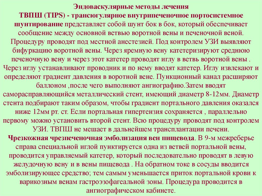
Эндоваскулярные методы леченияТВПШ (TIPS) - трансюгулярное внутрипеченочное портосистемное
шунтирование представляет собой шунт бок в бок, который обеспечивает
сообщение между основной ветвью воротной вены и печеночной веной.
Процедуру проводят под местной анестезией. Под контролем УЗИ выявляют
бифуркацию воротной вены. Через яремную вену катетеризируют среднюю
печеночную вену и через этот катетер проводят иглу в ветвь воротной вены .
Через иглу устанавливают проводник и по нему вводят катетер. Иглу извлекают и
определяют градиент давления в воротной вене. Пункционный канал расширяют
баллоном ,после чего выполняют ангиографию.Затем вводят
саморасправляющийся металлический стент, имеющий диаметр 8-12мм. Диаметр
стента подбирают таким образом, чтобы градиент портального давления оказался
ниже 12мм рт. ст. Если портальная гипертензия сохраняется , параллельно
первому можно установить второй стент. Всю процедуру проводят под контролем
УЗИ. ТВПШ не мешает в дальнейшем трансплантации печени.
Чрезкожная чрезпеченочная эмболизация вен пищевода. В 9-м межреберье
справа специальной иглой пунктируется одна из ветвей портальной вены,
проводится управляемый катетер, который последовательно проводят в левую
желудочную вену и в вены пищевода . На обратном токе в сосуды вводится
эмболизирующее средство; тем самым уменьшается приток портальной крови к
варикозным венам гастроэзофагеальной зоны. Процедура проводится в
ангиографическом кабинете.
52.
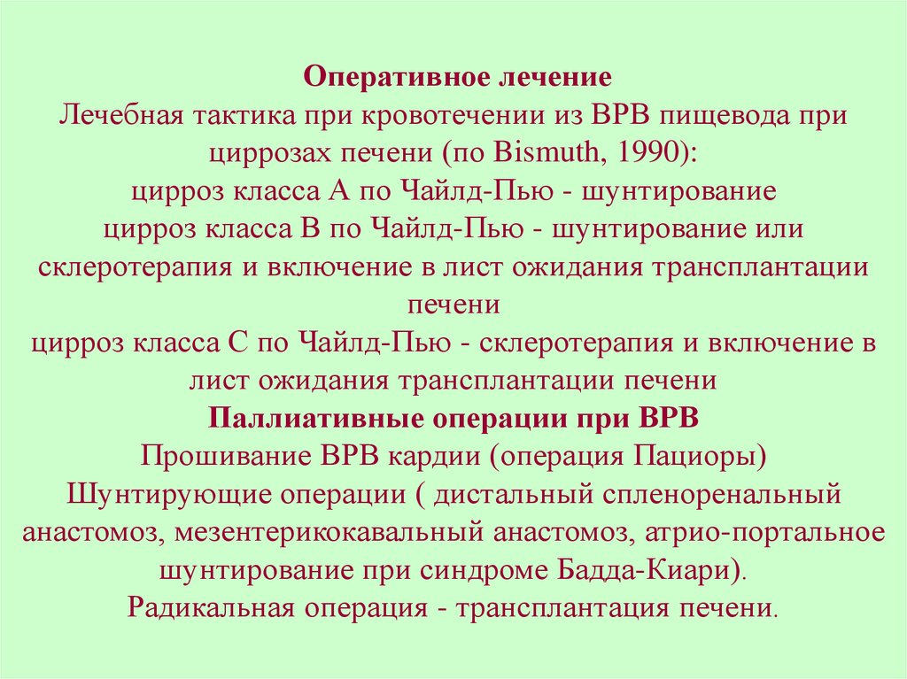
Оперативное лечениеЛечебная тактика при кровотечении из ВРВ пищевода при
циррозах печени (по Bismuth, 1990):
цирроз класса А по Чайлд-Пью - шунтирование
цирроз класса В по Чайлд-Пью - шунтирование или
склеротерапия и включение в лист ожидания трансплантации
печени
цирроз класса С по Чайлд-Пью - склеротерапия и включение в
лист ожидания трансплантации печени
Паллиативные операции при ВРВ
Прошивание ВРВ кардии (операция Пациоры)
Шунтирующие операции ( дистальный спленоренальный
анастомоз, мезентерикокавальный анастомоз, атрио-портальное
шунтирование при синдроме Бадда-Киари).
Радикальная операция - трансплантация печени.
53.
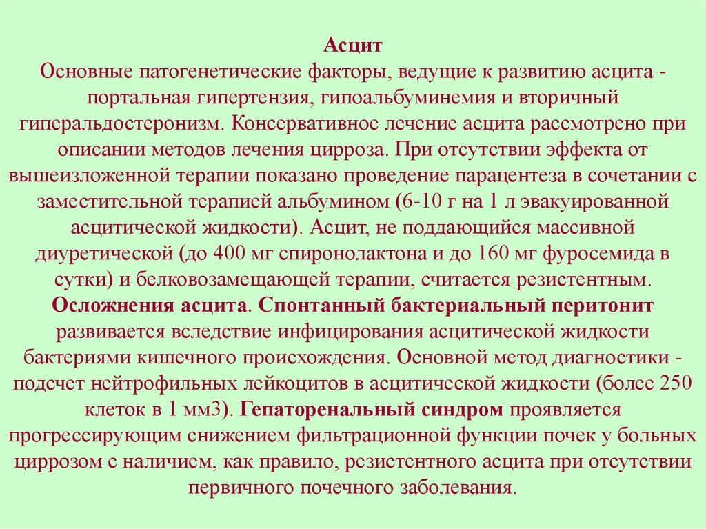
АсцитОсновные патогенетические факторы, ведущие к развитию асцита портальная гипертензия, гипоальбуминемия и вторичный
гиперальдостеронизм. Консервативное лечение асцита рассмотрено при
описании методов лечения цирроза. При отсутствии эффекта от
вышеизложенной терапии показано проведение парацентеза в сочетании с
заместительной терапией альбумином (6-10 г на 1 л эвакуированной
асцитической жидкости). Асцит, не поддающийся массивной
диуретической (до 400 мг спиронолактона и до 160 мг фуросемида в
сутки) и белковозамещающей терапии, считается резистентным.
Осложнения асцита. Спонтанный бактериальный перитонит
развивается вследствие инфицирования асцитической жидкости
бактериями кишечного происхождения. Основной метод диагностики подсчет нейтрофильных лейкоцитов в асцитической жидкости (более 250
клеток в 1 мм3). Гепаторенальный синдром проявляется
прогрессирующим снижением фильтрационной функции почек у больных
циррозом с наличием, как правило, резистентного асцита при отсутствии
первичного почечного заболевания.
54.
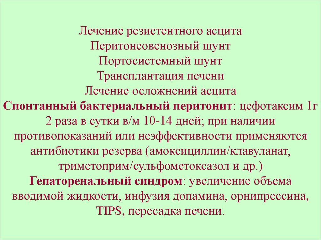
Лечение резистентного асцитаПеритонеовенозный шунт
Портосистемный шунт
Трансплантация печени
Лечение осложнений асцита
Спонтанный бактериальный перитонит: цефотаксим 1г
2 раза в сутки в/м 10-14 дней; при наличии
противопоказаний или неэффективности применяются
антибиотики резерва (амоксициллин/клавуланат,
триметоприм/сульфометоксазол и др.)
Гепаторенальный синдром: увеличение объема
вводимой жидкости, инфузия допамина, орнипрессина,
TIPS, пересадка печени.
55.
Трансплантация печениТрансплантация печени в настоящее время является единственным радикальным методом
лечения больных с терминальными заболеваниями печени различного генеза. Только в
США ежегодно выполняется до 5000 тыс. трансплантаций печени. Необходимость
выполнения трансплантации печени возникает ежегодно у 10-20 человек на 1 миллион
населения. С учетом этой цифры несложно высчитать потребности различных стран в этой
операции.
Показания
Необратимое заболевание печени с прогнозом жизни менее 12 мес.
Хроническое заболевание печени, значительно снижающее качество жизни и
трудоспособность
Прогрессирующее заболевание печени с ожидаемой продолжительностью жизни меньшей,
чем в случае трансплантации печени (в течение 1 года после трансплантации печени живет
85% реципиентов, в течение 5 лет- 70%, в течение 20 лет- 40%).
Противопоказания
Активная ВИЧ-инфекция
Внепеченочное распространение злокачественных опухолей
Внутрипеченочная холангиокарцинома
Сепсис (кроме билиарного)
Тяжелое кардиореспираторное заболевание
Активный алкоголизм
56.
Фульминантная печеночная недостаточность(ФПН)ФПН развивается в результате острого некроза большей части
гепатоцитов, или резкого ухудшения функции печени у пациентов, не
имеющих предшедствующего заболевания печени. Одним из облигатных
проявлений ФПН является печеночная энцефалопатия. О ФПН можно
говорить только в том случае, если энцефалопатия развивается в пределах
8 недель от появления первых симптомов острой печеночно-клеточной
недостаточности. Если острая печеночная энцефалопатия развивается в
сроки от 8 до 24 недель от появления первых симптомов поражения
печени, то следует говорить о подострой (субфульминантной) печеночной
недостаточности. Летальность при ФПН достигает 50-90%.
Причиной ФПН в 30-80% являются вирусные гепатиты, в 30-50% химические реагенты и лекарства, в 5% - яды, в 5% - ишемия и гипоксия
печени, в 5-10%- метаболические аномалии.
Трансплантация печени в связи с ФПН у взрослых выполняется в 5,4%, у
детей- в 10,5% случаев
57.
Нехолестатические циррозы печениНехолестатические циррозы печени составляют большую группу заболеваний,
которые приводят к необходимости трансплантации печени. Так, по поводу
алкогольного цироза печени выполняется 21,6% всех трансплантаций у взрослого
населения, цирроза печени, обусловленного HCV - 19,5%, HВV - 6,1%,
криптогенного цирроза - 12%, аутоиммунного цирроза - 5%.
Холестатические циррозы печени
Другую большую группу составляют холестатические циррозы печени
(первичный билиарный цирроз, вторичный билиарный цирроз, нередко
возникающий на фоне болезни Кароли или кисты холедоха, первичный
склерозирующий холангит). Так, по поводу первичного билиарного цирроза
выполняется 10,9% трансплантаций у взрослых, первичного склерозирующего
холангита - 9,9%. 55% детских трансплантаций печени выполняется по поводу
билиарной атрезии.
Метаболические заболевания
15,4% детских трансплантаций и 3,3% взрослых выполняется по поводу
врожденных дефектов метаболизма печени. К таким заболеваниям относятся
недостаточность альфа1-антирипсина, болезнь Вильсона-Коновалова,
гемохроматоз, тирозинемия, гипероксалурия, гликогенозы I и II типа,
гиперлипидемия II типа.
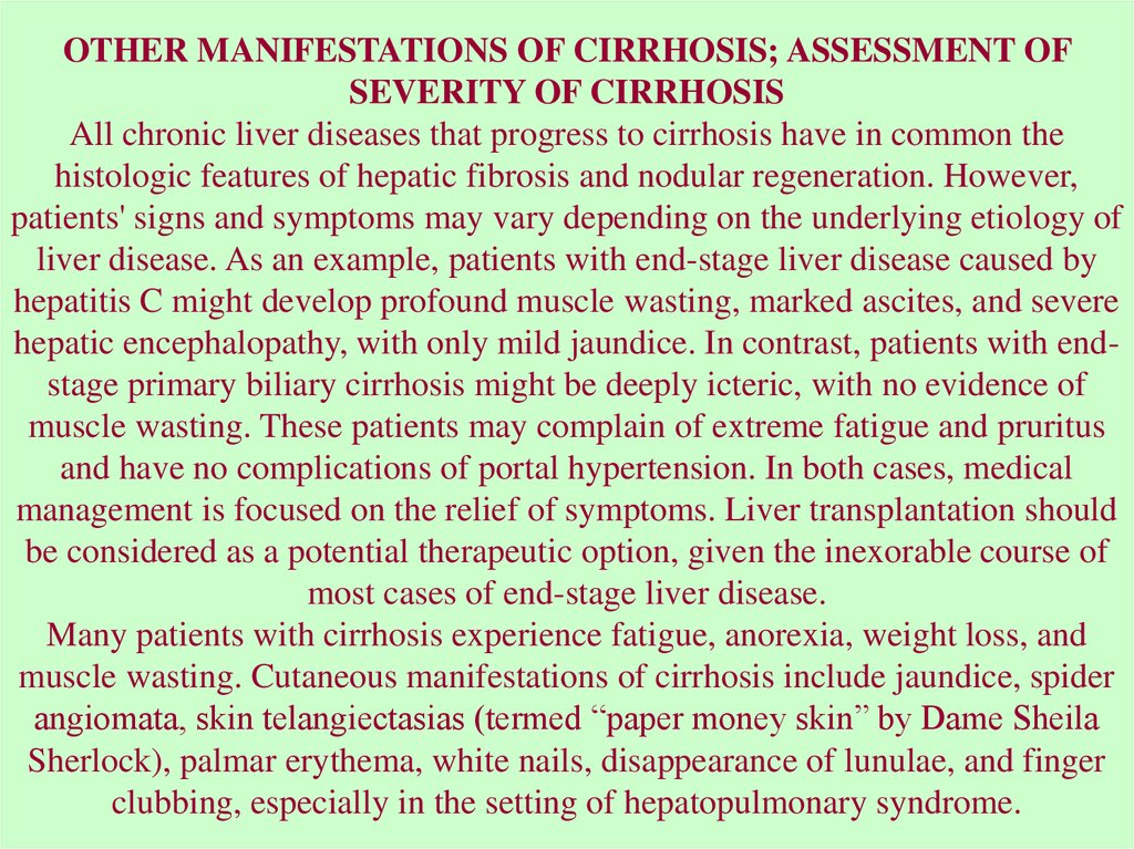
58.
Cirrhosis represents the final common histologic pathway for a widevariety of chronic liver diseases. The term cirrhosis was first
introduced by Laennec in 1826. It is derived from the Greek term
scirrhus and is used to describe the orange or tawny surface of the
liver seen at autopsy.
Many forms of liver injury are marked by fibrosis. Fibrosis is defined
as an excess deposition of the components of extracellular matrix (ie,
collagens, glycoproteins, proteoglycans) within the liver. This
response to liver injury potentially is reversible. In contrast, in most
patients, cirrhosis is not a reversible process.
Cirrhosis is defined histologically as a diffuse hepatic process
characterized by fibrosis and the conversion of normal liver
architecture into structurally abnormal nodules. The progression of
liver injury to cirrhosis may occur over weeks to years. Indeed,
patients with hepatitis C may have chronic hepatitis for as long as 40
years before progressing to cirrhosis.
59.
Often a poor correlation exists between histologicfindings and the clinical picture. Some patients
with cirrhosis are completely asymptomatic and
have a reasonably normal life expectancy. Other
individuals have a multitude of the most severe
symptoms of end-stage liver disease and have a
limited chance for survival. Common signs and
symptoms may stem from decreased hepatic
synthetic function (eg, coagulopathy), decreased
detoxification capabilities of the liver (eg, hepatic
encephalopathy), or portal hypertension (eg,
variceal bleeding).
60.
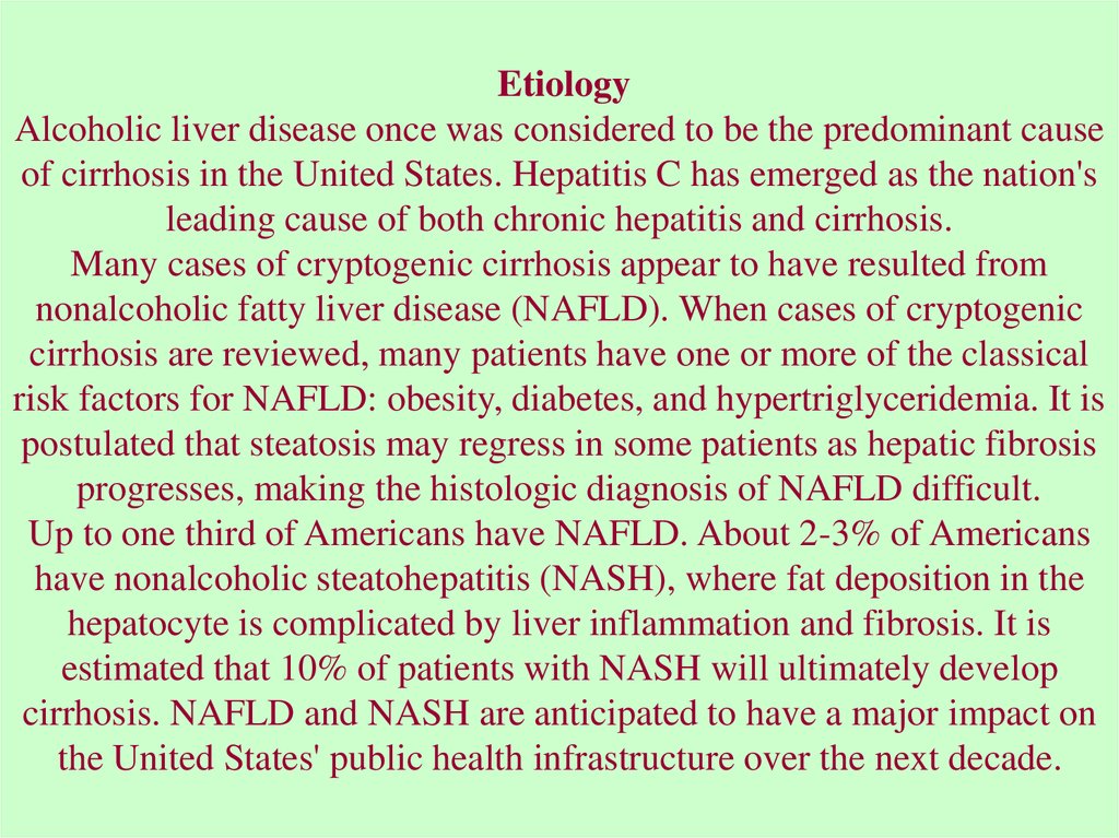
EtiologyAlcoholic liver disease once was considered to be the predominant cause
of cirrhosis in the United States. Hepatitis C has emerged as the nation's
leading cause of both chronic hepatitis and cirrhosis.
Many cases of cryptogenic cirrhosis appear to have resulted from
nonalcoholic fatty liver disease (NAFLD). When cases of cryptogenic
cirrhosis are reviewed, many patients have one or more of the classical
risk factors for NAFLD: obesity, diabetes, and hypertriglyceridemia. It is
postulated that steatosis may regress in some patients as hepatic fibrosis
progresses, making the histologic diagnosis of NAFLD difficult.
Up to one third of Americans have NAFLD. About 2-3% of Americans
have nonalcoholic steatohepatitis (NASH), where fat deposition in the
hepatocyte is complicated by liver inflammation and fibrosis. It is
estimated that 10% of patients with NASH will ultimately develop
cirrhosis. NAFLD and NASH are anticipated to have a major impact on
the United States' public health infrastructure over the next decade.
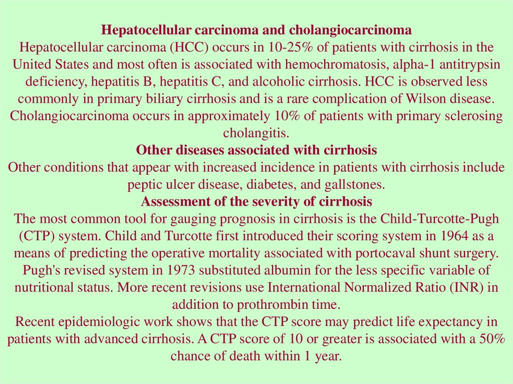
61.
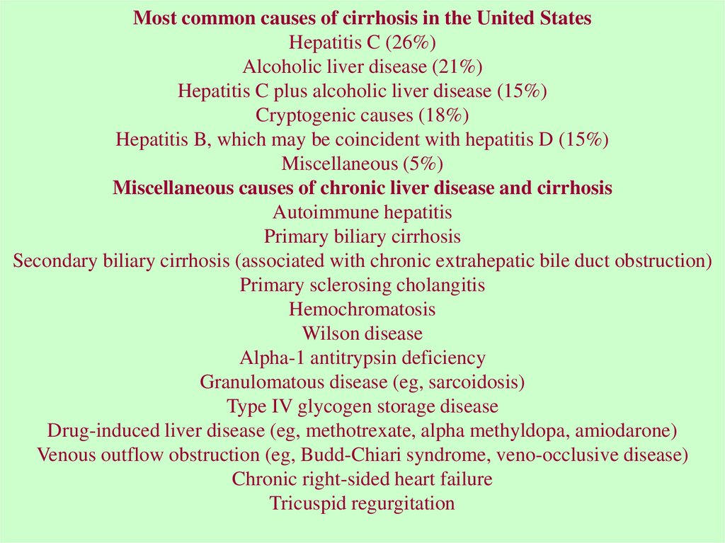
Most common causes of cirrhosis in the United StatesHepatitis C (26%)
Alcoholic liver disease (21%)
Hepatitis C plus alcoholic liver disease (15%)
Cryptogenic causes (18%)
Hepatitis B, which may be coincident with hepatitis D (15%)
Miscellaneous (5%)
Miscellaneous causes of chronic liver disease and cirrhosis
Autoimmune hepatitis
Primary biliary cirrhosis
Secondary biliary cirrhosis (associated with chronic extrahepatic bile duct obstruction)
Primary sclerosing cholangitis
Hemochromatosis
Wilson disease
Alpha-1 antitrypsin deficiency
Granulomatous disease (eg, sarcoidosis)
Type IV glycogen storage disease
Drug-induced liver disease (eg, methotrexate, alpha methyldopa, amiodarone)
Venous outflow obstruction (eg, Budd-Chiari syndrome, veno-occlusive disease)
Chronic right-sided heart failure
Tricuspid regurgitation
62.
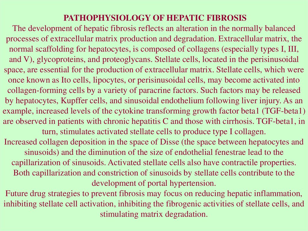
PATHOPHYSIOLOGY OF HEPATIC FIBROSISThe development of hepatic fibrosis reflects an alteration in the normally balanced
processes of extracellular matrix production and degradation. Extracellular matrix, the
normal scaffolding for hepatocytes, is composed of collagens (especially types I, III,
and V), glycoproteins, and proteoglycans. Stellate cells, located in the perisinusoidal
space, are essential for the production of extracellular matrix. Stellate cells, which were
once known as Ito cells, lipocytes, or perisinusoidal cells, may become activated into
collagen-forming cells by a variety of paracrine factors. Such factors may be released
by hepatocytes, Kupffer cells, and sinusoidal endothelium following liver injury. As an
example, increased levels of the cytokine transforming growth factor beta1 (TGF-beta1)
are observed in patients with chronic hepatitis C and those with cirrhosis. TGF-beta1, in
turn, stimulates activated stellate cells to produce type I collagen.
Increased collagen deposition in the space of Disse (the space between hepatocytes and
sinusoids) and the diminution of the size of endothelial fenestrae lead to the
capillarization of sinusoids. Activated stellate cells also have contractile properties.
Both capillarization and constriction of sinusoids by stellate cells contribute to the
development of portal hypertension.
Future drug strategies to prevent fibrosis may focus on reducing hepatic inflammation,
inhibiting stellate cell activation, inhibiting the fibrogenic activities of stellate cells, and
stimulating matrix degradation.
63.
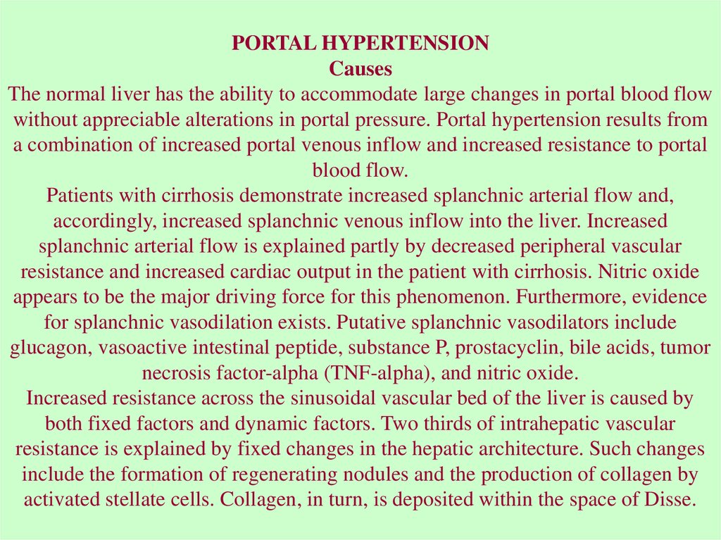
PORTAL HYPERTENSIONCauses
The normal liver has the ability to accommodate large changes in portal blood flow
without appreciable alterations in portal pressure. Portal hypertension results from
a combination of increased portal venous inflow and increased resistance to portal
blood flow.
Patients with cirrhosis demonstrate increased splanchnic arterial flow and,
accordingly, increased splanchnic venous inflow into the liver. Increased
splanchnic arterial flow is explained partly by decreased peripheral vascular
resistance and increased cardiac output in the patient with cirrhosis. Nitric oxide
appears to be the major driving force for this phenomenon. Furthermore, evidence
for splanchnic vasodilation exists. Putative splanchnic vasodilators include
glucagon, vasoactive intestinal peptide, substance P, prostacyclin, bile acids, tumor
necrosis factor-alpha (TNF-alpha), and nitric oxide.
Increased resistance across the sinusoidal vascular bed of the liver is caused by
both fixed factors and dynamic factors. Two thirds of intrahepatic vascular
resistance is explained by fixed changes in the hepatic architecture. Such changes
include the formation of regenerating nodules and the production of collagen by
activated stellate cells. Collagen, in turn, is deposited within the space of Disse.
64.
Dynamic factors account for one third of intrahepatic vascular resistance. Stellate cells serve ascontractile cells for adjacent hepatic endothelial cells. The nitric oxide produced by the
endothelial cells, in turn, controls the relative degree of vasodilation or vasoconstriction produced
by the stellate cells. In cirrhosis, decreased local production of nitric oxide by endothelial cells
permits stellate cell contraction, with resulting vasoconstriction of the hepatic sinusoid. (This
contrasts with the peripheral circulation where there are high circulating levels of nitric oxide in
cirrhosis.) Increased local levels of vasoconstricting chemicals, like endothelin, may also
contribute to sinusoidal vasoconstriction.
The portal hypertension of cirrhosis is caused by the disruption of hepatic sinusoids. However,
portal hypertension may be observed in a variety of noncirrhotic conditions. Prehepatic causes
include splenic vein thrombosis and portal vein thrombosis. These conditions commonly are
associated with hypercoagulable states and with malignancy (eg, pancreatic cancer).
Intrahepatic causes of portal hypertension are divided into presinusoidal, sinusoidal, and
postsinusoidal conditions.
The classic form of presinusoidal disease is caused by the deposition of Schistosoma oocytes in
presinusoidal portal venules, with the subsequent development of granulomata and portal fibrosis.
Schistosomiasis is the most common noncirrhotic cause of variceal bleeding worldwide.
Schistosoma mansoni infection is described in Puerto Rico, Central and South America, the
Middle East, and Africa. Schistosoma japonicum is described in the Far East. Schistosoma
hematobium, observed in the Middle East and Africa, can produce portal fibrosis but more
commonly is associated with urinary tract deposition of eggs.
65.
The classic sinusoidal cause of portal hypertension is cirrhosis.The classic postsinusoidal condition is an entity known as veno-occlusive disease. Obliteration of
the terminal hepatic venules may result from ingestion of pyrrolizidine alkaloids in Comfrey tea
or Jamaican bush tea and following the high-dose chemotherapy that precedes bone marrow
transplantation.
Posthepatic causes of portal hypertension may include chronic right-sided heart failure and
tricuspid regurgitation and obstructing lesions of the hepatic veins and inferior vena cava. These
latter conditions, and the symptoms they produce, are termed Budd-Chiari syndrome.
Predisposing conditions include hypercoagulable states, tumor invasion into the hepatic vein or
inferior vena cava, and membranous obstruction of the inferior vena cava. Inferior vena cava
webs are observed most commonly in South and East Asia and are postulated to be due to
nutritional factors.
Symptoms of Budd-Chiari syndrome are attributed to decreased outflow of blood from the liver,
with resulting hepatic congestion and portal hypertension. These symptoms include
hepatomegaly, abdominal pain, and ascites. Cirrhosis only ensues later in the course of disease.
Differentiating Budd-Chiari syndrome from cirrhosis by history or physical examination may be
difficult. Thus, Budd-Chiari syndrome must be included in the differential diagnosis of conditions
that produce ascites and varices. Hepatic vein patency is checked most readily by performing an
abdominal ultrasound with Doppler examination of the hepatic vessels. Abdominal CT scan with
intravenous contrast, abdominal MRI, and visceral angiography also may provide information
regarding the patency of hepatic vessels.
66.
ASCITESAscites is defined as an accumulation of excessive fluid
within the peritoneal cavity and may be a complication of
both hepatic and nonhepatic diseases. The 4 most
common causes of ascites in North America and Europe
are cirrhosis, neoplasm, congestive heart failure, and
tuberculous peritonitis.
In the past, ascites was classified as being a transudate or
an exudate. In transudative ascites, fluid was said to cross
the liver capsule because of an imbalance in Starling
forces. In general, ascites protein was less than 2.5 g/dL.
Classic causes of transudative ascites are portal
hypertension secondary to cirrhosis and congestive heart
failure.
67.
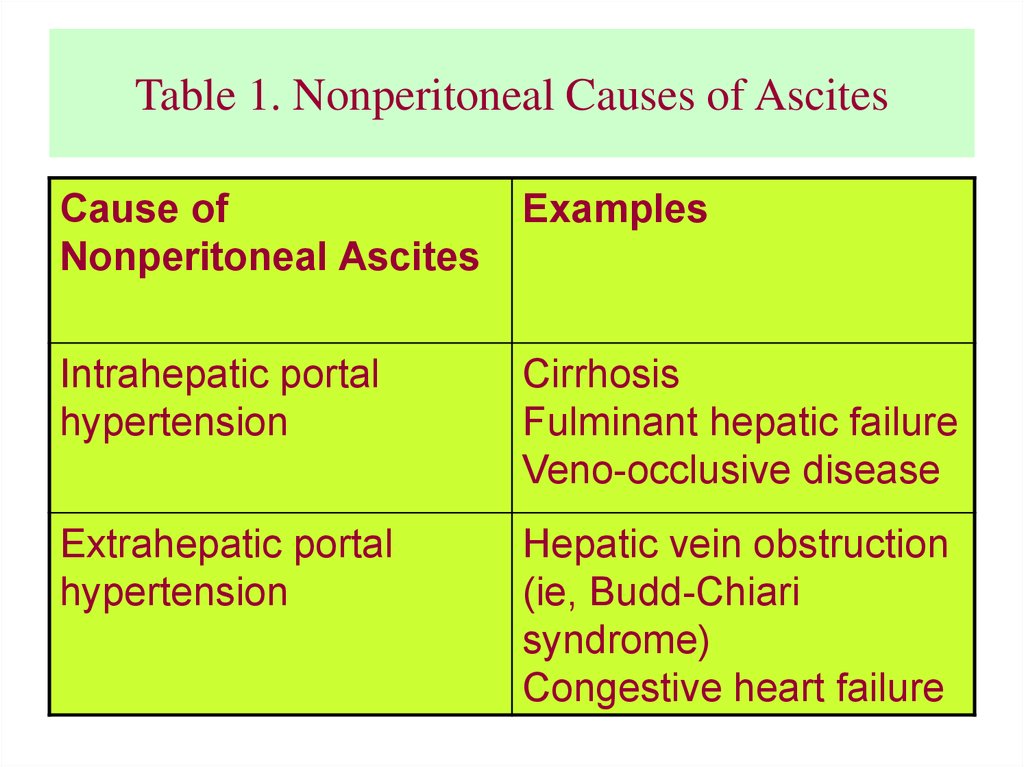
Table 1. Nonperitoneal Causes of AscitesCause of
Nonperitoneal Ascites
Examples
Intrahepatic portal
hypertension
Cirrhosis
Fulminant hepatic failure
Veno-occlusive disease
Extrahepatic portal
hypertension
Hepatic vein obstruction
(ie, Budd-Chiari
syndrome)
Congestive heart failure
68.
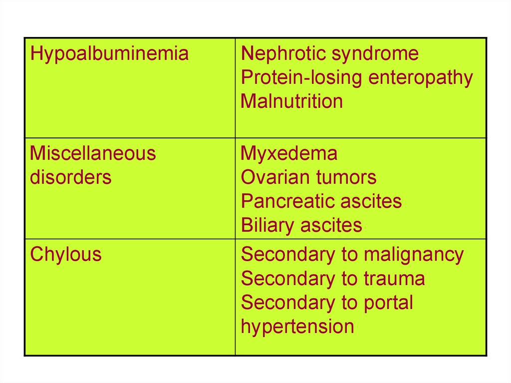
HypoalbuminemiaNephrotic syndrome
Protein-losing enteropathy
Malnutrition
Miscellaneous
disorders
Myxedema
Ovarian tumors
Pancreatic ascites
Biliary ascites
Secondary to malignancy
Secondary to trauma
Secondary to portal
hypertension
Chylous
69.
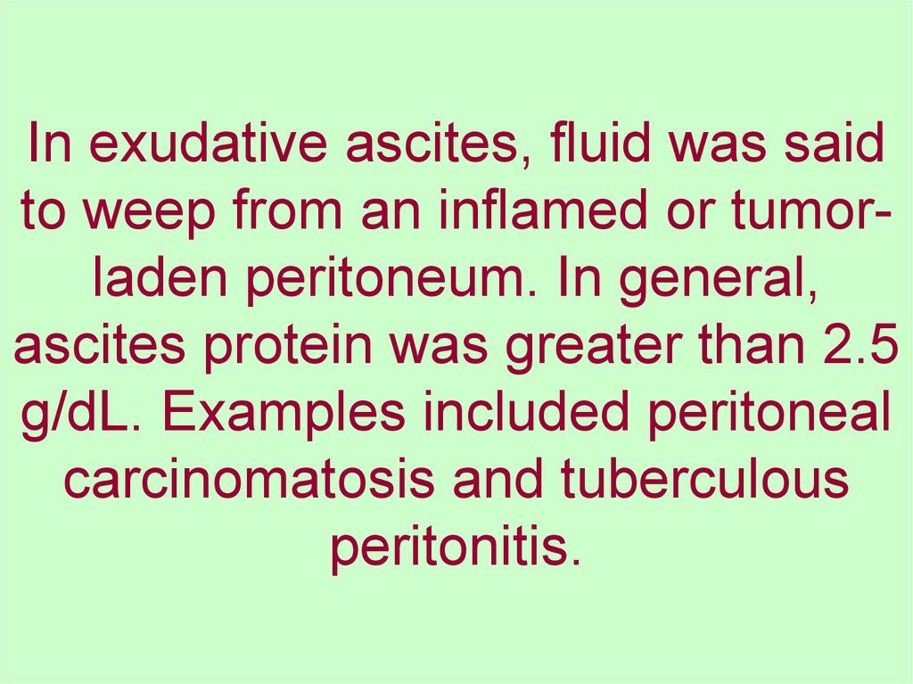
In exudative ascites, fluid was saidto weep from an inflamed or tumorladen peritoneum. In general,
ascites protein was greater than 2.5
g/dL. Examples included peritoneal
carcinomatosis and tuberculous
peritonitis.
70.
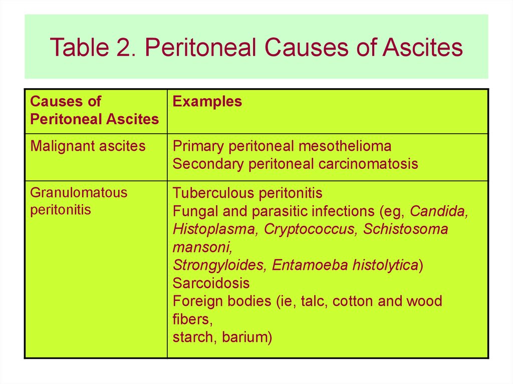
Table 2. Peritoneal Causes of AscitesCauses of
Examples
Peritoneal Ascites
Malignant ascites
Primary peritoneal mesothelioma
Secondary peritoneal carcinomatosis
Granulomatous
peritonitis
Tuberculous peritonitis
Fungal and parasitic infections (eg, Candida,
Histoplasma, Cryptococcus, Schistosoma
mansoni,
Strongyloides, Entamoeba histolytica)
Sarcoidosis
Foreign bodies (ie, talc, cotton and wood
fibers,
starch, barium)
71.
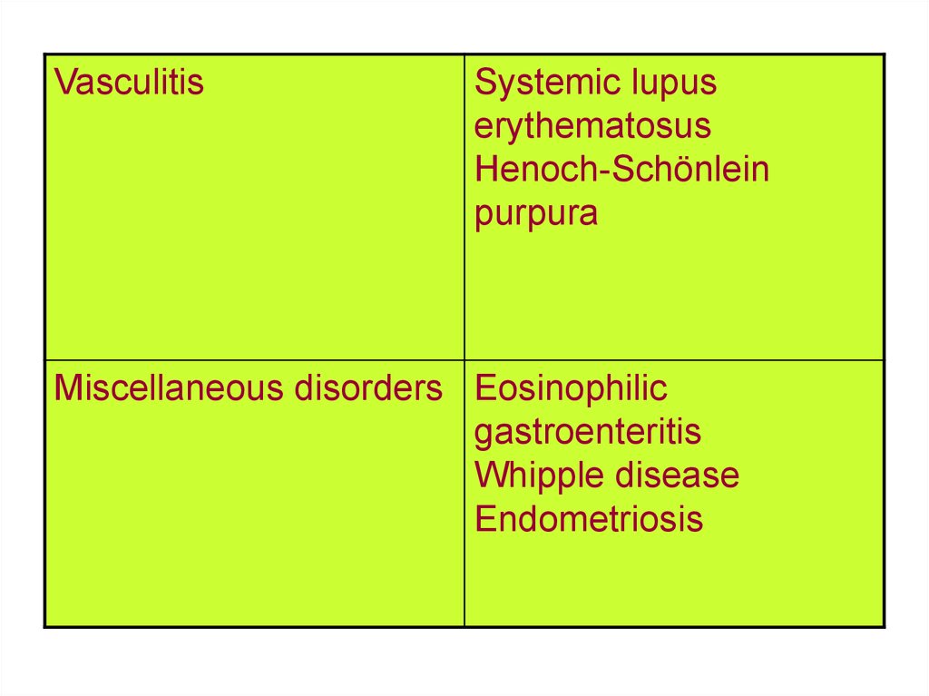
VasculitisSystemic lupus
erythematosus
Henoch-Schönlein
purpura
Miscellaneous disorders Eosinophilic
gastroenteritis
Whipple disease
Endometriosis
72.
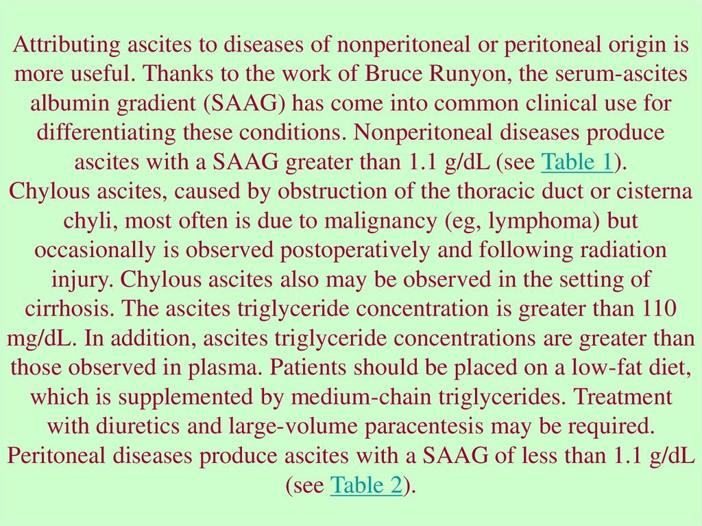
Attributing ascites to diseases of nonperitoneal or peritoneal origin ismore useful. Thanks to the work of Bruce Runyon, the serum-ascites
albumin gradient (SAAG) has come into common clinical use for
differentiating these conditions. Nonperitoneal diseases produce
ascites with a SAAG greater than 1.1 g/dL (see Table 1).
Chylous ascites, caused by obstruction of the thoracic duct or cisterna
chyli, most often is due to malignancy (eg, lymphoma) but
occasionally is observed postoperatively and following radiation
injury. Chylous ascites also may be observed in the setting of
cirrhosis. The ascites triglyceride concentration is greater than 110
mg/dL. In addition, ascites triglyceride concentrations are greater than
those observed in plasma. Patients should be placed on a low-fat diet,
which is supplemented by medium-chain triglycerides. Treatment
with diuretics and large-volume paracentesis may be required.
Peritoneal diseases produce ascites with a SAAG of less than 1.1 g/dL
(see Table 2).
73.
The role of portal hypertension in the pathogenesis of cirrhoticascites
The formation of ascites in cirrhosis depends on the presence of
unfavorable Starling forces within the hepatic sinusoid and on some
degree of renal dysfunction. Patients with cirrhosis are observed to
have increased hepatic lymphatic flow.
Fluid and plasma proteins diffuse freely across the highly permeable
sinusoidal endothelium into the space of Disse. Fluid in the space of
Disse, in turn, enters the lymphatic channels that run within the portal
and central venous areas of the liver.
Because the transsinusoidal oncotic gradient is approximately zero,
the increased sinusoidal pressure that develops in portal hypertension
increases the amount of fluid entering the space of Disse. When the
increased hepatic lymph production observed in portal hypertension
exceeds the ability of the cisterna chyli and thoracic duct to clear the
lymph, fluid crosses into the liver interstitium. Fluid may then
extravasate across the liver capsule into the peritoneal cavity.
74.
The role of renal dysfunction in the pathogenesis of cirrhoticascites
Patients with cirrhosis experience sodium retention, impaired
free water excretion, and intravascular volume overload. These
abnormalities may occur even in the setting of a normal
glomerular filtration rate. To some extent, these abnormalities
are due to increased levels of renin and aldosterone.
The peripheral arterial vasodilation hypothesis states that
splanchnic arterial vasodilation, driven by high nitric oxide
levels, leads to intravascular underfilling. This leads to
stimulation of the renin-angiotensin system and the sympathetic
nervous system and to antidiuretic hormone release. These
events are followed by an increase in sodium and water
retention, by an increase in plasma volume, and by the overflow
of ascites into the peritoneal cavity.
75.
Hepatorenal syndromeThis syndrome represents a continuum of renal dysfunction that
may be observed in patients with cirrhosis and is caused by the
vasoconstriction of large and small renal arteries and the
impaired renal perfusion that results. The syndrome may
represent an imbalance between renal vasoconstrictors and
vasodilators. Plasma levels of a number of vasoconstricting
substances are elevated in patients with cirrhosis and include
angiotensin, antidiuretic hormone, and norepinephrine. Renal
perfusion appears to be protected by vasodilators, including
prostaglandins E2 and I2 and atrial natriuretic factor.
Nonsteroidal anti-inflammatory drugs (NSAIDs) inhibit
prostaglandin synthesis. They may potentiate renal
vasoconstriction, with a resulting drop in glomerular filtration.
Thus, the use of NSAIDs is contraindicated in patients with
decompensated cirrhosis.
76.
Most patients with hepatorenal syndrome are noted to haveminimal histological changes in the kidneys. Kidney function
usually recovers when patients with cirrhosis and hepatorenal
syndrome undergo liver transplantation. In fact, a kidney
donated by a patient dying from hepatorenal syndrome functions
normally when transplanted into a renal transplant recipient.
Hepatorenal syndrome progression may be slow (type II) or
rapid (type I). Type I disease frequently is accompanied by
rapidly progressive liver failure. Hemodialysis offers temporary
support for such patients. These individuals are salvaged only by
performance of liver transplantation. Exceptions to this rule are
the patients with FHF or severe alcoholic hepatitis who
spontaneously recover both liver and kidney function. In type II
hepatorenal syndrome, patients may have stable or slowly
progressive renal insufficiency. Many such patients develop
ascites that is resistant to management with diuretics.
77.
Hepatorenal syndrome is diagnosed when a creatinine clearance less than 40 mL/min ispresent or when a serum creatinine greater than 1.5 mg/dL, urine volume less than 500
mL/d, and a urine sodium less than 10 mEq/L is present. Urine osmolality is greater
than plasma osmolality. In hepatorenal syndrome, renal dysfunction cannot be
explained by preexisting kidney disease, prerenal azotemia, the use of diuretics, or
exposure to nephrotoxins. Clinically, the diagnosis may be reached if central venous
pressure is determined to be normal or if no improvement of renal function occurs
following the infusion of at least 1.5 L of a plasma expander.
Nephrotoxic medications, including aminoglycoside antibiotics, should be avoided in
patients with cirrhosis. Patients with early hepatorenal syndrome may be salvaged by
aggressive expansion of intravascular volume with albumin and fresh frozen plasma
and by avoidance of diuretics. Administration of oral prostaglandins may be beneficial,
but this point is controversial. Use of renal-dose dopamine is not effective.
Recently, a number of investigators have employed systemic vasoconstrictors in an
attempt to reverse the effects of nitric oxide on peripheral arterial vasodilation. In
Europe, administration of intravenous terlipressin (an analog of vasopressin not
available in the United States) improved the renal dysfunction of patients with
hepatorenal syndrome. A combination of midodrine (an oral alpha agonist),
subcutaneous octreotide, and albumin infusion also improved renal function in a small
series of patients with hepatorenal syndrome.
78.
Clinical features of ascitesAscites is suggested by the presence of a number of findings
upon physical examination, which are abdominal distention,
bulging flanks, shifting dullness, and elicitation of a "puddle
sign" in patients in the knee-elbow position. A fluid wave may
be elicited in patients with massive tense ascites. However,
physical examination findings are much less sensitive than
performing abdominal ultrasonography, which can detect as
little as 30 mL of fluid. Furthermore, ultrasound with Doppler
can help assess the patency of hepatic vessels. Factors
associated with worsening of ascites include excess fluid or
salt intake, malignancy, venous occlusion (eg, Budd-Chiari
syndrome), progressive liver disease, and spontaneous
bacterial peritonitis (SBP).
79.
Spontaneous bacterial peritonitisSBP is observed in 15-26% of patients hospitalized with ascites. The syndrome arises most commonly
in patients whose low-protein ascites (<1 g/dL) contains low levels of complement, resulting in
decreased opsonic activity. SBP appears to be caused by the translocation of GI tract bacteria across the
gut wall and also by the hematogenous spread of bacteria. The most common causative organisms are
Escherichia coli, Streptococcus pneumoniae, Klebsiella species, and other gram-negative enteric
organisms.
Classic SBP is diagnosed by the presence of neutrocytosis, which is defined as greater than 250
polymorphonuclear (PMN) cells per mm3 of ascites, in the setting of a positive ascites culture. Culturenegative neutrocytic ascites is observed more commonly. Both conditions represent serious infections
that carry a 20-30% mortality rate.
The most commonly used regimen in the treatment of SBP is a 5-day course of cefotaxime at 1-2 g
intravenously every 8 hours. Alternatives include oral ofloxacin and other intravenous antibiotics with
activity against gram-negative enteric organisms. Many authorities advise repeat paracentesis in 48-72
hours in order to document a decrease in the ascites PMN count to less than 250 cells/mm3 and to
assure the efficacy of therapy.
Once SBP develops, patients have a 70% chance of redeveloping the condition within 1 year.
Prophylactic antibiotic therapy can reduce the recurrence rate of SBP to 20%. Some of the regimens
used in the prophylaxis of SBP include norfloxacin at 400 mg orally every day and trimethoprimsulfamethoxazole at 1 double-strength tablet 5 days per week.
Therapy with norfloxacin at 400 mg orally twice per day for 7 days can reduce serious bacterial
infection in patients with cirrhosis who have GI bleeding. One study noted that the 37% incidence of
serious bacterial infection was reduced to 10% when treatment with norfloxacin was instituted.
Furthermore, it can be argued that all patients with low-protein ascites should undergo prophylactic
therapy (eg, with norfloxacin 400 mg/d PO) at the time of hospital admission, given the high incidence
of hospital-acquired SBP.
80.
Other complications of massive ascitesPatients with massive ascites may experience abdominal discomfort, depressed
appetite, and decreased oral intake. Diaphragmatic elevation may lead to
symptoms of dyspnea. Pleural effusions may result from the passage of ascitic
fluid across channels in the diaphragm.
Umbilical and inguinal hernias are common in patients with moderate and
massive ascites. The use of an elastic abdominal binder may protect the skin
overlying a protruding umbilical hernia from maceration and may help prevent
rupture and subsequent infection. Timely large-volume paracentesis also may
help to prevent this disastrous complication. Umbilical hernias should not
undergo elective repair unless patients are significantly symptomatic or their
hernias are irreducible. As with all other surgeries in patients with cirrhosis,
herniorrhaphy carries multiple potential risks such as intraoperative bleeding,
postoperative infection, and liver failure because of anesthesia-induced
reductions in hepatic blood flow. However, these risks become acceptable in
patients with severe symptoms from their hernia. Urgent surgery is necessary in
the patient whose hernia has been complicated by bowel incarceration.
81.
Paracentesis in the diagnosis of ascitesParacentesis is essential in determining whether ascites is
caused by portal hypertension or by another process.
Ascites studies also are used to rule out infection and
malignancy. Paracentesis should be performed in all
patients with either new onset of ascites or worsening
ascites. Paracentesis also should be performed when SBP
is suggested by the presence of abdominal pain, fever,
leukocytosis, or worsening hepatic encephalopathy. Some
argue that paracentesis should be performed in all patients
with cirrhosis who have ascites at the time of
hospitalization, given the significant possibility of
asymptomatic SBP.
82.
Table 3. Ascites TestsRoutine
Optional
Special
Cell count
Total protein
Cytology
Albumin
Glucose
TB smear and
culture
Lactic
Lactic
Triglycerides
dehydrogenase dehydrogenase
83.
Gram stainBilirubin
Amylase
84.
Ascitic fluid with more than 250 PMNs/mm3 defines neutrocyticascites and SBP. Many cases of ascites fluid with more than 1000
PMNs/mm3 (and certainly >5000 PMNs/mm3) are associated with
appendicitis or a perforated viscus with resulting bacterial peritonitis.
Appropriate radiologic studies must be performed in such patients to
rule out surgical causes of peritonitis. Lymphocyte-predominant
ascites raises concerns about the possibility of underlying malignancy
or tuberculosis. Similarly, grossly bloody ascites may be observed in
malignancy and tuberculosis. Bloody ascites is seen infrequently in
uncomplicated cirrhosis. A common clinical dilemma is how to
interpret the ascites PMN count in the setting of bloody ascites. This
author recommends subtraction of 1 PMN for every 250 RBCs in
ascites in order to ascertain a corrected PMN count.
The yield of ascites culture studies may be increased by directly
inoculating 10 mL of ascites into aerobic and anaerobic culture bottles
at the patient's bedside.
85.
Medical treatment of ascitesTherapy for ascites should be tailored to the patient's needs. Some
patients with mild ascites respond to sodium restriction or diuretics
taken once or twice per week. Other patients require aggressive
diuretic therapy, careful monitoring of electrolytes, and occasional
hospitalization to facilitate even more intensive diuresis.
Sodium restriction
Salt restriction is the first line of therapy. In general, patients begin
with a diet containing less than 2000 mg sodium per day. Some
patients with refractory ascites require a diet containing less than
500 mg sodium per day. However, ensuring that patients do not
construct diets that might place them at risk for calorie and protein
malnutrition is important. Indeed, the benefit of commercially
available liquid nutritional supplements (which often contain
moderate amounts of sodium) often exceeds the risk of slightly
increasing the patient's salt intake.
86.
DiureticsDiuretics should be considered the second line of therapy. Spironolactone (Aldactone)
blocks the aldosterone receptor at the distal tubule. It is dosed at 50-300 mg once per day.
Although the drug has a relatively short half-life, its blockade of the aldosterone receptor
lasts for at least 24 hours. Adverse effects of spironolactone include hyperkalemia,
gynecomastia, and lactation. Other potassium-sparing diuretics, including amiloride and
triamterene, may be used as alternative agents, especially in patients complaining of
gynecomastia.
Furosemide (Lasix) may be used as a solo agent or in combination with spironolactone. The
drug blocks sodium reuptake in the loop of Henle. It is dosed at 40-240 mg per day in 1-2
divided doses. Patients infrequently need potassium repletion when furosemide is dosed in
combination with spironolactone.
Aggressive diuretic therapy in hospitalized patients with massive ascites can safely induce a
0.5- to 1-kg weight loss per day, providing that patients undergo careful monitoring of renal
function. Diuretic therapy should be held in the event of electrolyte disturbances, azotemia,
or induction of hepatic encephalopathy. Thus far, evidence-based medicine has not
supported the use of albumin as an aid to diuresis in the patient with cirrhosis who is
hospitalized. However, the author's anecdotal experience suggests that albumin may increase
the efficacy and safety of diuretics. The author's practice in patients who are
hypoalbuminemic is to administer intravenous furosemide following intravenous infusion of
albumin at 25 g twice per day, in addition to ongoing therapy with spironolactone.
87.
Large-volume paracentesisAggressive diuretic therapy is ineffective in controlling ascites in
approximately 5-10% of patients. Such patients with massive ascites may need
to undergo large-volume paracentesis in order to receive relief from symptoms
of abdominal discomfort, anorexia, or dyspnea. The procedure also may help
reduce the risk of umbilical hernia rupture.
Large-volume paracentesis was first used in ancient times. It fell out of favor
from the 1950s through the 1980s with the advent of diuretic therapy and
following a handful of case reports describing paracentesis-induced azotemia.
In 1987, Gines and colleagues demonstrated that large-volume paracentesis
could be performed with minimal or no impact on renal function. This and
other studies showed that 5-15 L of ascites could be removed safely at one time.
Large-volume paracentesis is thought to be safe in patients with peripheral
edema and in patients not currently treated with diuretics. Debate exists
whether colloid infusions (eg, with 5-10 g albumin per 1 L ascites removed) are
necessary to prevent intravascular volume depletion in patients who are
receiving ongoing diuretic therapy or in patients with mild or moderate
underlying renal insufficiency.
88.
Peritoneovenous shuntsLeVeen shunts and Denver shunts are devices that permit the
return of ascites fluid and proteins to the intravascular space.
Plastic tubing inserted subcutaneously under local anesthesia
connects the peritoneal cavity to the internal jugular vein or
subclavian vein via a pumping chamber. These devices are
successful at relieving ascites and reversing protein loss in some
patients. However, serious complications are observed in 10% of
the recipients of these devices. Complications include peritoneal
infection, sepsis, disseminated intravascular coagulation, and
congestive heart failure. Shunts may clot and require
replacement in an additional 30% of patients. However,
peritoneovenous shunts may be a reasonable form of therapy for
patients with refractory ascites who are not candidates for TIPS
or liver transplantation.
89.
Portosystemic shunts and transjugular intrahepatic portosystemic shuntsThe prime indication for portocaval shunt surgery is the management of refractory variceal
bleeding. However, since 1945, the medical field has recognized that portocaval shunts, by
decompressing the hepatic sinusoid, may improve ascites. The performance of a side-to-side
portocaval shunt for ascites management must be weighed against the approximate 5% mortality
rate associated with this surgery and the chance (as high as 30%) of inducing hepatic
encephalopathy.
TIPS is an effective tool in managing massive ascites in some patients. Ideally, TIPS placement
produces a decrease in sinusoidal pressure and a decrease in plasma renin and aldosterone levels,
with subsequent improved urinary sodium excretion. In one study, 74% of patients with
refractory ascites achieved complete remission of ascites within 3 months of TIPS placement.
However, patient selection for the procedure is important. Creation of TIPS has the potential to
worsen preexisting hepatic encephalopathy and exacerbate liver dysfunction in patients with
severe underlying liver failure.
In the author's opinion, TIPS use should be reserved for patients with Child class B cirrhosis or
patients with Child class C cirrhosis without severe coagulopathy or encephalopathy. Previously,
shunt stenosis was observed in one half of cases within 1 year of placement, necessitating
angiographic revision. Although the advent of coated stents appears to be reducing the incidence
of shunt stenosis, patients must still be willing to return to the hospital for Doppler and
angiographic follow-up of TIPS patency.
Liver transplantation
Patients with massive ascites have a less than 50% 1-year survival rate. Liver transplantation
should be considered as a potential means of salvaging the patient prior to the onset of intractable
liver failure or hepatorenal syndrome.
90.
HEPATIC ENCEPHALOPATHYDefinition
Hepatic encephalopathy is a syndrome observed in some patients with cirrhosis
that is marked by personality changes, intellectual impairment, and a depressed
level of consciousness. The diversion of portal blood into the systemic
circulation appears to be a prerequisite for the syndrome. Indeed, hepatic
encephalopathy may develop in patients who do not have cirrhosis who
undergo portocaval shunt surgery.
Pathogenesis
A number of theories have been postulated to explain the pathogenesis of
hepatic encephalopathy in patients with cirrhosis. Patients may have altered
brain energy metabolism and increased permeability of the blood-brain barrier.
The latter may facilitate the passage of neurotoxins into the brain. Putative
neurotoxins include short-chain fatty acids, mercaptans, false neurotransmitters
(eg, tyramine, octopamine, and beta-phenylethanolamines), ammonia, and
gamma-aminobutyric acid (GABA).
91.
Clinical features of hepatic encephalopathyThe symptoms of hepatic encephalopathy may range from mild to severe and
may be observed in as many as 70% of patients with cirrhosis. Symptoms are
graded on the following scale:
Grade 0 - Subclinical; normal mental status, but minimal changes in memory,
concentration, intellectual function, coordination
Grade 1 - Mild confusion, euphoria or depression, decreased attention, slowing
of ability to perform mental tasks, irritability, disorder of sleep pattern (ie,
inverted sleep cycle)
Grade 2 - Drowsiness, lethargy, gross deficits in ability to perform mental tasks,
obvious personality changes, inappropriate behavior, intermittent disorientation
(usually for time)
Grade 3 - Somnolent but arousable, unable to perform mental tasks,
disorientation to time and place, marked confusion, amnesia, occasional fits of
rage, speech is present but incomprehensible
Grade 4 - Coma, with or without response to painful stimuli
Patients with mild and moderate hepatic encephalopathy demonstrate decreased
short-term memory and concentration on mental status testing. Findings upon
physical examination include asterixis and fetor hepaticus.
92.
Laboratory abnormalities in hepatic encephalopathyAn elevated arterial or free venous serum ammonia level is the classic
laboratory abnormality reported in patients with hepatic encephalopathy.
This finding may aid in the assignment of a correct diagnosis to a patient
with cirrhosis who presents with altered mental status. However, serial
ammonia measurements are inferior to clinical assessment in gauging
improvement or deterioration in patients under therapy for hepatic
encephalopathy. No utility exists for checking the ammonia level in a
patient with cirrhosis who does not have hepatic encephalopathy.
Some patients with hepatic encephalopathy have the classic but nonspecific
electroencephalogram (EEG) changes of high-amplitude low-frequency
waves and triphasic waves. EEG may be helpful in the initial workup of a
patient with cirrhosis and altered mental status when ruling out seizure
activity may be necessary.
CT scan and MRI studies of the brain may be important in ruling out
intracranial lesions when the diagnosis of hepatic encephalopathy is in
question.
93.
Management of hepatic encephalopathyNonhepatic causes of altered mental function must be excluded in patients with
cirrhosis who have worsening mental function. A check of the blood ammonia level
may be helpful in such patients. Medications that depress CNS function, especially
benzodiazepines, should be avoided. Precipitants of hepatic encephalopathy should be
corrected (eg, metabolic disturbances, GI bleeding, infection, constipation).
Lactulose is helpful in patients with the acute onset of severe encephalopathy symptoms
and in patients with milder, chronic symptoms. This nonabsorbable disaccharide
stimulates the passage of ammonia from tissues into the gut lumen and inhibits
intestinal ammonia production. Initial lactulose dosing is 30 mL orally once or twice
daily. Dosing is increased until the patient has 2-4 loose stools per day. Dosing should
be reduced if the patient complains of diarrhea, abdominal cramping, or bloating.
Higher doses of lactulose may be administered via either a nasogastric tube or rectal
tube to hospitalized patients with severe encephalopathy. Other cathartics, including
colonic lavage solutions that contain polyethylene glycol (PEG) (eg, Go-Lytely), also
may be effective in patients with severe encephalopathy.
Neomycin and other antibiotics (eg, metronidazole, oral vancomycin, paromomycin,
oral quinolones) serve as second-line agents. They work by decreasing the colonic
concentration of ammoniagenic bacteria. Neomycin dosing is 250-1000 mg orally 2-4
times daily.
94.
Rifaximin (Xifaxan, Salix Pharmaceuticals, Inc, Morrisville, NC) is a nonabsorbableantibiotic that received approval by the US Food and Drug Administration (FDA) in
2004 for the treatment of travelers' diarrhea. Experience in Europe over the last 2
decades suggests that rifaximin can decrease colonic levels of ammoniagenic bacteria,
with resulting improvement in hepatic encephalopathy symptoms. Typical rifaximin
dosing in European hepatic encephalopathy trials was two 200 mg tablets taken orally 3
times daily. Work is being done to determine if lower doses of the medication can
effectively treat hepatic encephalopathy.
Other chemicals capable of decreasing blood ammonia levels are L-ornithine Laspartate (available in Europe) and sodium benzoate.
Low-protein diets were recommended routinely in the past for patients with cirrhosis.
High levels of aromatic amino acids contained in animal proteins were believed to lead
to increased blood levels of the false neurotransmitters tyramine and octopamine, with
resulting worsening of encephalopathy symptoms. In this author's experience, the vast
majority of patients can tolerate a protein-rich diet (>1.2 g/kg/d) including well-cooked
chicken, fish, vegetable protein, and, if needed, protein supplements. Although protein
restriction may play a role in the management of the patient with an acute flare of
hepatic encephalopathy, it rarely is necessary in patients with chronic encephalopathy
symptoms. Furthermore, many patients with cirrhosis have protein-calorie malnutrition
at baseline. The routine restriction of dietary protein intake increases their risk for
worsening malnutrition.
95.
OTHER MANIFESTATIONS OF CIRRHOSIS; ASSESSMENT OFSEVERITY OF CIRRHOSIS
All chronic liver diseases that progress to cirrhosis have in common the
histologic features of hepatic fibrosis and nodular regeneration. However,
patients' signs and symptoms may vary depending on the underlying etiology of
liver disease. As an example, patients with end-stage liver disease caused by
hepatitis C might develop profound muscle wasting, marked ascites, and severe
hepatic encephalopathy, with only mild jaundice. In contrast, patients with endstage primary biliary cirrhosis might be deeply icteric, with no evidence of
muscle wasting. These patients may complain of extreme fatigue and pruritus
and have no complications of portal hypertension. In both cases, medical
management is focused on the relief of symptoms. Liver transplantation should
be considered as a potential therapeutic option, given the inexorable course of
most cases of end-stage liver disease.
Many patients with cirrhosis experience fatigue, anorexia, weight loss, and
muscle wasting. Cutaneous manifestations of cirrhosis include jaundice, spider
angiomata, skin telangiectasias (termed “paper money skin” by Dame Sheila
Sherlock), palmar erythema, white nails, disappearance of lunulae, and finger
clubbing, especially in the setting of hepatopulmonary syndrome.
96.
Patients with cirrhosis may experience increased conversion of androgenic steroids into estrogensin skin, adipose tissue, muscle, and bone. Males may develop gynecomastia and impotence. Loss
of axillary and pubic hair is noted in both men and women. Hyperestrogenemia also may explain
spider angiomata and palmar erythema.
Hematologic manifestations
Anemia may result from folate deficiency, hemolysis, or hypersplenism. Thrombocytopenia
usually is secondary to hypersplenism and decreased levels of thrombopoietin. Coagulopathy
results from decreased hepatic production of coagulation factors. If cholestasis is present,
decreased micelle entry into the small intestine leads to decreased vitamin K absorption, with
resulting reduction in hepatic production of factors II, VII, IX, and X. Patients with cirrhosis also
may experience fibrinolysis and disseminated intravascular coagulation.
Pulmonary and cardiac manifestations
Patients with cirrhosis may have impaired pulmonary function. Pleural effusions and the
diaphragmatic elevation caused by massive ascites may alter ventilation-perfusion relations.
Interstitial edema or dilated precapillary pulmonary vessels may reduce pulmonary diffusing
capacity.
Patients also may have hepatopulmonary syndrome (HPS). In this condition, pulmonary
arteriovenous anastomoses result in arteriovenous shunting. Classic HPS is marked by the
symptom of platypnea and the finding of orthodeoxia, but the syndrome must be considered in
any patient with cirrhosis who has evidence of oxygen desaturation. HPS most readily is detected
by echocardiographic visualization of late-appearing bubbles in the left atrium following the
injection of agitated saline. Some cases of HPS may be corrected by liver transplantation.
Pulmonary hypertension is observed in approximately 1% of patients with cirrhosis. Its etiology
is unknown.
97.
Hepatocellular carcinoma and cholangiocarcinomaHepatocellular carcinoma (HCC) occurs in 10-25% of patients with cirrhosis in the
United States and most often is associated with hemochromatosis, alpha-1 antitrypsin
deficiency, hepatitis B, hepatitis C, and alcoholic cirrhosis. HCC is observed less
commonly in primary biliary cirrhosis and is a rare complication of Wilson disease.
Cholangiocarcinoma occurs in approximately 10% of patients with primary sclerosing
cholangitis.
Other diseases associated with cirrhosis
Other conditions that appear with increased incidence in patients with cirrhosis include
peptic ulcer disease, diabetes, and gallstones.
Assessment of the severity of cirrhosis
The most common tool for gauging prognosis in cirrhosis is the Child-Turcotte-Pugh
(CTP) system. Child and Turcotte first introduced their scoring system in 1964 as a
means of predicting the operative mortality associated with portocaval shunt surgery.
Pugh's revised system in 1973 substituted albumin for the less specific variable of
nutritional status. More recent revisions use International Normalized Ratio (INR) in
addition to prothrombin time.
Recent epidemiologic work shows that the CTP score may predict life expectancy in
patients with advanced cirrhosis. A CTP score of 10 or greater is associated with a 50%
chance of death within 1 year.
98.
Table 4. Child-Turcotte-Pugh Scoring System for Cirrhosis (Childclass A=5-6 points, Child class B=7-9 points, Child class C=10-15
points)
Clinical
variable
1 points
2 points 3 points
Encephalopathy None
Stage 1- Stage 3-4
2
Ascites
Slight
Moderate
2-3
>3
Absent
Bilirubin (mg/dL) <2
99.
Bilirubin in PBCor
PSC (mg/dL)
<4
4-10
10
Albumin (g/dL)
>3.5
2.8-3.5
<2.8
Prothrombin
time
(seconds
prolonged
or INR)
<4 s or
INR <1.7
4-6 s or
>6 s or
INR 1.7-2.3 INR
>2.3
100.
TREATMENT OF CIRRHOSISSpecific medical therapies may be applied to many liver diseases in an effort to
diminish symptoms and prevent or forestall the development of cirrhosis. Examples
include prednisone and azathioprine for autoimmune hepatitis, interferon and other
antiviral agents for hepatitis B and C, phlebotomy for hemochromatosis,
ursodeoxycholic acid for primary biliary cirrhosis, and zinc and penicillamine for
Wilson disease. These therapies become progressively less effective if chronic liver
disease evolves into cirrhosis. Once cirrhosis develops, treatment is aimed at the
management of complications as they arise. Certainly variceal bleeding, ascites, and
hepatic encephalopathy are among the most serious complications experienced by
patients with cirrhosis. However, attention also must be paid to patients' chronic
constitutional complaints.
Nutrition
Many patients complain of anorexia, which may be compounded by the direct
compression of ascites on the GI tract. Care should be taken to assure that patients
receive adequate calories and protein in their diets. Patients frequently benefit from the
addition of commonly available liquid and powdered nutritional supplements to the
diet. Only rarely can patients not tolerate proteins in the form of chicken, fish,
vegetables, and nutritional supplements. Institution of a low-protein diet in the fear that
hepatic encephalopathy might develop places the patient at risk for the development of
profound muscle wasting.
101.
Adjunctive therapiesZinc deficiency commonly is observed in patients with cirrhosis. Treatment
with zinc sulfate at 220 mg orally twice daily may improve dysgeusia and can
stimulate appetite. Furthermore, zinc is effective in the treatment of muscle
cramps and is adjunctive therapy for hepatic encephalopathy.
Pruritus is a common complaint in both cholestatic liver diseases (eg, primary
biliary cirrhosis) and in noncholestatic chronic liver diseases (eg, hepatitis C).
Although increased serum bile acid levels once were thought to be the cause of
pruritus, endogenous opioids are more likely to be the culprit pruritogens. Mild
itching complaints may respond to treatment with antihistamines.
Cholestyramine is the mainstay of therapy for the pruritus of liver disease. Care
should be taken to avoid coadministration of this organic anion binder with any
other medication, to avoid compromising GI absorption. Other medications that
may provide relief against pruritus include ursodeoxycholic acid, ammonium
lactate 12% skin cream (Lac-Hydrin, Westwood-Squibb Pharmaceuticals, Inc,
Princeton, NJ), naltrexone (an opioid antagonist), rifampin, gabapentin, and
ondansetron.
102.
Some male patients suffer from hypogonadism. Patients with severe symptomsmay undergo therapy with topical testosterone preparations, although their
safety and efficacy is not well studied. Similarly, the utility and safety of
growth hormone therapy remains unclear.
Patients with cirrhosis may develop osteoporosis. Supplementation with
calcium and vitamin D is important in patients at high risk for osteoporosis,
especially patients with chronic cholestasis, patients with primary biliary
cirrhosis, and patients receiving corticosteroids for autoimmune hepatitis. The
discovery of decreased bone mineralization upon bone densitometry studies
also may prompt institution of therapy with an aminobisphosphonate (eg,
alendronate sodium).
Regular exercise, including walking and even swimming, should be encouraged
in patients with cirrhosis, lest the patient slip into a vicious cycle of inactivity
and muscle wasting. Debilitated patients frequently benefit from formal
exercise programs supervised by a physical therapist. Patients with chronic liver
disease should receive vaccination to protect them against hepatitis A. Other
protective measures include vaccination against hepatitis B, pneumococci, and
influenza.
103.
Drug hepatotoxicity in the patient with cirrhosisThe institution of any new medical therapy warrants the performance of
more frequent liver chemistries. Indeed, patients with liver disease can ill
afford to have drug-induced liver disease superimposed on their
condition. Medications frequently associated with drug-induced liver
disease include NSAIDs, isoniazid, valproic acid, erythromycin,
amoxicillin/clavulanate, ketoconazole, and chlorpromazine.
NSAID use may predispose patients with cirrhosis to develop GI
bleeding. Patients with decompensated cirrhosis are at risk for NSAIDinduced renal insufficiency, presumably because of prostaglandin
inhibition and worsening of renal blood flow. Other nephrotoxic agents
such as aminoglycoside antibiotics also should be avoided.
Low-dose estrogens and progesterone appear to be safe in the setting of
liver disease. Hepatic 3-methylglutaryl coenzyme A (HMG Co-A)
reductase inhibitors frequently are associated with mild elevations of
alanine aminotransferase (ALT). However, severe hepatotoxicity is
reported infrequently. Their use should be monitored carefully in patients
with liver disease.
104.
Surgery in the patient with cirrhosisSurgery and general anesthesia carry increased risks in the patient with
cirrhosis. Anesthesia reduces cardiac output, induces splanchnic
vasodilation, and causes a 30- to 50%-reduction in hepatic blood flow. This
places the cirrhotic liver at additional risk for decompensation. Surgery is
said to be safe in the setting of mild chronic hepatitis. Its risk in patients
with severe chronic hepatitis is unknown. Patients with well-compensated
cirrhosis have an increased but acceptable risk of morbidity and mortality. A
study of nonshunt abdominal surgeries demonstrated a 10% mortality rate
for patients with Child class A cirrhosis as opposed to a 30% mortality rate
for patients with Child class B cirrhosis and a 75% mortality rate for
patients with Child class C cirrhosis. Thus, unless absolutely necessary,
surgery should be avoided in the patient with cirrhosis. Although
cholecystectomy was among the riskier surgeries noted, several recent
reports have described the successful performance of laparoscopic
cholecystectomy in patients with Child class A and B cirrhosis.
105.
Monitoring the patient with cirrhosisPatients with cirrhosis should undergo routine follow-up monitoring of their
complete blood count, renal and liver chemistries, and prothrombin time. The
author's policy is to monitor stable patients 3-4 times per year. The author performs
routine diagnostic endoscopy to determine whether the patient has asymptomatic
esophageal varices. Follow-up endoscopy is performed in 2 years if varices are not
present. If varices are present, treatment is initiated with a nonselective betablocker (eg, propranolol, nadolol), aiming for a 25% reduction in heart rate. Such
therapy offers effective primary prophylaxis against the new onset of variceal
bleeding.
This author encourages the screening of patients to rule out the interval
development of HCC. The author's practice is to perform abdominal
ultrasonography and alpha-fetoprotein testing twice yearly, although the clinical
utility and cost-effectiveness of this strategy remains controversial. In the past,
clinical suspicion for HCC mandated the performance of a confirmatory biopsy, by
either ultrasound or CT guidance. However, guided biopsy is accompanied by a
significant false-negative rate. Biopsy may be complicated either by hemorrhage or
by the tracking of tumor cells in the needle tract. Increasingly, patients with clinical
diagnoses of cirrhosis and HCC are monitored in the setting of liver transplant
programs.
106.
Many hepatologists and surgeons now rely on noninvasive testing whenit comes to making a diagnosis of HCC. In many transplant programs,
the presence of a suspicious lesion on both triple-phase CT scan and
MRI or the combination of a suspicious finding on radiologic study and
an alpha-fetoprotein (AFP) level of greater than 400 ng/mL is felt to have
the same or an even greater diagnostic power than guided liver biopsy.
Patients with a diagnosis of HCC and no evidence of extrahepatic
disease, as determined by chest and abdominal CT scans and by bone
scan, should be offered curative therapy. Commonly, this therapy entails
liver resection surgery for patients with Child Class A cirrhosis and an
accelerated course to liver transplantation for patients with Child Class B
and C cirrhosis. Patients who await liver transplantation are often offered
minimally invasive therapies in an effort to keep tumors from spreading.
These therapies include percutaneous injection therapy with ethanol,
radiofrequency thermal ablation, and chemoembolization.
107.
LIVER TRANSPLANTATIONLiver transplantation has emerged as an important strategy in
the management of patients with decompensated cirrhosis.
Patients should be referred for consideration of liver
transplantation after the first signs of hepatic decompensation.
Advances in surgical technique, organ preservation, and
immunosuppression have resulted in dramatic improvements
in postoperative survival over the last 2 decades. In the early
1980s, the percentage of patients surviving 1 year and 5 years
after liver transplant were only 70% and 15%, respectively.
Now, patients can anticipate a 1-year survival rate of 85-90%
and a 5-year survival rate of higher than 70%. Quality of life
after liver transplant is good or excellent in most cases.
108.
Contraindications for liver transplantation include severe cardiovascular or pulmonarydisease, active drug or alcohol abuse, malignancy outside the liver, sepsis, or
psychosocial problems that might jeopardize patients' abilities to follow their medical
regimens after transplant. The presence of HIV in the bloodstream also is a
contraindication to transplant. However, successful liver transplantations are now being
performed in patients with no detectable HIV viral load due to antiretroviral therapy.
Additional clinical study is required before liver transplantation can be offered routinely
to such patients.
Organ allocation
Approximately 5000 liver transplants are performed in the United States each year.
Although an increasing number of patients' lives are saved each year by transplant, the
rising number of diagnosed cases of cirrhosis, fueled in part by the hepatitis C
epidemic, has resulted in a dramatic increase in the number of patients listed as
candidates for liver transplantation.
Approximately 12-15% of patients listed as candidates die while waiting because of the
relatively static number of organ donations. Strategies to improve the current donor
organ shortage include programs to increase public awareness of the importance of
organ donation, increased use of living donor liver transplantation for pediatric and
adult recipients, organ donation after cardiac death, and the use of extended criteria
donors (ECD).