


 Английский язык
Похожие презентации:


Introduction to the Practice of Medicine - II
1. Introduction to the Practice of Medicine - II
Examination of the AbdomenTuesday, January 28, 2003
Michael J. Klamut, M.D.
2. Examination of the Abdomen
Session Objectives:Describe relevant anatomy and physiology as
it pertains to the examination of the abdomen
Demonstrate the steps in examining the
abdomen using illustrations and a SP
Review common abnormalities encountered
on the Physical Examination of the abdomen
3. Examination of the Abdomen
Introduction:The Medical History is an account of the
events in the pt’s life that have relevance
to the mental/physical health of the pt.
Accurate information is essential before
undertaking the PE of the abdomen.
4. Examination of the Abdomen
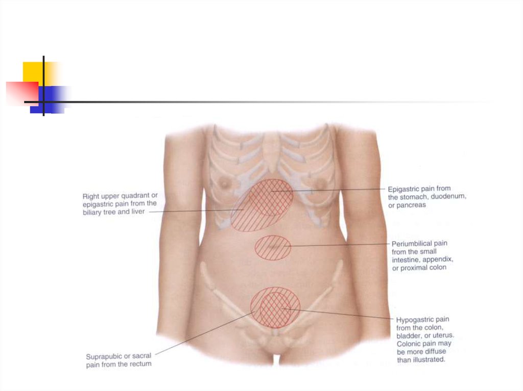
Pain is a common symptom of diseasesof the abdomen It is important to
assess different aspects of a pt’s
abdominal pain so that a reasonable
Differential Diagnosis can be formulated
5. Examination of the Abdomen
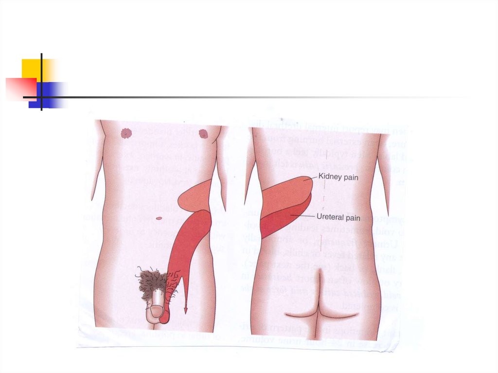
Important aspects of abdominal pain:Location and radiation of pain
Character of pain (cramping, sharp, dull,
burning, constant)
Timing of the pain
Exacerbating/alleviating features
Relationship to food intake
Relationship to defecation
6. Examination of the Abdomen
Important related symptoms/signs inpatients with abdominal pain:
Fever/rigors/sweats
Nausea/vomiting
Weight loss
Change in bowel habits
Evidence of GI blood loss (hematemesis,
melena,hematochezia, occult loss)
7. Examination of the Abdomen
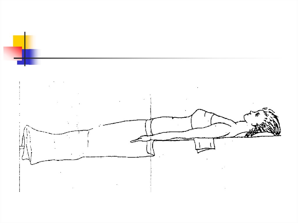
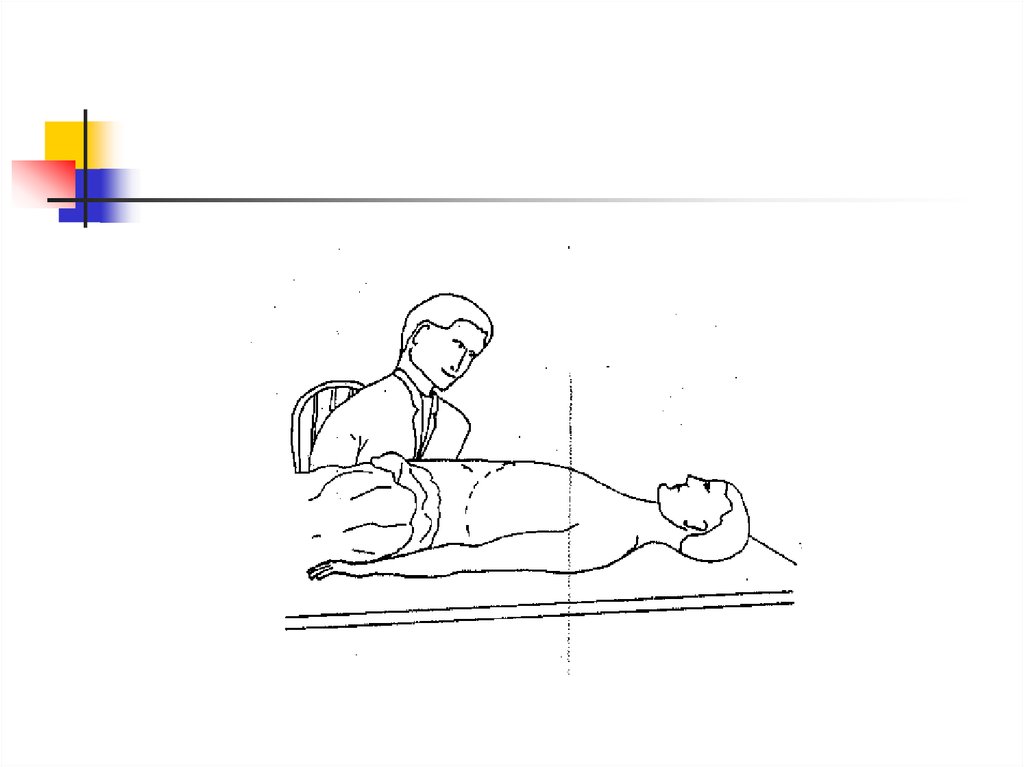
Physical Examination:The PE of the abdomen must be performed
in an organized, systematic fashion in
order to yield accurate and consistent
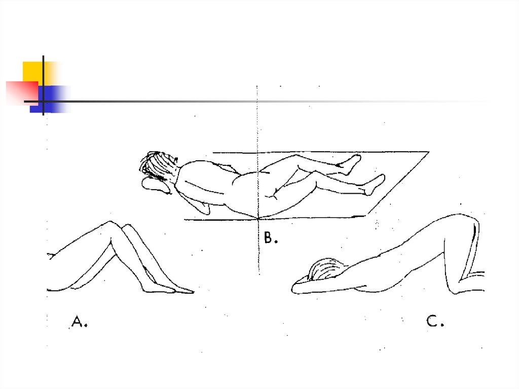
results.Pt should be properly prepared. Pt
should be lying supine, relaxed, draped,
with hands at sides or crossed on chest.
Quiet room/temp. Relaxed, confident
examiner.
8. Examination of the Abdomen
Physical Examinationof the Abdomen isconducted in four parts
Inspection/observation
Auscultation
Percussion
Palpation
9.
10.
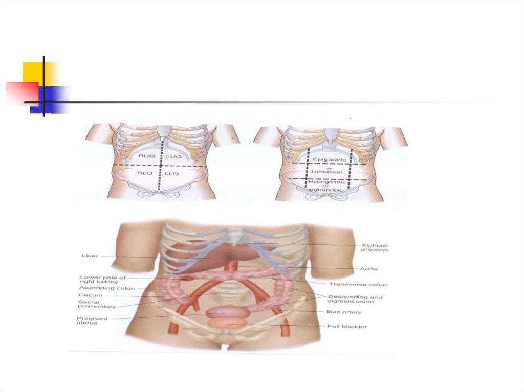
11. Examination of the Abdomen
For descriptive purposes, the abdomenis divided into four quadrants
RUQ,LUQ,RLQ,LLQ
Epigastric,umbilical, periumbilical,
suprapubic are terms also used by
clinicians to describe symptoms and
findings in those specific regions
12.
13.
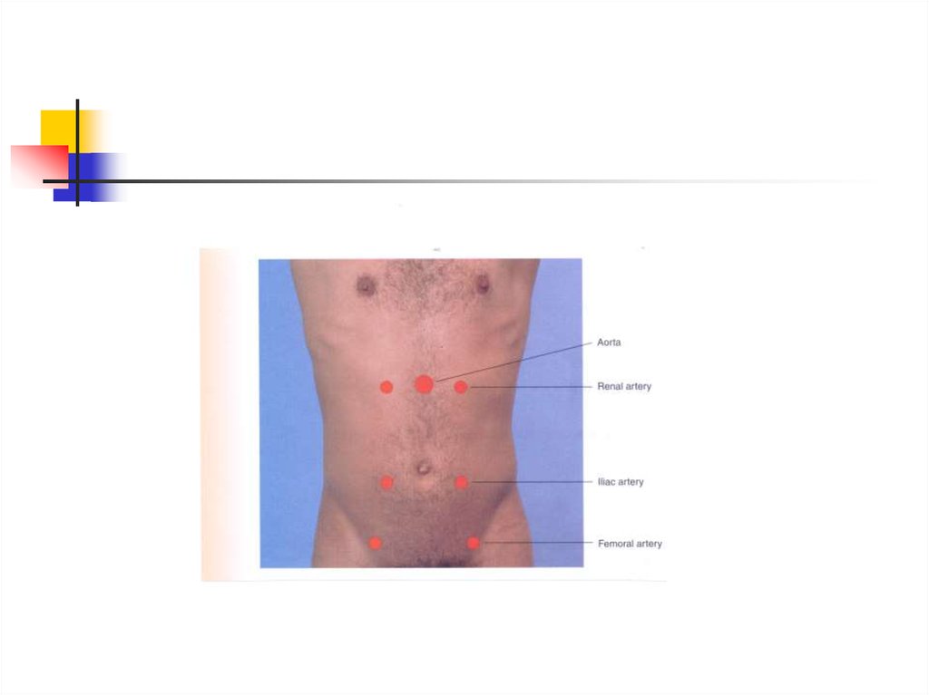
14. Examination of the Abdomen
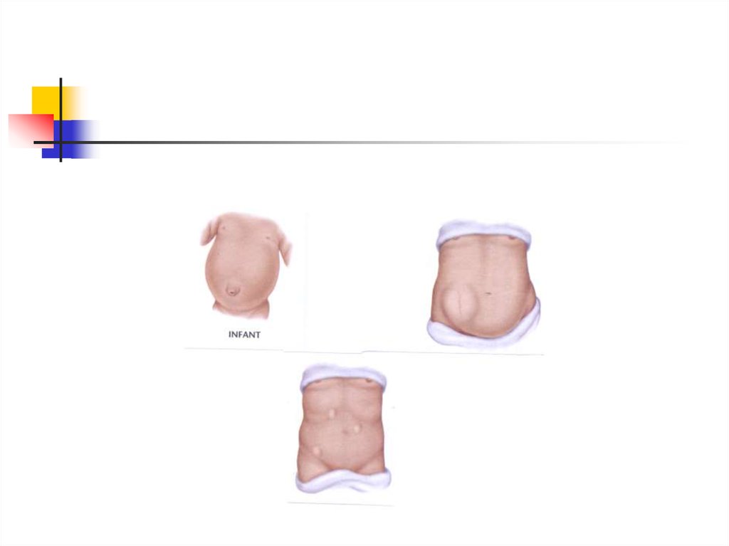
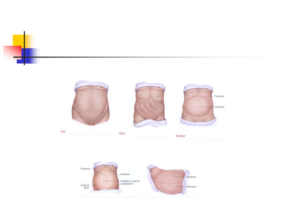
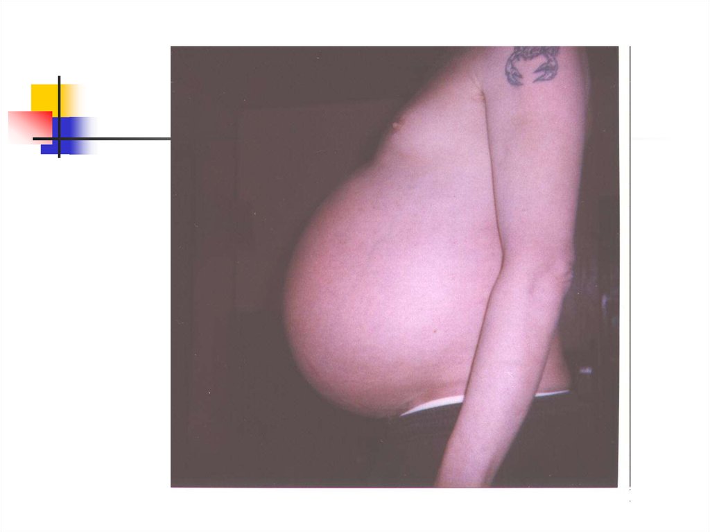
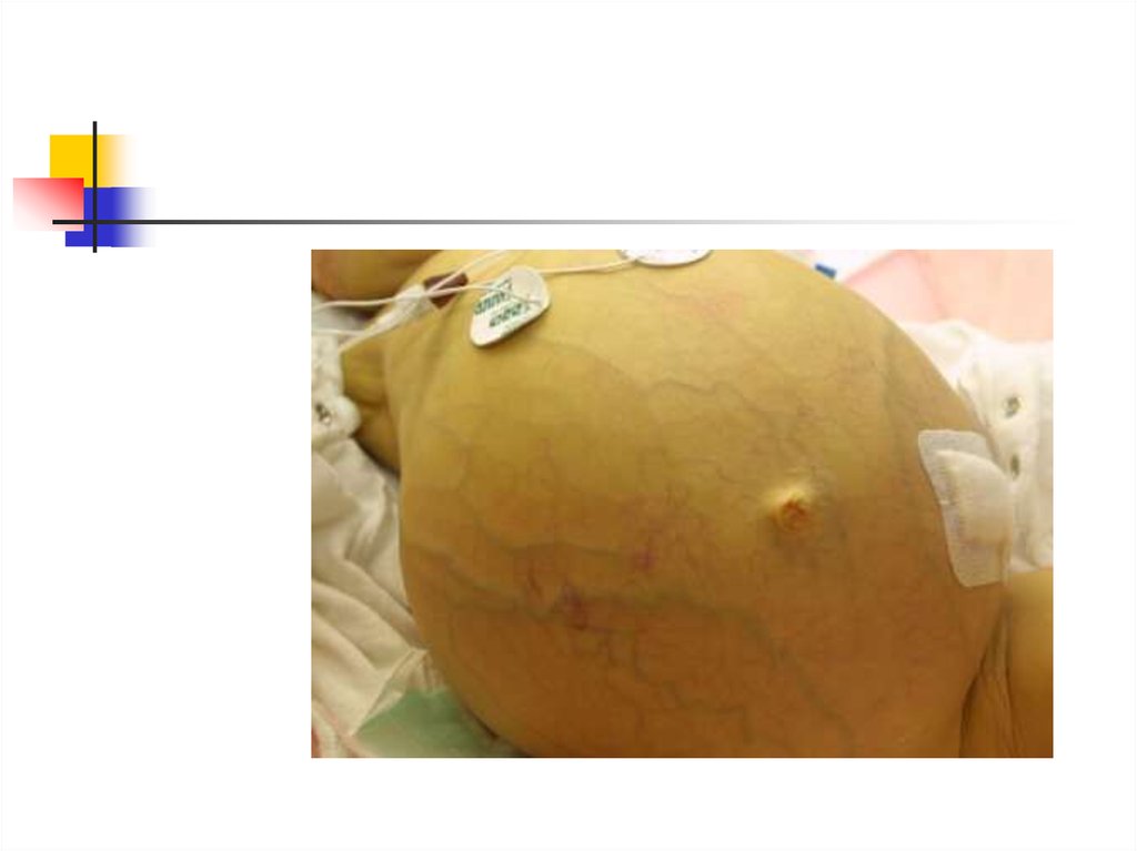
Inspection/Observation (#40)Inspect the contour of the abdomen. It
may be flat, rounded, protuberant, or
scaphoid
Are there any visible pulsations/masses?
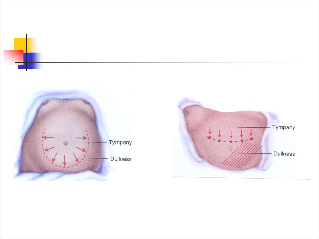
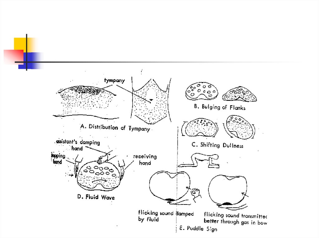
Do the flanks bulge (ascites)?
Inspect skin (scars,striae,veins,rashes)
Inspect umbilicus
15.
16. Examination of the Abdomen
Auscultation (#41)Useful in assessing bowel motility and
vascular bruits
Note frequency/character of the bowel
sounds (borborygmi) with stethoscope.
Listen in one spot. Listen for bruits.
No particular bowel sound is diagnostic but
rushes and high pitched tinkles suggest
obstructed gut.
17.
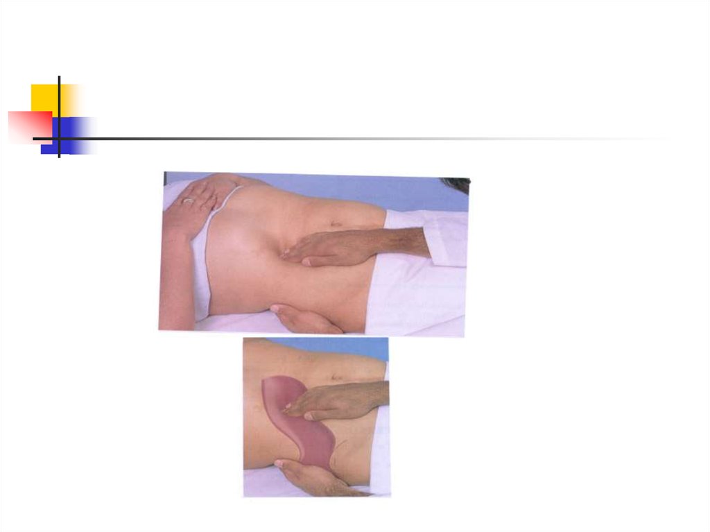
18. Examination of the Abdomen
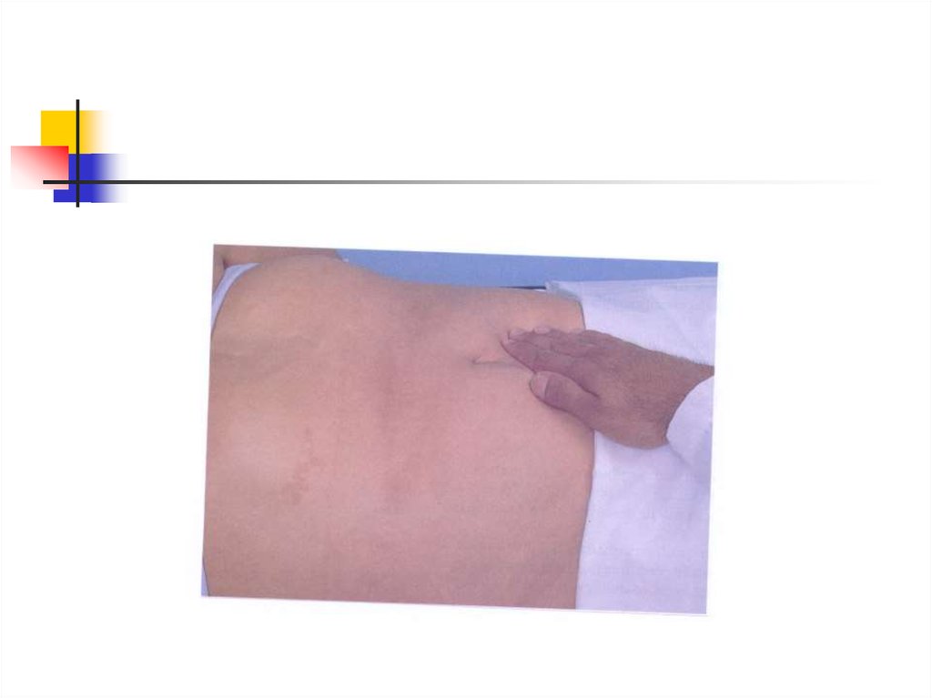
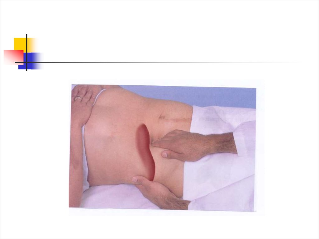
Palpation (#43-#50)Palpate lightly then deeply in all four
quadrants
Differentiate between voluntary and
involuntary guarding
If a mass is detected note its location,
size, shape, consistency, tenderness,
pulsation, and mobility
19.
20.
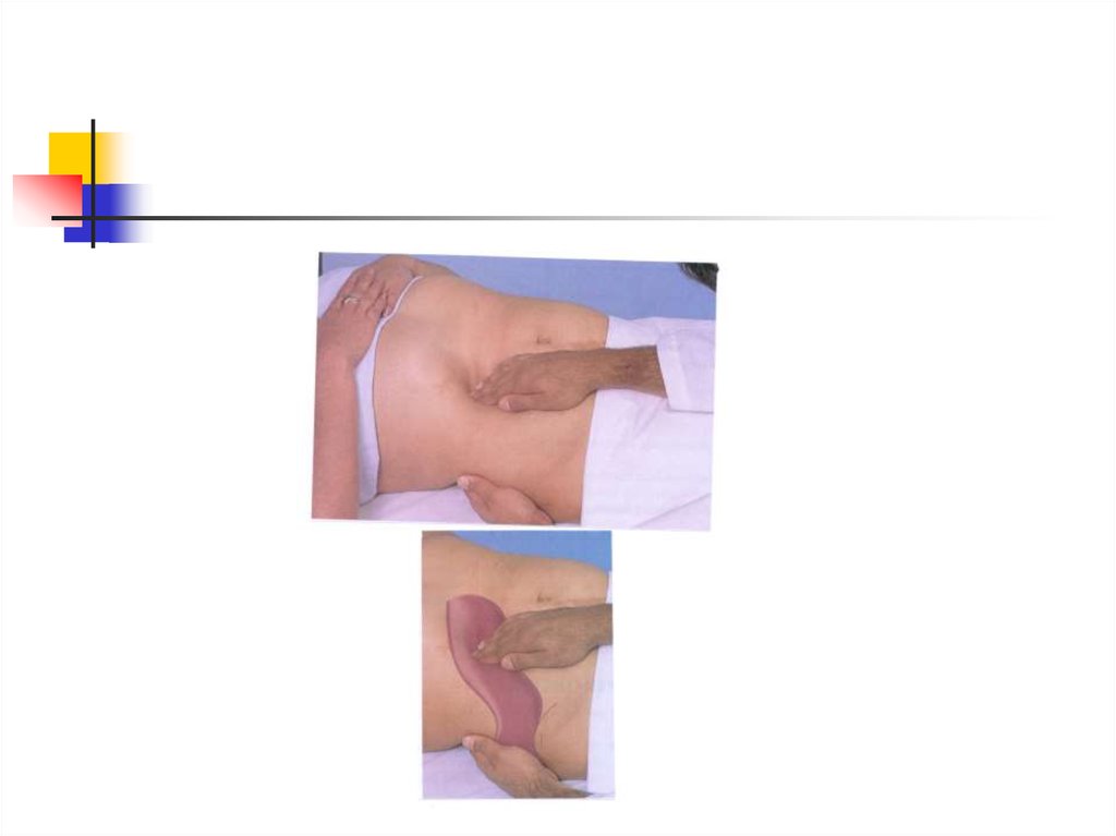
21. Examination of the Abdomen
Palpation (#43-#50) cont’dAssess peritoneal irritation and rebound
tenderness
Palpate liver, spleen, inguinal and femoral
lymph nodes
22.
23.
24.
25.
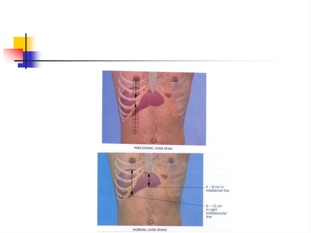
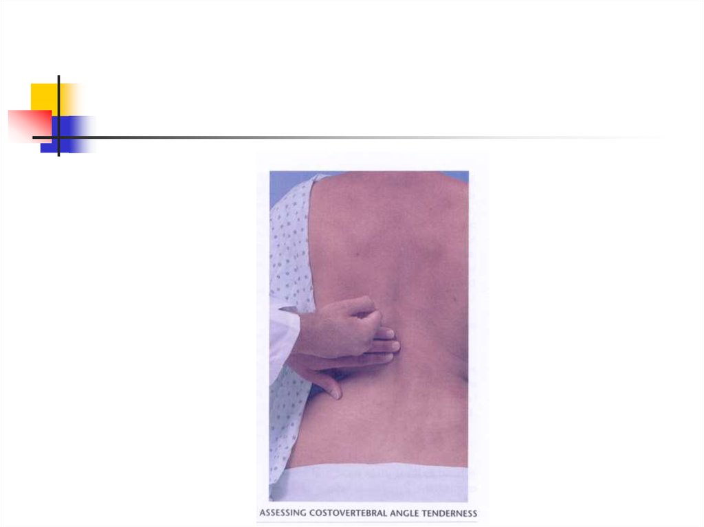
26. Examination of the Abdomen
Percussion (#48)Percuss the liver in mid-clavicular line.
Assess size by percussing upper and lower
borders. In COPD, normal sized livers are
frequently palpated and lower border may
be displaced downward.
In lean pts, spleen may be percussed
27.
28. Examination of the Abdomen
Rectal examination and stool specimenfor FOBT
Last step of the physical examination.
Stool sample retained for FOBT
29.
30.
31.
32.
33.
34.
35.
36.
37.
38.
39.
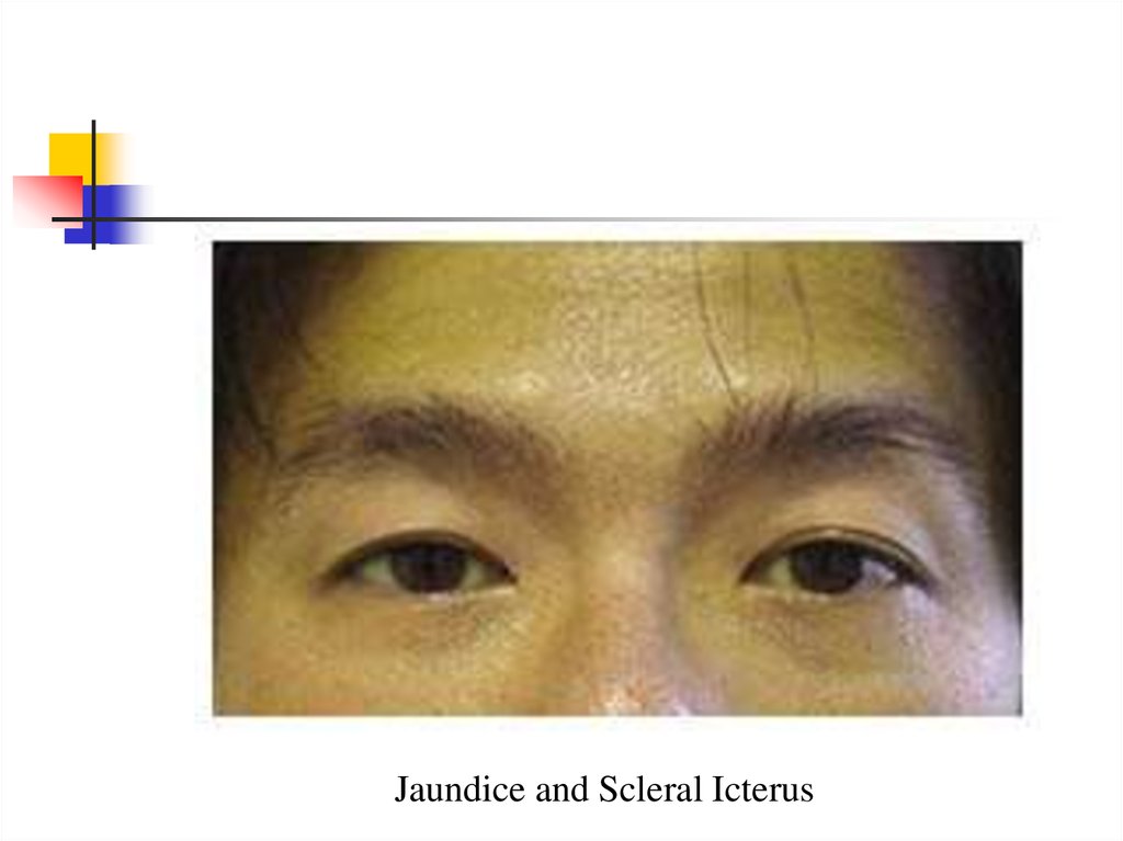
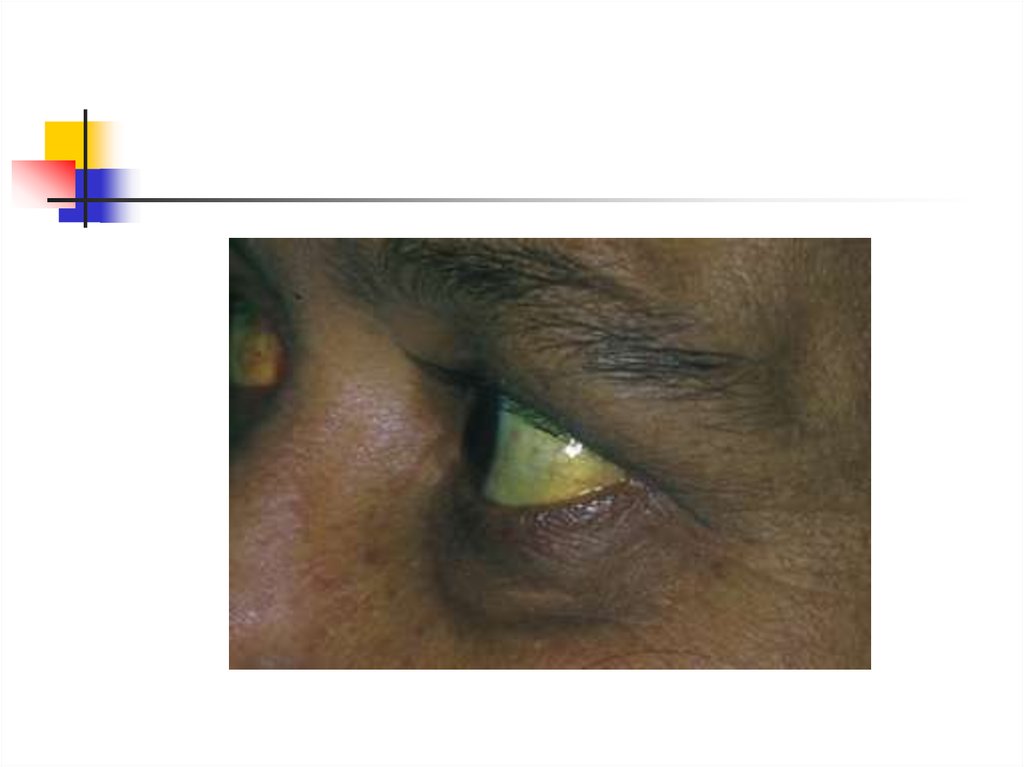
Jaundice and Scleral Icterus40.
41.
Gynaecomastia or enlargement of breast tissue inmen may occur either bilaterally or unilaterally.
42.
Palmar Erythema is charactarized by a prominent rim ofcolour beginning on the hypothenar border of the hand but
also in some individuals involving the thenar eminence and
even the fingertips. Similar changes nay be observed on
the soles of the feet.
43.
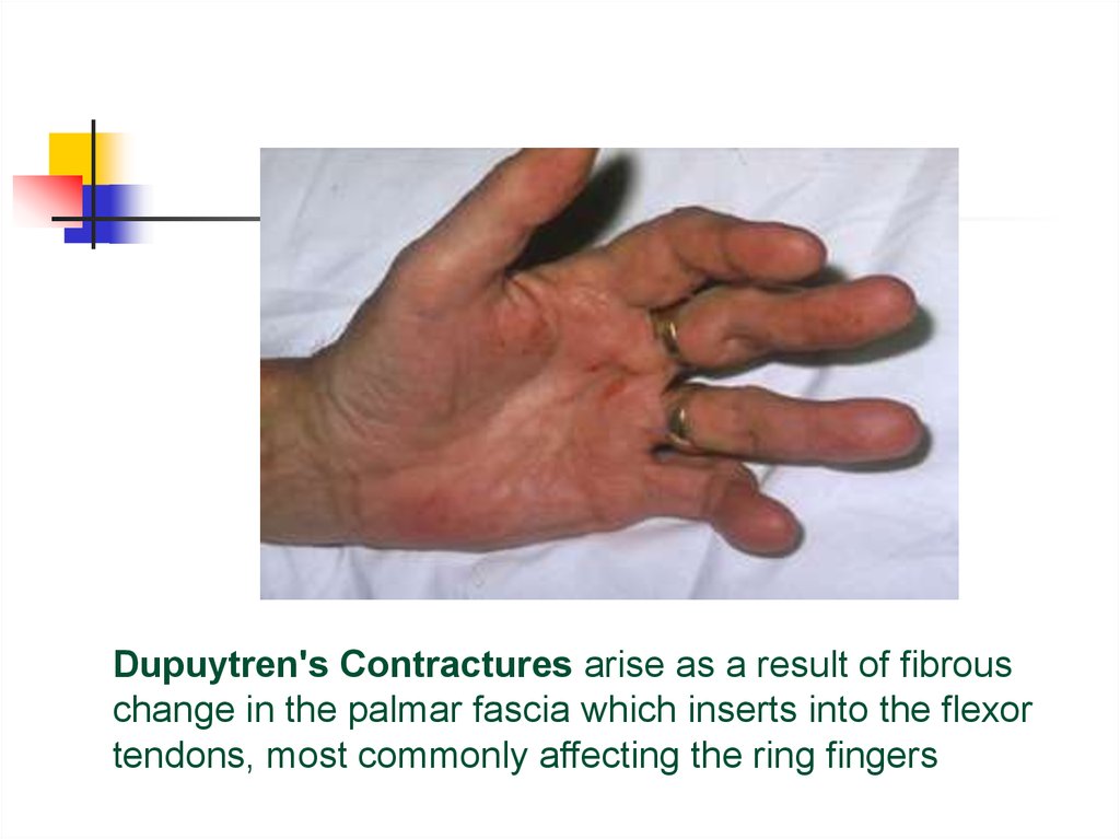
Dupuytren's Contractures arise as a result of fibrouschange in the palmar fascia which inserts into the flexor
tendons, most commonly affecting the ring fingers
44.
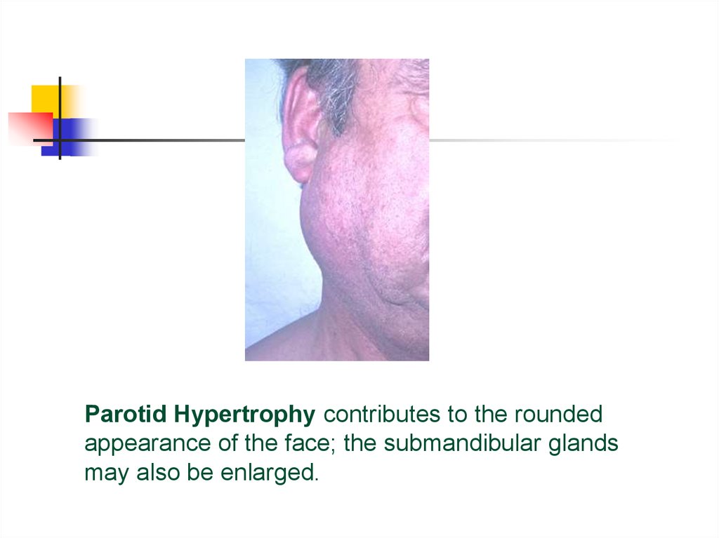
Parotid Hypertrophy contributes to the roundedappearance of the face; the submandibular glands
may also be enlarged.
45.
Spider Naevi are found only in the distribution of thesuperior vena cava, most commonly on the face and the
anterior chest wall. They comprise an enlarged central
arteriole from which vessels radiate in a spoke-like
manner.
46.
47.
48.
49.
50.
51.
52.
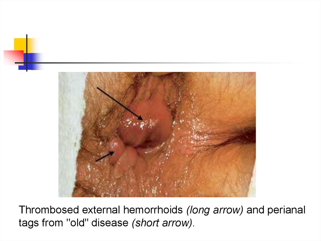
Thrombosed external hemorrhoids (long arrow) and perianaltags from "old" disease (short arrow).
53.
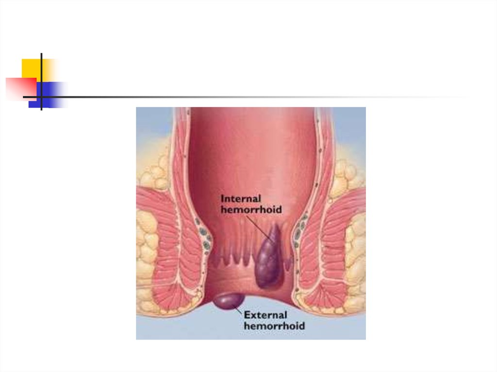
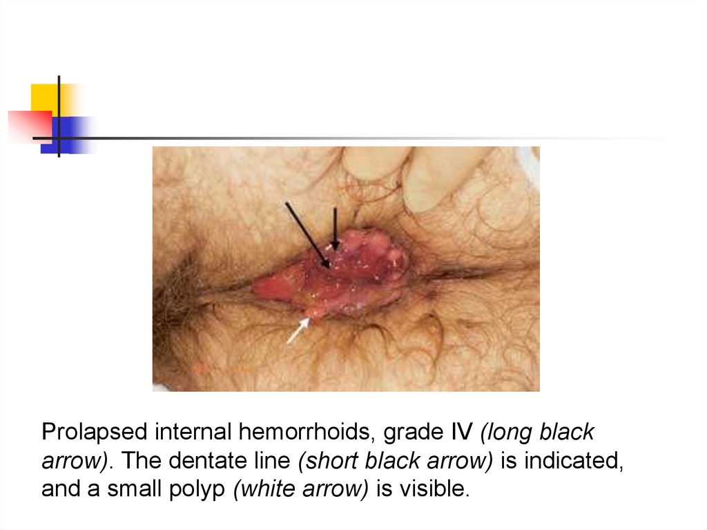
Prolapsed internal hemorrhoids, grade IV (long blackarrow). The dentate line (short black arrow) is indicated,
and a small polyp (white arrow) is visible.
54.
Extensive perianal condyloma acuminata (arrow). Thiscondition is generally caused by infection with human
papillomavirus 6 or 11.
55.
Acute posterior fissure (arrow). Anterior and posterior fissures are mostcommon. Fissures can often be identified by merely spreading the glutei but
generally require anoscopy. When fissures are found laterally, syphilis,
tuberculosis, occult abscesses, leukemic infiltrates, carcinoma, herpes,
acquired immunodeficiency syndrome (AIDS) or inflammatory bowel disease
should be considered as causes.
56.
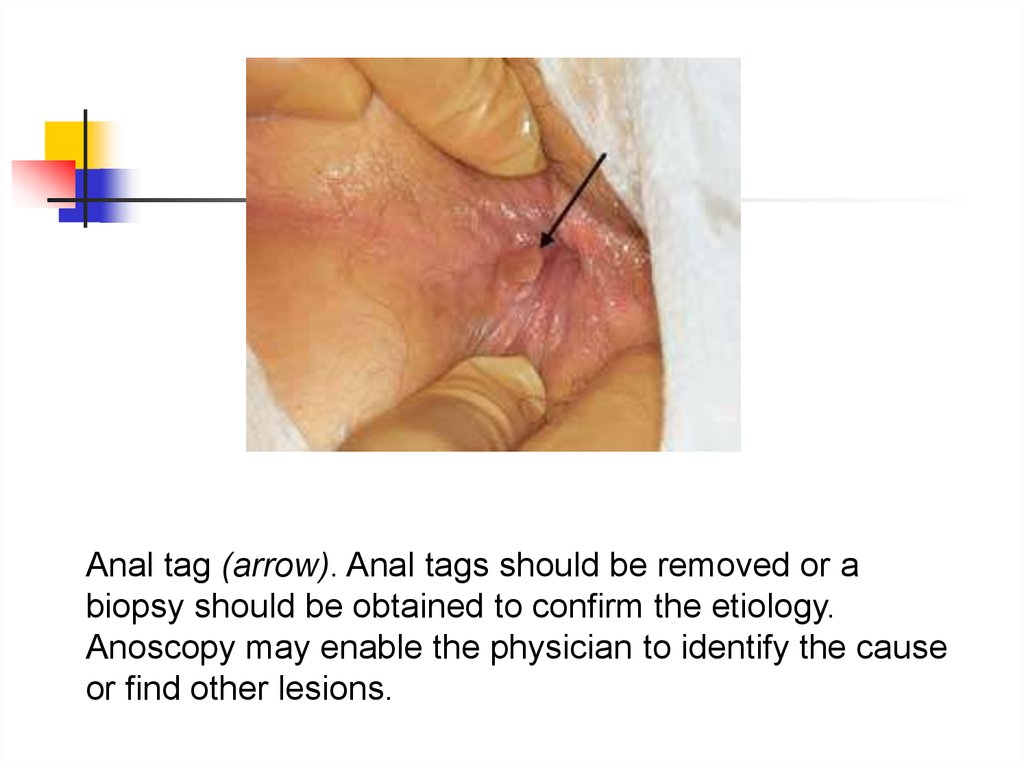
Anal tag (arrow). Anal tags should be removed or abiopsy should be obtained to confirm the etiology.
Anoscopy may enable the physician to identify the cause
or find other lesions.
57.
Anal cancer (arrow). This anal cancer had been treated forthree months with steroid suppositories although the
patient had never had a physical examination. Simple
inspection of the external anal area allowed the physician
to identify this aggressive tumor.
58.
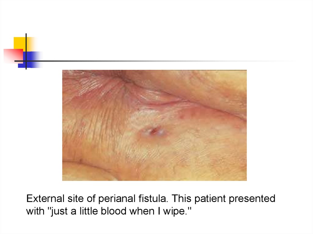
External site of perianal fistula. This patient presentedwith "just a little blood when I wipe."
59.
The wooden end of a cotton-tipped applicator was inserted 3cm (see Figure 5), confirming a fistula. Blood on the end of a
cotton-tipped applicator being withdrawn from a fistula that
could easily have been missed.