


")


 Медицина
Похожие презентации:


 infections")

Phylogenetic disorders of respiratory system
1. Medical Academy named after S.I. Georgievsky of Vernadsky CFU
2.
PHYLOGENETIC DISORDERSOF RESPIRATORY SYSTEM
REPRESENTED BY :
DHRUV MANGAL
195 b (LA-2)
SUPERVISOR:
ANNA ZHUKOVA
3. Respiratory syncytial virus
(RSV) is a major cause of lowerrespiratory tract infection in young children in both the
community and hospital setting.
4. BACKGROUND OF RSV
Respiratory syncytial virus (RSV) is an important cause ofbronchiolitis and pneumonia in infants and young children
[1]. Globally, it is estimated that RSV causes over 30 million
new acute lower respiratory tract infection (LRTI) episodes
annually, resulting in more than 3.4 million hospital
admissions and199,000 deaths in children younger than 5
years of age [1]. One-third of RSV-related deaths occur in the
first year of life, with 96 % of these deaths occurring in low resource countries
5. Laboratory diagnosis of RSV
A commercial multiplex PCR assay (Seeplex RV7, Seegene,Seoul, South Korea) was used to screen for 7 respiratory
viruses (Inf luenza A, Inf luenza B, Metapneumovirus, RSV
A/B, Rhinovirus A, Parainf luenza 1/2/3, Adenovirus) on
nasopharyngeal aspirate or bronchoalveolar lavage
specimens. Bacterial and viral co-pathogens were identified
on blood, tracheal aspirate or urine specimens obtained at
the discretion of the attending clinicians.
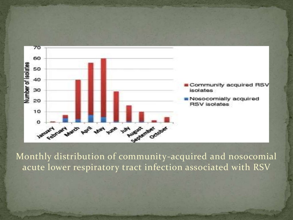
6. RESULTS OF RSV
Two-hundred and twenty-six children with PCR-confirmedRSV acute lower respiratory tract infection were identified
during the study period, January to October 2012. Figure 1
shows the monthly distribution of community-acquired and
nosocomial acute respiratory tract infection associated with
RSV. Case detection peaked in May.
7. Clinical characteristics, management and outcome
The median duration of symptoms preceding hospitalisationwas 2 days (IQR: 1–4 days). As shown in Table 3, the
commonest presenting symptoms were cough 196 (86.7 %),
difficulty in breathing (tight chest) 115 (50.9 %) and fever 91
(41.6 %). Wheezing was present in 20 (8.8 %) of the cases.
With regards to the management of the patients, 170 (75.2 %)
cases received supplemental oxygen, 89 (39.4 %) were
admitted either in the intensive care or high care units while
59 (26.1 %) required assisted ventilation by continuous
positive airway pressure (CPAP) or mechanical ventilation.
8. Genotype distribution and pattern of RSV infection
RSV A and RSV B accounted for 181 (80.1 %) and 45 (19.9 %)of the infections, respectively. There were no mixed
infections with both A and B groups. The prevalent
genotypes were NA1 (n = 127, 70.1 %), ON1 (n = 45, 24.9 %)
and NA2 (n = 9, 5.0 %) for groupA, while the only circulating
RSV B genotype was BA4. Age, gender, need for assisted
ventilation, HIV status and presence of co-pathogens were
not associated with the RSV genotypes.
9.
Monthly distribution of community-acquired and nosocomialacute lower respiratory tract infection associated with RSV
10. Factors associated with nosocomial RSV infection
Factors significantly associated with nosocomial infection onunivariate analysis included age 6 months or older and preexisting conditions. However, on multivariate analysis age 6
months or older was the only factor independently associated
with nosocomial infection with RSV (Table 6). The odds of
nosocomial infection was 3.35 times higher in infants and
children aged 6 months or older, compared to younger
infants (adjusted OR = 3.35 (1.20–9.36); p = 0.02).
11. HUMAN RHINOVIRUS
In order to evaluate the circulation of the different humanrhinovirus (HRV) species and genotypes in Italian children
with radiographically confirmed community-acquired
pneumonia (CAP), a nasopharyngeal swab was obtained from
643 children admitted to hospital because of CAP during five
consecutive winter and early spring seasons (2007-2012).
Real-time reverse transcriptase polymerase chain reaction
(RT-PCR) was used to identify HRV, and the HRV-positive
samples were used for sequencing analysis and to reconstruct
the phylogenetic tree
12. Study population and samples
The study was carried out in Pediatric Clinic 1 of the Department of Pathophysiology andTransplantation of the University of Milan during five consecutive years. The enrolment
occurred between November 1 and April 30 in the years 2007 -2008, 2008-2009, 2009-2010
and 2011-2011 and between November 1 and June 30 in 2011 -2012. It was approved by the
Institutional Review Board of the Fondazione IRCCS Ca’ Granda, Ospedale Maggiore
Policlinico, Milan, Italy. The written informed consent of a parent or legal guardian was
required, and the older children were asked to give their assent .
13. Nucleic acid extraction and real-time reverse transcriptase polymerase chain reaction (RT-PCR)
Viral nucleic acids were extracted from the nasopharyngealswabs using a Nuclisens EasyMAG automated extraction
system (Biomeriéux, Craponne, France), and the extracts
were tested for respiratory viruses using the RVP Fast assay
(Luminex Molecular Diagnostics Inc., Toronto, Canada) in
accordance with the manufacturer’s instructions. The RVP
Fast assay consists of a single multiplex polymerase chain
reaction (PCR) with labelled primers, followed by the singlestep hybridization of the PCR products with the f luorescent
bead array and incubation with reporter reagents.
14. Sequencing analysis, phylogeny and classification
The hypervariable part of the 5' NCR (non-coding region),the entire VP4 gene and the 5' terminus of the VP2 gene in
the HRV-positive samples were amplified by means of a RTPCR as previously described [6,14]. The PCR products were
purified using the Wizard SV Gel and PCR Clean-Up System
(Promega, Milan, Italy), and then sequenced in both
directions using the same forward and reverse primers as
those used in the PCR. The nucleotide sequences were
obtained by means of automated DNA sequencing using an
ABI PRISM 3730 genetic analyser (Applied Biosystems, Foster
City, CA)
15. Genetic variations and clustering of strains
The HRV sequences showed marked genetic diversity. TheHRV-C sequences were the most heterogenous, with an intraspecies nucleotide p-distance of 0.25, which was greater than
the p-distance within HRV-A (0.20) or HRV-B (0.21).
Nucleotide variability was 37% between HRV-A and HRV-B,
37.3% between HRV-A and HRV-C, and 39.9% between HRVB and HRV-C. Figure 2 shows the phylogenetic tree
constructed on the basis of the VP4/VP2 region of the 151
sequences from this study and the references sequences.
16. Pasteurella multocida
is a leading cause of respiratorydiseases in many host species. To understand the genetic
characteristics of P. multocida strains isolated from different
host species, we sequenced the genomic DNA of P. multocida
isolated from pigs and analyzed the genetic characteristics of
strains from avian species, bovine species, pigs, and rabbits
using whole genome sequence (WGS) data. Our results found
that a capsular: lipopolysaccharide (LPS): multilocus
sequence typing (MLST) genotype A: L1: ST129 (43.75%) was
predominant in avian P. multocida; while genotypes B: L2:
ST122 (60.00%) and A: L3: ST79 (30.00%) were predominate
in bovine P. multocida; genotype D: L6: ST50 (37.50%) in
porcine P.
17. P. multocida Strains and Whole Genome Sequencing
A total of 47 P. multocida strains were selected for wholegenome sequencing in this study (Table 1). Most of these
strains were isolated from pigs with respiratory disorders in
China (Peng et al., 2018), with the exception of strain HN04,
which is a capsular type B isolate from a swine haemorrhagic
septicaemia case. The capsular types and LPS genotypes of
these 47 P. multocida isolates were determined through PCR
assays, as described previously
18. Phylogenetic Trees
he phylogenetic relationship between P. multocida strainsfrom different host species was predicted by analyzing the
whole-genome single nucleotide polymorphisms (WG-SNPs)
as well as the set of SNPs present in all single-copy core genes
across genomes (CG-SNPs). The WG-SNPs were identified by
comparing each of the WGSs against the reference P.
multocida ATCC 43137 genome sequence (GenBank accession
NO. CP008918) using MAFFT (version 7.222) software (Katoh
and Standley, 2013).
19.
20. Results
Whole genome sequencing yielded approximately796.25~1823.87 Mbp raw data for the 47 porcine P. multocida
isolates. The data filter strategy produced approximately
701.86~1589.23 Mb clean data for assembly. Sequences
assembled using SPAdes v3.9.0 (Bankevich et al., 2012)
generated approximately 18~66 contigs for the 47 porcine P.
multocida isolates, with an average of 25 contigs for each
strain.