


 immunity.")


")
 on a cell - target.")
")


 Медицина
Похожие презентации:


")


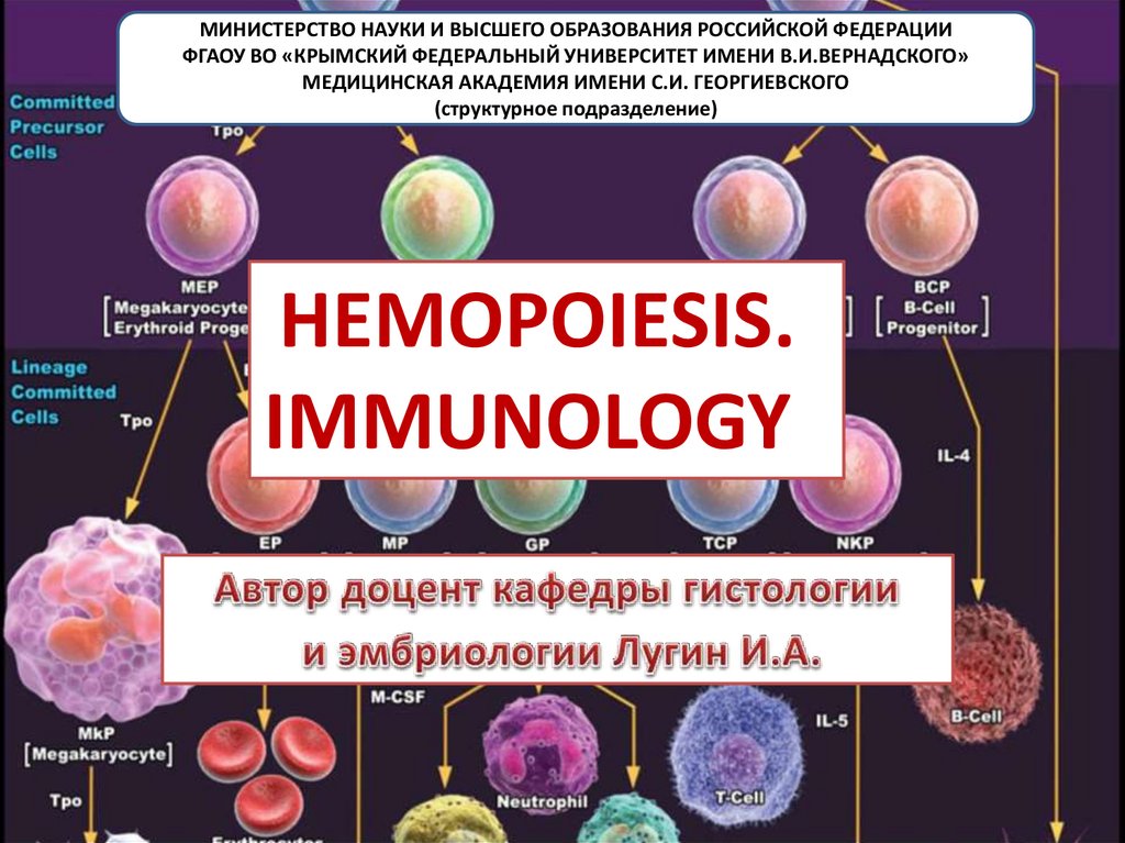
Hemopoiesis. Immunology
1.
МИНИСТЕРСТВО НАУКИ И ВЫСШЕГО ОБРАЗОВАНИЯ РОССИЙСКОЙ ФЕДЕРАЦИИФГАОУ ВО «КРЫМСКИЙ ФЕДЕРАЛЬНЫЙ УНИВЕРСИТЕТ ИМЕНИ В.И.ВЕРНАДСКОГО»
МЕДИЦИНСКАЯ АКАДЕМИЯ ИМЕНИ С.И. ГЕОРГИЕВСКОГО
(структурное подразделение)
HEMOPOIESIS.
IMMUNOLOGY
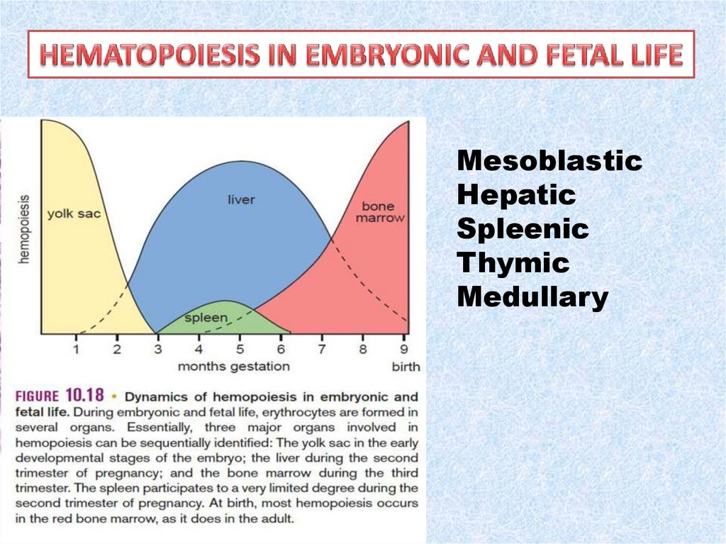
2. HEMOPOIESIS.
• The formation of blood cells in the penatal life is namedHemopoiesis (Gr. haima, blood + poiesis, a making).
Mature blood cells have a relatively short life span and
must be continuously replaced with new cells from
precursors developing In the early embryo these blood
cells arise in the yolk sac mesoderm. In the second
trimester, hemopoiesis (also called hematopoiesis) occurs
primarily in the developing liver, with the spleen playing a
minor role
• Skeletal elements begin to ossify and bone marrow
develops in their medullary cavities, so in the thirdtrimester marrow of specific bones becomes the major
hemopoietic organ.
3.
MesoblasticHepatic
Spleenic
Thymic
Medullary
4. Theories of hematopoiesis
• The monophyletic theorysuggests that a pluripotent
stem cell (CFU-S) can form all
mature blood cell types.
• The several polyphyletic
theories suggest that each
mature blood cell type is
derived from a distinct stem
cell.
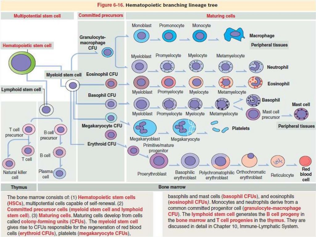
5. Hemopoietic Stem Cells
All blood cells arise from a single type of pluripotent
hemopoietic stem cell in the bone marrow that can
give rise to all the blood cell types. These
pluripotent stem cells are rare, proliferate slowly,
and give rise to two major lineages of progenitor
cells with restricted potentials (committed to
produce specific blood cells): one for lymphoid cells
(lymphocytes) and another for myeloid cells (Gr.
myelos, marrow), which develop in bone marrow.
Myeloid cells include granulocytes, monocytes,
erythrocytes, and megakaryocytes.
The immune system, the lymphoid progenitor cells
migrate from the bone marrow to the thymus or
the lymph nodes, spleen, and other lymphoid
structures, where they proliferate and
differentiate.
6. Progenitor & Precursor Cells
Progenitor & Precursor Cells• The progenitor cells for blood cells are often called colonyforming units (CFUs), because they give rise to colonies of
only one cell type when cultured in vitro or injected into a
spleen.
• There are four major types of progenitor cells/CFUs:
• Erythroid lineage of erythrocytes
• Thrombocytic lineage of megakaryocytes for platelet
formation
• Granulocyte-monocyte lineage of all three granulocytes
and monocytes
• Lymphoid lineage of B lymphocytes, T lymphocytes, and
natural killer cells
7.
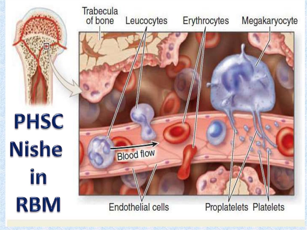
8. Hematopoietic stem cell niche
• This event requires a special environment,termed the hematopoietic stem cell niche, which
provides the protection and signals necessary to
carry out the differentiation of cells from HSC
progenitors. This niche relocates from the yolk
sac to eventually rest in the bone marrow of
mammals. Many pathological states can arise
from disturbances in this niche environment,
highlighting its importance in maintaining
hematopoiesis.
9.
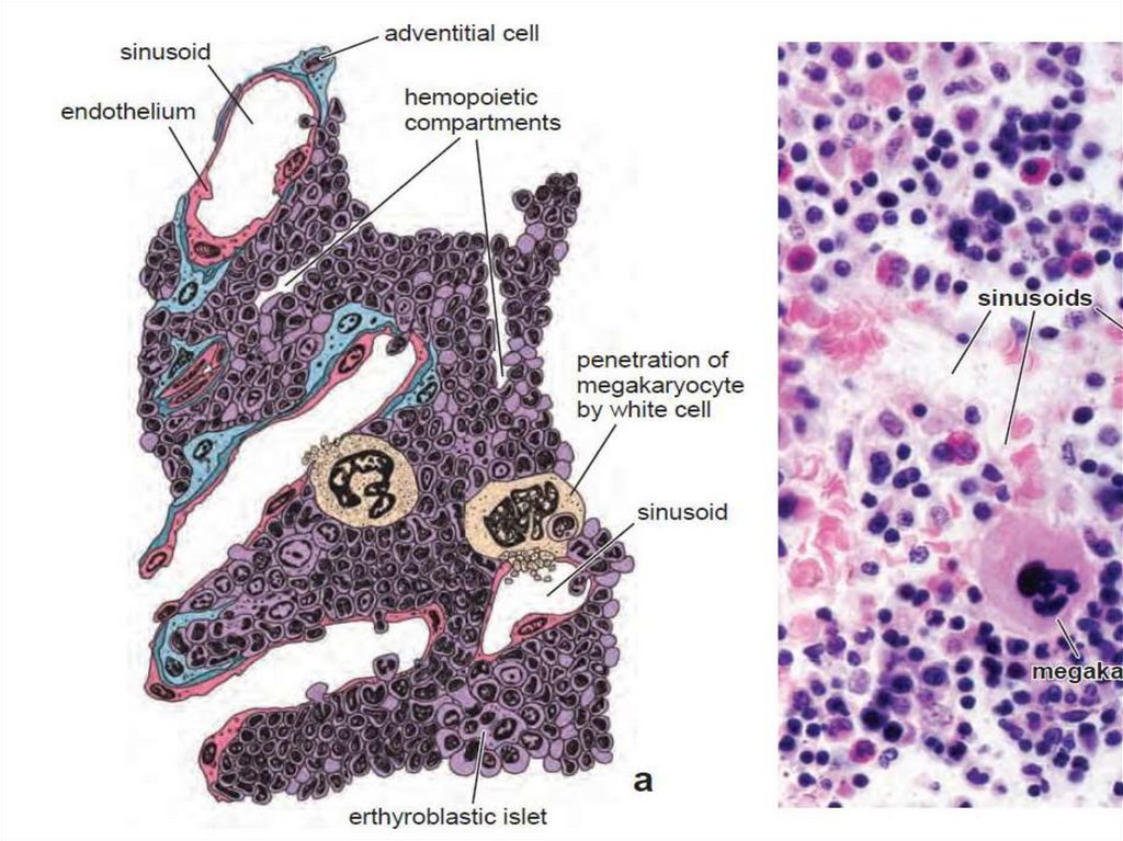
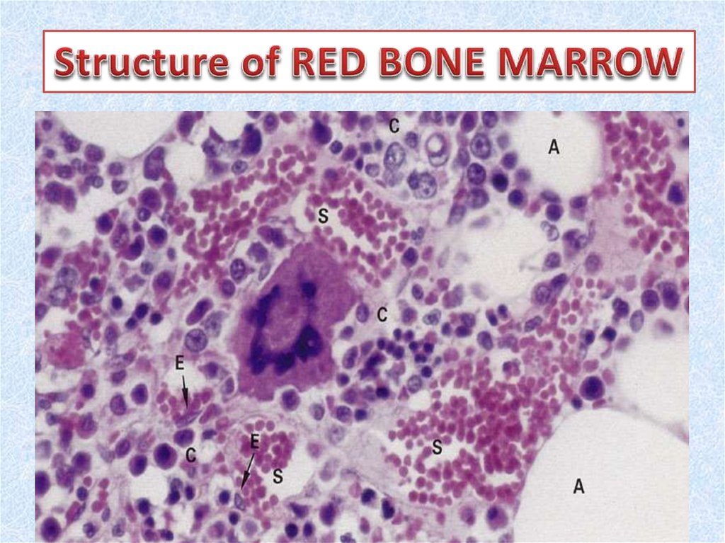
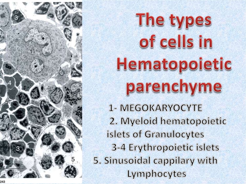
10. Red bone marrow
• Red bone marrow contains a reticular connective tissue stroma(Gr. stroma, bed), hemopoietic cords or islands of cells, and
sinusoidal capillaries.
• The stroma is a meshwork of specialized fibroblastic cells
called stromal cells (also called reticular or adventitial cells)
and a delicate web of reticular fibers supporting the
hemopoietic cells and macrophages.
• The matrix of bone marrow also contains collagen type I,
proteoglycans, fibronectin, and laminin, the latter
glycoproteins interacting with integrins to bind cells to the
matrix. Red marrow is also a site where older, defective
erythrocytes undergo phagocytosis by macrophages, which
then reprocess heme-bound iron for delivery to the
differentiating erythrocytes.
11.
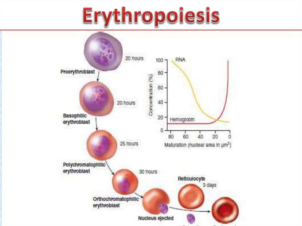
12. Erythropoiesis
• Erythropoiesis. In healthy adults, erythropoiesis (red bloodcell formation) occurs exclusively in bone marrow.
Erythrocytes derive from CFU-Es, which in turn derive from
CFU-Ss. The differentiation of erythrocytes from stem cells
is commonly described by naming cell types at specific
stages in the process according to their histologic
characteristics. Cellular changes that occur during erythroid
differentiation include (1) decrease in cell size, (2)
condensation of nuclear chromatin, (3) decrease in nuclear
diameter, (4) accumulation of hemoglobin in the cytoplasm
(increased acidophilia), (5) decline in the number of
ribosomes in the cytoplasm (decreased basophilia) and (6)
ejection of the nucleus.
13.
14.
15. Erythrocyte maturation
• is commonly divided into 6 stages. Cells at these stages(class of cells) are identified by examining their overall
diameter, the size and chromatin pattern of their
nuclei, and the staining properties of their cytoplasm.
Cells in transition between these stages are commonly
found ill bone marrow smears. Cell division occurs
throughout the early stages, but once cells reach the
normoblast stage they generally lose their ability to
divide. tarts with the least mature cells; the sixth stage
is the mature erythrocyte (privious pages). f.
Proerythroblasts are large (14—19 um in diameter) and
contain a large, centrally ocated, pale-staining nucleus
with one or 2 large nucleoli
16. Erythrocyte maturation
• The small amount of :ytoplasm (about 20% of cellvolume) contains polyribosomes actively involved in
lemoglobin synthesis. The resulting cytoplasmic
basophilia allows these cells to be listinguished from
myeloblasts, with which they are most easily confused,
'roerythroblasts are capable of multiple mitoses and
may be considered unipoten- ialstem cells. 2.
Basophilic erythroblasts are slightly smaller than
proerythroblasts, vith a diameter of 13—16 um. They
have slightly smaller nuclei with patchy chromatin.
Their nucleoli are difficult to distinguish
17. Erythrocyte maturation
• The cytoplasm is more intensely >asophilic,typically staining a deep royal blue. A prominent,
clear, juxtanuclear ;ytocenter is often visible.
Basophilic erythroblasts continue hemoglobin
synthesis il a high rate and are capable of mitosis.
3. Polychromatophilic erythroblasts are mailer
yet (12-15 um in diameter), with significant
amounts of hemoglobin begin- ling to accumulate
in their cytoplasm. The conflicting staining
affinities of the )olyribosomes (basophilic) and
hemoglobin (acidophilic) give the cytoplasm a
trayish appearance.
18. Erythropoiesis
• The nucleus is smaller than in less mature cells,with more :ondensed chromatin that forms a
checkerboard pattern. These cells can still synhesize hemoglobin and divide. 4. Normoblasts
(orthochromatophilic erythroblasts) ire easily
identified because of their small size (8—10 um in
diameter); acidophilic :ytoplasm with only traces
of basophilia; and small, eccentrically placed
nuclei ivith chromatin so condensed that it
appears black. Although early normoblasts
19.
20. Leukopoiesis
• Leukopoiesis (white blood cell formation) encompassesboth granulopoiesis and agranulopoiesis. Leukopoietic
CFUs that have been identified include CFU-GM (forms
both granulocytes and macrophages), CFU-G (forms all
granulocyte types), CFU-M (forms macrophages), and
CFU-Eo (forms only eosinophils). All these CFUs with
limited capabilities derive from the pluripotential CFU-S.
• Granulopoiesis occurs in the bone marrow of healthy
adults. The three types of granulocytes — neutrophils,
basophils, and eosinophils — may all derive from a single precursor (CFU-G).
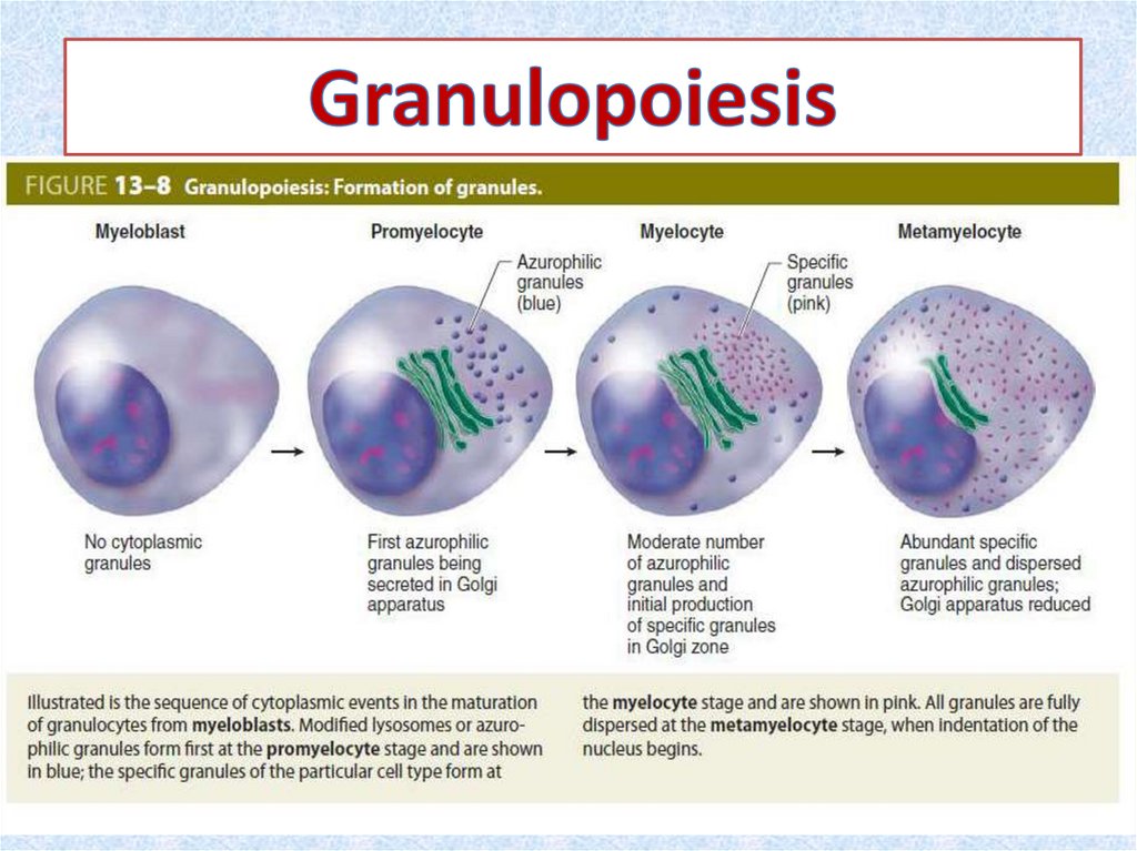
21. Maturation of Granulocytes
• The structural changes include (1) decrease in cellsize, (2) condensation of nuclear chromatin, (3)
changes in nuclear
• shape (flattening ► indentation ► lobulation, a
progression resembling the gradual deflation of a
balloon), and (4) accumulation of cytoplasmic
granules. Granulocyte maturation is commonly
divided into 6 stages. These stages are identified
by examining overall diameter; size, shape, and
chromatin pattern in the nuclei; and type and
number of specific granules in the cytoplasm
22.
23.
1. Myeloblasts, the earliestrecognizable granulocyte
precursors, are about 15
um in diameter.
2. Promyelocytes are larger
than myeloblasts (15-24
um in diameter) and their
chromatin is slightly more
condensed
3. Myelocytes are typically
smaller than promyelocytes (10-16 um in
diameter). This is the first
stage at which sufficient
numbers of specific
granules accumulate in the
cytoplasm to allow one to
distinguish the 3 immature
granulocyte types —
neutrophilic
24.
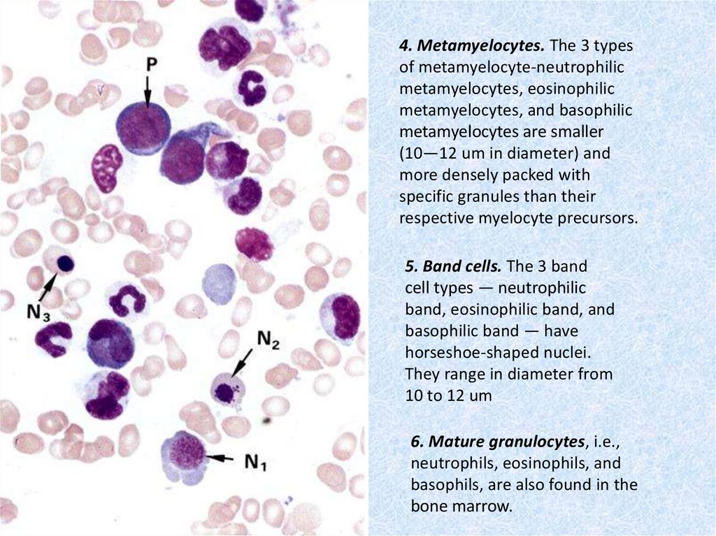
4. Metamyelocytes. The 3 typesof metamyelocyte-neutrophilic
metamyelocytes, eosinophilic
metamyelocytes, and basophilic
metamyelocytes are smaller
(10—12 um in diameter) and
more densely packed with
specific granules than their
respective myelocyte precursors.
5. Band cells. The 3 band
cell types — neutrophilic
band, eosinophilic band, and
basophilic band — have
horseshoe-shaped nuclei.
They range in diameter from
10 to 12 um
6. Mature granulocytes, i.e.,
neutrophils, eosinophils, and
basophils, are also found in the
bone marrow.
25. Agranulopoiesis
• Agranulopoiesis: agranulocytes (monocytesand lymphocytes), like the other blood cell
types, derive from CFU-Ss. The morphologic
changes during maturation include a decrease
in overall cell diameter, a decrease in nuclear
diameter and an increase in nuclear
heterochromatin content. However, the
morphologic characteristics of agranulocytes
at immature stages are much less distinct than
those of erythrocytes and granulocytes
26. Monocytopoiesis
• 1. Monocytopoiesis. The CFU derivatives thatgive rise to monocytes are called monoblasts and
are difficult to identify in bone marrow smears. A
product of the monoblast, the promonocyte, is
only slightly easier to identify and serves as the
immediate precursor of monocytes.
Promonocytes are larger (10-20 um in diameter)
than monocytes and have pale staining nuclei and
basophilic cytoplasm. The similarity between
monocyte precursors and other stem cells in the
bone marrow makes identification difficult.
27. Lymphopoiesis
• 2. Lymphopoiesis. In adults, lymphopoiesis occursmainly in lymphoid tissues and organs and to a lesser
extent in bone marrow. Prior to division, the precursor,
or lymphoblast, is usually much larger than the typical
circulating lymphocyte .
• However, many circulating lymphocytes can respond to
antigenic stimulation by blasting (enlarging to assume
the typical lymphoblast morphology), indicating that
they are dormant stem cells. Some of these cells,
called null cells, are neither T nor B cells and may
represent a circulating form of the CFU-Ss.
28. Thrombopoiesis
• Thrombopoiesis. Platelet (thrombocyte)production is carried out in the bone marrow
by unusually large cells (100 um in diameter)
called megakaryocytes. Immature
megakaryocytes, called megakaryoblasts,
derive from CFU-Megs, which in turn derive
from CFU-Ss. Megakaryoblasts undergo
successive incomplete mitoses involving
repeated DNA replications without cellular or
nuclear division
29. Maturation of Megakaryocyte
• The result of this process, called endomitosis, is asingle large megakaryocyte with a single, large,
multilobed, polyploid (up to 64n) nucleus.
Maturation involves lobulation of the nucleus and
development of an elaborate demarcation
membrane system that subdivides the peripheral
cytoplasm, outlining cytoplasmic fragments
destined to become platelets. As the demarcation
membranes fuse to form the plasma membranes
of the platelets, ribbon like groups of platelets are
shed from the megakaryocyte periphery into the
marrow sinusoids to enter the circulation
30.
31.
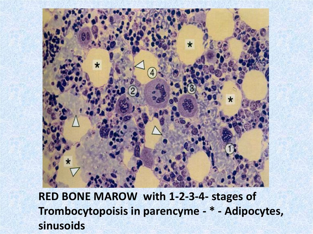
RED BONE MAROW with 1-2-3-4- stages ofTrombocytopoisis in parencyme - * - Adipocytes,
sinusoids
32. Regulation of hematopoiesis
• involves specific colony-stimulating factors (CSFs) suchas erythropoietin, leukopoietin and thrombopoietin.
These hormones act at various steps in hematopoiesis
to enhance proliferation and differentiation of CFUs.
• Some growth factors—principally three interleukins
(IL-1, IL-3, IL-6)—stimulate proliferation of
pluripotential and multipotential stem cells, thus
maintaining their populations. Additional cytokines,
granulocyte colony-stimulating factor (G-CSF), IL-3, IL7, IL-8, IL-11, IL-12, macrophage inhibitory protein- ,
and erythropoietin, are believed to be responsible for
the mobilization and differentiation of these cells into
unipotential progenitor cells
33. Erythropoietin/ Thrombopoietin
• CSFs are also responsible for the stimulation of cell divisionand for the differentiation of unipotential cells of the
granulocytic and monocytic series. Erythropoietin activates
cells of the erythrocytic series, whereas thrombopoietin
stimulates platelet production. Steel factor (stem cell
factor), which acts on pluripotential, multipotential, and
unipotential stem cells, is produced by stromal cells of the
bone marrow and is inserted into their cell membranes.
Stem cells must come in contact with these stromal cells
before they can become mitotically active. It is believed
that hemopoiesis cannot occur without the presence of
cells that express stem cell factors, which is why postnatal
blood cell formation is restricted to the bone marrow (and
liver and spleen, if necessary).
34. Cell lineages
• Hemopoiesis is initiated in an apparentrandom manner when individual stem cells
begin to differentiate into one of the blood
cell lineages. Stem cells have surface
receptors for specific cytokines and growth
factors that influence and direct their
proliferation and maturation into a specific
lineage
35. INTERACTION OF IMMUNE CELLS
• The immune system of an organism consist oftwo basic ingredients: organs of a
hemopoiesis and lymphoid organs (a red bone
marrow, a thymus gland, a spleen, a lymph
nodes) and immune cells, or immunocytes.
• Main function of immunocytes is to provide
organism responses on a specific discernment
and destruction (elimination) of an antigen.
36. lymphoid organs
• Typical immunocytes are Т-and B-lymphocytes,macrophages and plasmocytes. The leading part in
responses of artificial immunity belongs to
lymphocytes as only they can specificly recognize a
concrete antigen.
• All lymphoid tissues and organs produce lymphocytes.
In peripheral lymphoid organs (lymph nodes, spleen,
tonsils) and unencapsulated lymphatic aggregates,
lymphocyte production is antigen-dependent and
provides committed immunocompetent cells that
respond to specific antigens.
37. Central lymphoid organs
• In central lymphoid organs (thymus, bonemarrow, bursa of Fabricius [in birds]), lymphocyte
production is antigen-independent and supplies
uncommitted T-lymphocyte (thymus) or Blymphocyte (bone marrow, bursa) precursors that
subsequently move to peripheral organs and
tissues. Mounting effective immune responses to
new antigens requires ongoing production of
uncommitted lymphocytes by the central
lymphoid organs.
38. Cellular (cell-mediated) immunity.
• Activated Т lymphocytes differentiate intospecialized cell types, some of which (CD8+)
contact and kill intruding cells, and some of
which (CD4+) release cytokines, substances
that enhance various aspects of the immune
response. Cytokines are interleukins (IL): IL 1,
IL 4, IL 5, IL 6, interferons, the factor of a
necrosis of tumour.
39. Humoral immunity
• Activated В lymphocytes differentiate intoplasma cells that secrete antigen-binding
immunoglobulins (antibodies), which
circulate in the blood and lymph.
• Immunologic memory. Lymphoid function in
response to initial exposure to a particular
infection protects an organism during
subsequent exposure to the same infective
agent.
40. Specificity
• Specificity. An ability to respond to one type ofinfection (chicken pox) does not imply resistance
to another (tuberculosis).
• Tolerance. Antigen-disposal mechanisms directed
toward the body's own cells (as occurs
occasionally in autoimmunity) can be disastrous,
even fatal. Thus, a key aspect of immune function
is the ability to distinguish "self from "nonself"
antigens, and to tolerate the self.
41. Lymphocyte programming and activation
• This multistep process is outlined below.• 1. Cells of mesodermal origin are programmed in the
bone marrow or thymus as B- or T-lymphocyte
precursors, respectively.
• 2. These cells subsequently move to peripheral organs,
where each encounters a specific antigen to which it
becomes programmed (committed) to respond. The
concentration of antigens on the surfaces of antigenpresenting cells, or the delivery of processed antigens
to lymphocytes by macrophages, improves the
efficiency of this step over that available from random
lymphocyte-antigen collisions.
42.
43. Selectively stimulation
• 3. Not all lymphocytes can respond to allantigens. Our ability to respond to a variety of
antigens rests in the diversity of antigenbinding capabilities of virgin (preactivated)
lymphocytes. It is estimated that lymphocytes
able to bind more than a billion different
antigens are present prior to any antigenic
challenge. When such a challenge occurs, a
lymphocyte able to bind the antigen is
selectively stimulated to divide (activated).
44. Clonal expansion
• Activated cells enlarge and form lymphoblasts(blast transformation) and subsequently
undergo a series of divisions (clonal
expansion), forming a clone of cells
competent to recognize that antigen. This
process is termed clonal selection. Many
immunocompetent lymphocyte clones may be
generated in response to different parts of a
single antigen.
45.
46. Secondary immune response
• 4. The products of this initial clonal expansionundergo differentiation into two basic cell types;
effector cells, which immediately begin antigen
disposal (primary immune response), and
memory cells, which are held in reserve for
subsequent encounters with the antigen
(secondary immune response). T-lymphocyte
derivatives form three main effector cell types,
which enter the circulation and search the body
for their antigens, providing cellular immunity. Blymphocyte derivatives form
47.
Clonal expansion and differentiation of B-lymphocytesand Plasma cells
48.
• 5. When the same antigen is again encountered,memory cells generated during the initial clonal
selection and expansion (either Т or B) undergo
the same process—blast transformation, clonal
expansion and differentiation—that occurs during
the primary response, but more rapidly (with a
shorter lag time between exposure and response)
and more effectively (owing to the increased
number of responsive cells, and the greater
affinity of the antibodies) than before.
49. Antigens
• These are foreign (nonself) substances that are able toelicit an immune response (cellular, humoral, or both).
They can be entire cells (bacteria, tumor cells) or large
molecules (proteins, polysaccharides, nucleoproteins).
Their antigenicity is determined by several factors:
larger and more complex (branched or folded)
molecules are more potent antigens than smaller,
simpler ones; proteins are more antigenic than
carbohydrates; and lipids are nonantigenic unless
complexed with a more potent antigen. Particularly
potent antigens are said to be immunodominant. The
site of entry of an antigen into the body also can affect
its antigenicity.
50.
• The specific part of an antigen that elicits theimmune response (and to which the antibodies bind) is called an antigenic
determinant, or epitope; it can consist of a
monosaccharide or as few as four to six amino
acids. Thus a bacterium can have many
antigenic determinants and elicit many
cellular and humoral responses.
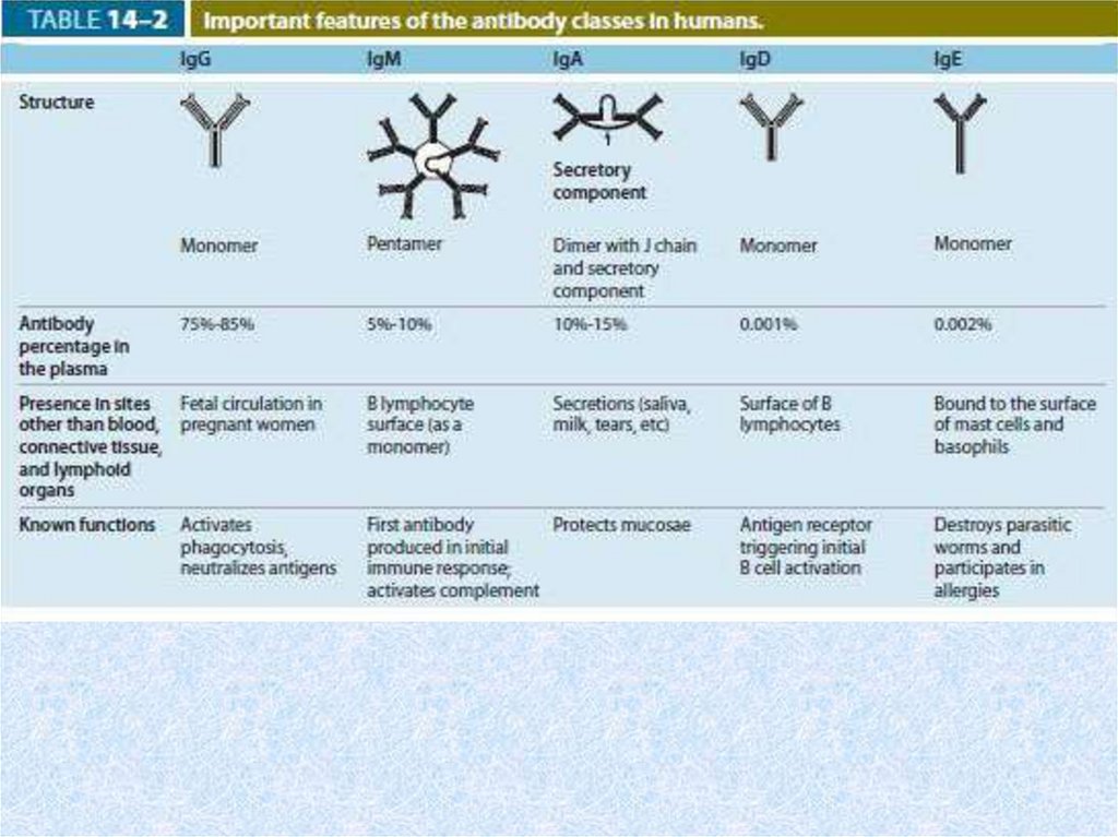
51. Immunoglobulins (Ig)
• These antibodies are proteins secreted by plasma cellsinto body fluids (blood, lymph, tissue fluid, saliva,
tears, milk, mucus) in response to antigenic
stimulation. They bind with high affinity to the
antigenic determinants that elicited their production
and make up most of the blood's gamma-globulins.
• Immunoglobulins (antibodies) are immune protective
proteins. Everyone Ig has rigorous specificity to
concrete antigen. Exist in two forms: а) as
membranous receptors of a B-lymphocytes; б) as the
antibodies loosely circulating in a blood plasma and a
lymph.
52.
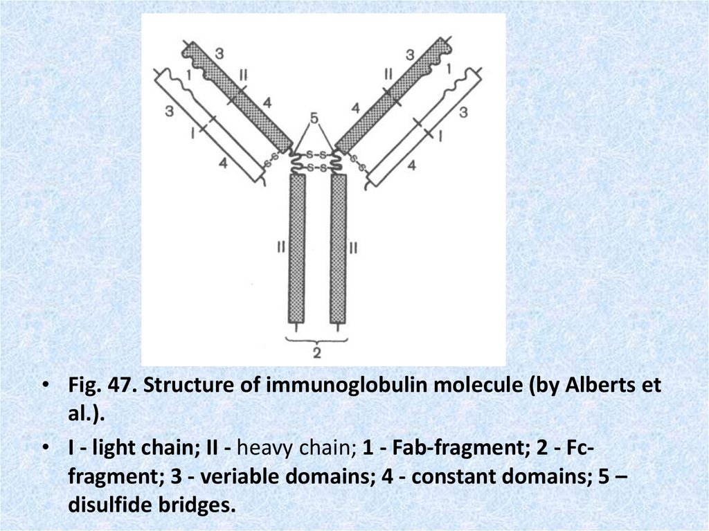
• Fig. 47. Structure of immunoglobulin molecule (by Alberts etal.).
• I - light chain; II - heavy chain; 1 - Fab-fragment; 2 - Fcfragment; 3 - veriable domains; 4 - constant domains; 5 –
disulfide bridges.
53.
54. The mechanism of cytolytic activity of the Т-killer (Т-cytotoxic lymphocyte) on a cell - target.
The mechanism of cytolytic activity of the Т-killer (Тcytotoxic lymphocyte) on a cell - target.• T - cytotoxic lymphocyte is effector of cellular immunodefence. Cytotoxic
(cytolytic) responses is effector immune mechanisms direct on elimination
of cells which are too large for a phagocytosis by routine phagocytes
(neutrophils). The cell of an antigen (a bacterium, a cancer cell, cell with
virusis) is a cell - target for the Т-killer. The cell with virus contains a
complex consisting of the MHC 1 and virus peptides on the plasmolemma.
The Т-killer with help of TCR recognize an antigenic peptide, and with the
help of receptor CD8 finds out a molecula of the MHC 1. Thus the Т-killer
forms with a cell - target strong communication. Then the Т-killer secrite
from the granules proteins perforin and granzims. Perforin invokes pores
and ion channels in a plasmolemma of a target cell. Through pores inside
of a cell - target water starts to come uncontrolledly and it bursts. Besides
through pores go to cytoplasm granzims (major of them granzim В). They
include in a nucleus of a cell - target the mechanism of apoptosis
(genetical programmed destruction of a cell). As a result of an apoptosis
activation the cell - target blasts itself. After a secretion of perforin and
granzim Т-cytotoxic lymphocyte is disconnected from a target and
searches for a new antigen to manufacture new cytolysis.
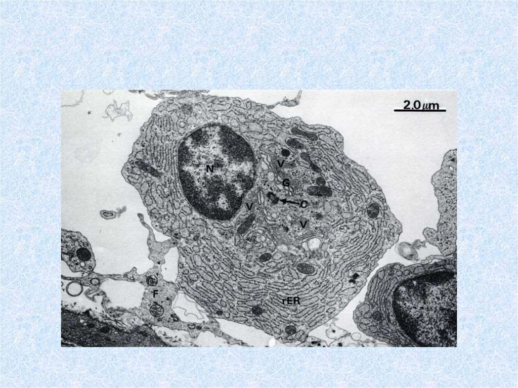
55. Plasma cells (plasmocytes)
• are differentiated B-lymphocyte effector cells secretethe Igs primarily responsible for humoral immunity.
Their morphology includes a "clock face" nucleus,
basophilic cytoplasm and abundant rouph endoplasmic
reticulum typical of protein-secreting cells. Plasma
cells, found in all lymphoid tissues and loose
connective tissue, occur in high concentration in the
medullary cords of lymph nodes, the red pulp cords in
the spleen, and the lamina propria under mucosal and
glandular epithelia. They are rare in the thymus,
occurring only in the medulla. Each plasma cell
secretes only one class of Ig that binds only one
antigen.