

 Медицина
Похожие презентации:


")


Amniotic fluid embolism
1.
AMNIOTIC FLUIDEMBOLISM
(AFE)
ASEEM GROVER
GROUP NO 163 (2)
2.
AMNIOTIC FLUID EMBOLISM• AFE is thought to occur when amniotic fluid , fetal cells,
hair, or other debris enter the maternal circulation.
• Ricardo Meyer (1926); reported the presence of fetal cellular
debris in the maternal circulation.
• Steiner and Luschbaugh (1941) described the autopsy
findings of eight cases of AFE.
• Until 1950, only 17 cases had been reported.
• AFE was not listed as a distinct heading in causes of
maternal mortality until 1957 when it was labeled as
obstetric shock.
• Since then more than 400 cases have been documented,
probably as a result of an increased awareness.
3.
AMNIOTIC FLUID EMBOLISM• Overall incidence ranges from 1 in 8,000 to 1 in 80,000
pregnancies.
• 10% of maternal deaths in USA &16% in U.K.
• The first well-documented case with ultimate survival was
published in 1976
(Resnik R, et al. Obstet Gynecol 1976;47:295-8).
• 75 % of survivors are expected to have long-term neurologic
deficits.
• If the fetus is alive at the time of the event, nearly 70 % will
survive the delivery but 50% of the survived neonates will incur
neurologic damage.
4.
AMNIOTIC FLUID EMBOLISM• Time of event:
- During labor.
- During C/S.
- After normal vaginal delivery.
- During second trimester TOP.
AFE syndrome has been reported to occur as
late as 48 hours following delivery.
5.
Risk factors of AFEAdvanced maternal age
Multiparity
Meconium
Cervical laceration
Intrauterine foetal death
Very strong frequent or uterine
tetanic contractions
Sudden foetal expulsion (short
labour)
Placenta accreta
Polyhydramnios
Uterine rupture
Maternal history of allergy or
atopy
Chorioamnionitis
Macrosomia
Male fetal sex
Oxytocin (controversial)
Nevertheless, these and other frequently cited risk factors
are not consistently observed and at the present time
Experts agree that this condition is not preventable.
6.
Experimental AFEThe cardiorespiratory effects of acute intravascular injection
of amniotic fluid have been studied in pregnant ewes :
The initial response was hypotension.
A 40 % decrease in mean arterial pressure was followed by a 100 %
increase in mean pulmonary artery pressure.
Little change occurred in the left atrial pressure or the pulmonary artery
wedge pressure.
A 40 percent fall in cardiac output was associated with the rapid rise in
pulmonary artery pressure.
These changes resulted in a two- to threefold increase in pulmonary
vascular resistance and a two- to threefold decrease in systemic
vascular resistance.
7.
Experimental AFE• Intravascular injection of amniotic fluid in rhesus
monkeys failed to produce cardiovascular
changes similar to the syndrome observed in
pregnant ewes or humans.
8.
Pathophysiology- Poorly understood.
- Cotton (1996), has proposed a biphasic model.
Phase 1:
Amniotic fluid and fetal cells enter the maternal
circulation biochemical mediators pulmonary artery
vasospasm pulmonary hypertension elevated right
ventricular pressure hypoxia myocardial and pulmonary
capillary damage, left heart failure acute respiratory
distress syndrome
Phase 2:
biochemical mediators DIC Hemorrhagic phase
characterized by massive hemorrhage and uterine
atony.
9.
Pathophysiology• The similar homodynamic derangements seen with AFE
syndrome , anaphylactic, and septic shock have led
investigators to postulate a substance in amniotic fluid
resulting in the release of primary and secondary
endogenous mediators (i.e. arachidonic acid metabolites)
which might also be responsible for the associated
coagulopathy in AFE.
• The prevention of fatal homodynamic collapse in
experimental AFE with inhibitors of leukotriene synthesis
would support an anaphylactic mechanism for AFE.
10.
Pathophysiology• Measurement of tryptase ( a degranulation
product of mast cells released with histamine
during anaphylactic reactions) levels to further
investigate the anaphylactic nature of AFE.
• The syndrome does not appear to be dependent
on the amount of fluid or particulate matter that
enters the vasculature.
11.
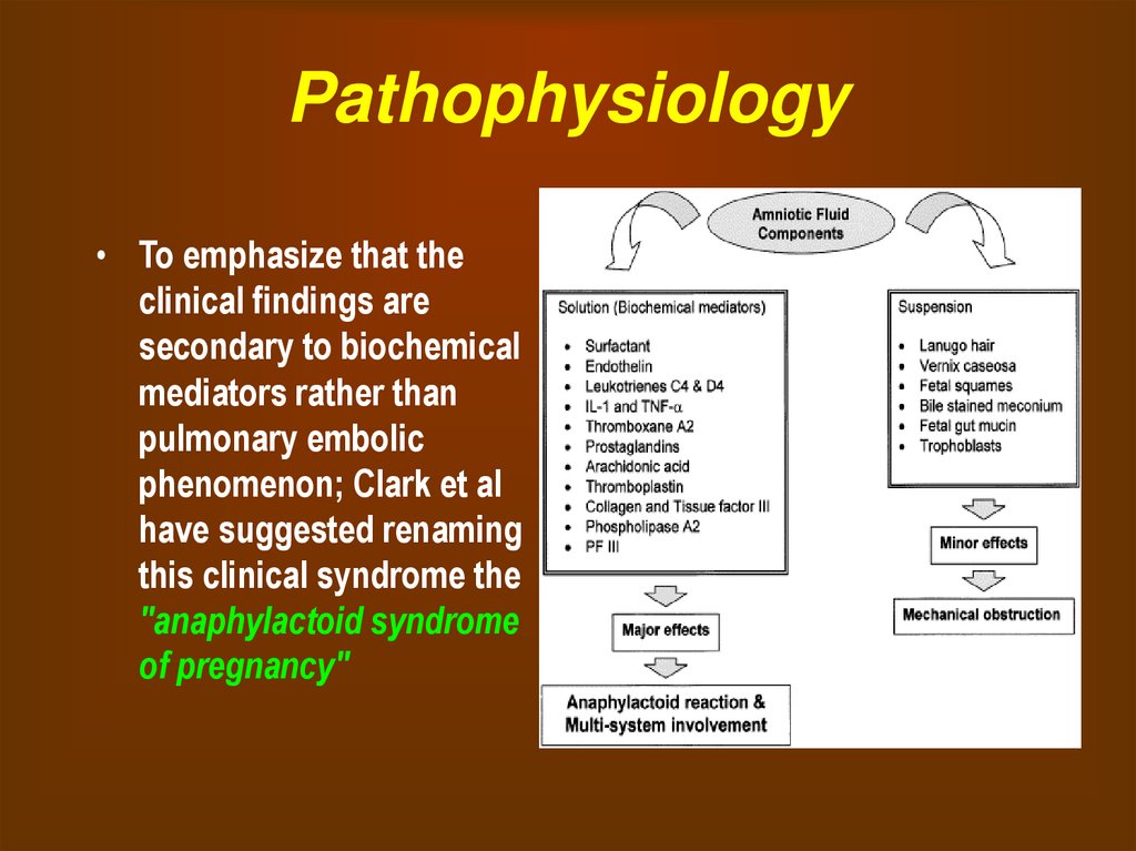
Pathophysiology• To emphasize that the
clinical findings are
secondary to biochemical
mediators rather than
pulmonary embolic
phenomenon; Clark et al
have suggested renaming
this clinical syndrome the
"anaphylactoid syndrome
of pregnancy"
12.
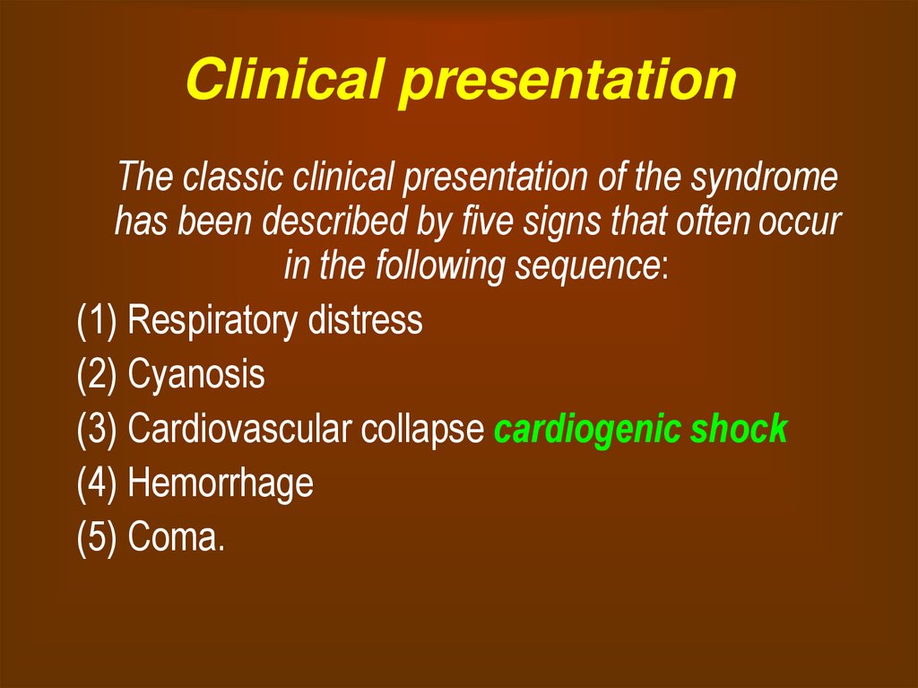
Clinical presentationThe classic clinical presentation of the syndrome
has been described by five signs that often occur
in the following sequence:
(1) Respiratory distress
(2) Cyanosis
(3) Cardiovascular collapse cardiogenic shock
(4) Hemorrhage
(5) Coma.
13.
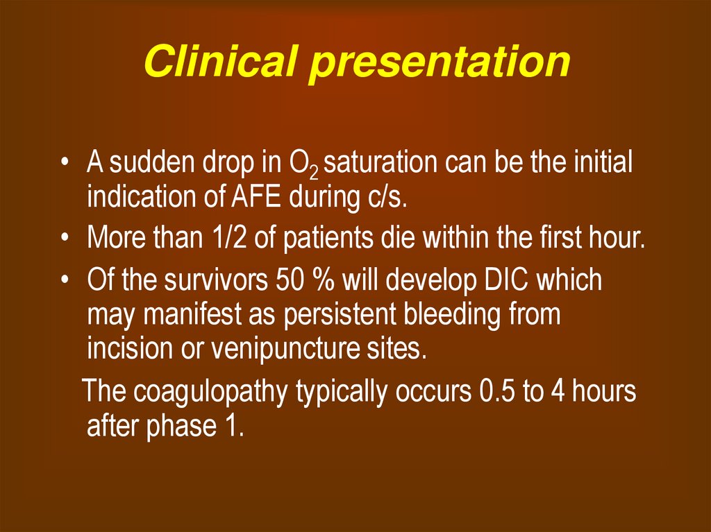
Clinical presentation• A sudden drop in O2 saturation can be the initial
indication of AFE during c/s.
• More than 1/2 of patients die within the first hour.
• Of the survivors 50 % will develop DIC which
may manifest as persistent bleeding from
incision or venipuncture sites.
The coagulopathy typically occurs 0.5 to 4 hours
after phase 1.
14.
Clinical presentation• 10-15% of patients will develop grand mal
seizures.
• CXR may be normal or show effusions, enlarged
heart, or pulmonary edema.
• ECG may show a right strain pattern with ST-T
changes and tachycardia.
15.
Diagnosis• In 1941, Steiner and Luschbaugh described histopathologic
findings in the pulmonary vasculature in 8 multiparous
women dying of sudden shock during labor.
• Findings included mucin, amorphous eosinophilic material ,
and in some cases squamous cells.
• The presence of squamous cells in the pulmonary
vasculature once considered pathognomonic for AFE is
neither sensitive nor specific (only 73% of patients dying from
AFE had this finding).
• The monoclonal antibody TKAH-2 may eventually prove more
useful in the rapid diagnosis of AFE.
16.
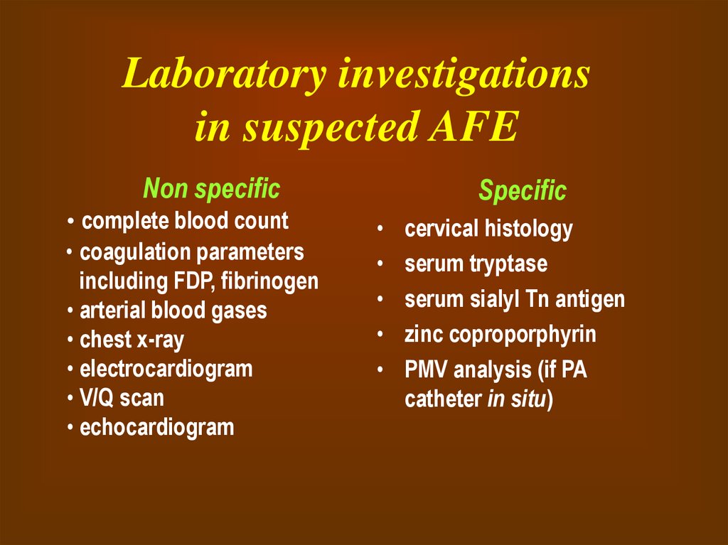
Laboratory investigationsin suspected AFE
Non specific
• complete blood count
• coagulation parameters
including FDP, fibrinogen
• arterial blood gases
• chest x-ray
• electrocardiogram
• V/Q scan
• echocardiogram
Specific
cervical histology
serum tryptase
serum sialyl Tn antigen
zinc coproporphyrin
PMV analysis (if PA
catheter in situ)
17.
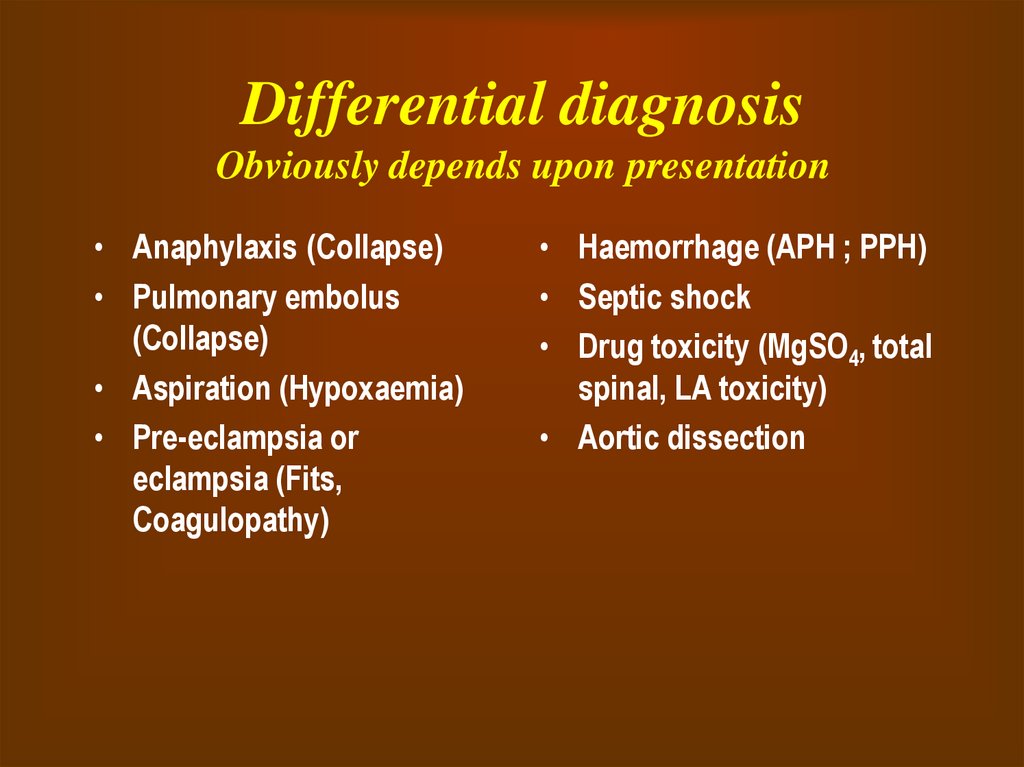
Differential diagnosisObviously depends upon presentation
• Anaphylaxis (Collapse)
• Pulmonary embolus
(Collapse)
• Aspiration (Hypoxaemia)
• Pre-eclampsia or
eclampsia (Fits,
Coagulopathy)
• Haemorrhage (APH ; PPH)
• Septic shock
• Drug toxicity (MgSO4, total
spinal, LA toxicity)
• Aortic dissection
18.
Management of AFEGOALS OF MANAGEMENT:
• Restoration of cardiovascular and pulmonary
equilibrium
- Maintain systolic blood pressure
>90 mm Hg.
- Urine output > 25 ml/hr
- Arterial pO2 > 60 mm Hg.
• Re-establishing uterine tone
• Correct coagulation abnormalities
19.
Management of AFE• As intubation and CPR may be required it is necessary
to have easy access to the patient, experienced help,
and a resuscitation tray with intubation equipment, DC
shock, and emergency medications.
• IMMEDIATE MEASURES :
- Set up IV Infusion, O2 administration.
- Airway control endotracheal intubation
maximal ventilation and oxygenation.
• LABS : CBC,ABG,PT,PTT,fibrinogen,FDP.
20.
Management of AFE• Treat hypotension, increase the circulating volume and
cardiac output with crystalloids.
• After correction of hypotension, restrict fluid therapy to
maintenance levels since ARDS follows in up to 40% to 70%
of cases.
• Steroids may be indicated (recommended but no evidence
as to their value)
• Dopamine infusion if patient remains hypotensive
(myocardial support).
• Other investigators have used vasopressor therapy such as
ephedrine or levarterenol with success (reduced systemic
vascular resistance)
21.
Management of AFEIn the ICU
• To assess the effectiveness of treatment and resuscitation, it
is prudent to continuously monitor ECG, pO2, CO2, and urine
output.
• There is support in literature for early placement of arterial,
central venous, and pulmonary artery catheters to provide
critical information and guide specific therapy.
22.
Management of AFEIn the ICU
• Central venous pressure monitoring is important to
diagnose right ventricular overload and guide fluid infusion
and vasopressor therapy. Blood can also be sampled from
the right heart for diagnostic purposes.
• Pulmonary artery and capillary wedge pressures and
echocardiography are useful to guide therapy and evaluate
left ventricular function and compliance.
• An arterial line is useful for repeated blood sampling and
blood gases to evaluate the efficacy of resuscitation.
23.
Management of AFECoagulopathy
• DIC results in the depletion of fibrinogen, platelets,
and coagulation factors, especially factors V, VIII,
and XIII. The fibrinolytic system is activated as well.
• Most patients will have hypofibrinogenemia,
abnormal PT and aPTT and low Platelet counts
• Treat coagulopathy with FFP for a prolonged aPTT,
cryoprecipitate for a fibrinogen level less than 100
mg/dL, and transfuse platelets for platelet counts
less than 20,000/mm3
24.
Restoration of uterine tone• Uterine atony is best treated with massage,
uterine packing, and oxytocin or prostaglandin
analogues.
• Improvement in cardiac output and uterine
perfusion helps restore uterine tone.
• Extreme care should be exercised when using
prostaglandin analogues in hypoxic patients, as
bronchospasm may worsen the situation.
25.
Sympathomimetic Vasopressor agentDopamine
• Dopamine increases myocardial contractility and systolic
BP with little increase in diastolic BP. Also dilates the renal
vasculature, increasing renal blood flow and GFR.
• DOSE: 2-5 mcg/kg/min IV; titrate to BP and cardiac output.
• Contraindications: ventricular fibrillation, hypovolemia,
pheochromocytoma.
• Precautions: Monitor urine flow, cardiac output, pulmonary
wedge pressure, and BP during infusion; prior to infusion,
correct hypovolemia with either whole blood or plasma, as
indicated; monitoring central venous pressure or left
ventricular filling pressure may be helpful
26.
Maternal Mortality in AFE• Maternal death usually occurs in one of three ways: (1)
sudden cardiac arrest, (2) hemorrhage due to coagulopathy,
or (3) initial survival with death due to acute respiratory
distress syndrome (ARDS) and multiple organ failure
• For women diagnosed as having AFE, mortality rates
ranging from 26% to as high as 86% have been reported.
• The variance in these numbers is explained by dissimilar
case definitions and possibly improvements in intensive
care management of affected patients.
27.
Further issues in theManagement
• Transfer:
Transfer to a level 3 hospital may be required once the
patient is stable.
• Deterrence/Prevention:
Amniotic fluid embolism is an unpredictable event.
• Risk of recurrence is unknown. The recommendation for
elective cesarean delivery during future pregnancies in an
attempt to avoid labor is controversial.
• Perimortem cesarean delivery:
After 5 minutes of unsuccessful CPR in arrested mothers,
abdominal delivery is recommended.
28.
Medical/Legal Pitfalls• Failure to respond emergently is a pitfall. AFE is a clinical
diagnosis. Steps must be taken to stabilize the patient as
soon as symptoms manifest.
• Failure to perform perimortem cesarean delivery in a timely
fashion is a pitfall.
• Failure to consider the diagnosis during legal abortion is a
pitfall. A review of the literature indicates that most case
reports of AFE have occurred during late second-trimester
abortions.
29.
SUMMARY• AFE is a sudden and unexpected rare but life
threatining complication of pregnancy.
• It has a complex pathogenesis and serious
implications for both mother and infant
• Associated with high rates of mortality and
morbidity.
• Diagnosis of exclusion.
• Suspect AFE when confronted with any pregnant
patient who has sudden onset of respiratory
distress, cardiac collapse, seizures, unexplained
fetal distress, and abnormal bleeding
• Obstetricians should be alert to the symptoms of
AFE and strive for prompt and aggressive treatment.