 Медицина
 Биографии
Похожие презентации:


 — Greek father of medicine")
")


")
William Osler Abbott (1902-1943)
1.
Penza State UniversityDepartment Medical Institute
Course paper in History of medicine.
William Osler Abbott (1902 – 1943)
Student :ASEEL AL-ZUBIRI
Group : 19lc4a
Year : 1st year
Professor : Gavrilova Tatiana.
2.
Contents:Fig 1………………………………………………………..2
Biography………………………………………………….2
Career.....…………………………………………………..3
Fig 2………………………………………………………...3
Awards……………………………………………………..4
Family ……………………………………………………..5
Death……………………………………………………….6
References …………………………………………………7
3.
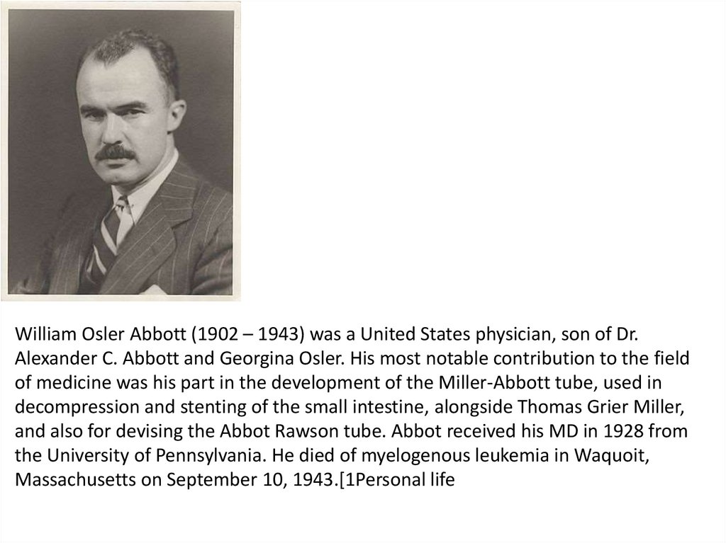
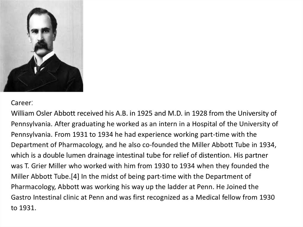
William Osler Abbott (1902 – 1943) was a United States physician, son of Dr.Alexander C. Abbott and Georgina Osler. His most notable contribution to the field
of medicine was his part in the development of the Miller-Abbott tube, used in
decompression and stenting of the small intestine, alongside Thomas Grier Miller,
and also for devising the Abbot Rawson tube. Abbot received his MD in 1928 from
the University of Pennsylvania. He died of myelogenous leukemia in Waquoit,
Massachusetts on September 10, 1943.[1Personal life
4.
William Osler Abbott was born July 26, 1902 in New Bedford,MA. He was given the nickname "pete" growing up.[2] His father,
Alexander Abbott, was a member of the resident staff at Johns
Hopkins Hospital and his mother, Georgina Osler, was a niece of
William Osler. His parents met while his mother was taking care
of Osler's home in Baltimore. His family would vacation at
Waquoit a lot in the summers, which is where he came to love
everything about the sea. At just the young age of 10, Abbott
would skin the fish in jars of water and put the bones and
cartilage together with fine wire and when he was only 15, he
could sail 30 miles at night from Waquoit to Nantucket. William
Osler Abbott married a young lady from Kansas City named Lucy
Waldo in 1928.[3] The newly married Abbotts spent their
honeymoon in an open dory sailing among Cape Cod's Elizabeth
Islands.[4] They had three children, Thomas William Osler, Ann
Gatewood, and Lucy Featherstone.[3
5.
Career:William Osler Abbott received his A.B. in 1925 and M.D. in 1928 from the University of
Pennsylvania. After graduating he worked as an intern in a Hospital of the University of
Pennsylvania. From 1931 to 1934 he had experience working part-time with the
Department of Pharmacology, and he also co-founded the Miller Abbott Tube in 1934,
which is a double lumen drainage intestinal tube for relief of distention. His partner
was T. Grier Miller who worked with him from 1930 to 1934 when they founded the
Miller Abbott Tube.[4] In the midst of being part-time with the Department of
Pharmacology, Abbott was working his way up the ladder at Penn. He Joined the
Gastro Intestinal clinic at Penn and was first recognized as a Medical fellow from 1930
to 1931.
6.
From 1931 to 1937 he was known as an instructor at Penn. Inthe year 1937, Abbott worked with a man named Arthur Joy
Rawson creating the Abbott Rawson Tube, which is a double
barreled Gastroenterostomy tube for use in postoperative
care.[4] From an instructor he was now named associate from
the year 1937 to 1941. Abbott was known as a professor of
medicine at Penn, however the following year he brought his
expertise to the U.S. Army. When Abbott joined the U.S. Army
he was already the rank of a major.
Shortly, being discharged due to his diagnosis of leukemia.[2]
After fabricating his double lumen tube, Abbott would initially
swallow the tube in the morning at his home on the Main Line
outside of Philadelphia, ride to work on the train with the
proximal end exiting his nose and curled around his ear or
leaving his mouth beside a pipe and residing in a coat pocket.
7.
Once in the hospital the intubation continued underfluoroscopic guidance. With the fundamentals of a
practical technique of intubation established, he
began his investigations of the absorptive capacity of
the gut and the effect of drugs on the intestine in
December 1932. On May 15, 1942 Abbott, a major in
the Medical Corps, left Philadelphia with the 20th
General Hospital for Camp Claiborne, Louisiana. Eight
days later while undergoing a physical examination a
large spleen was detected and blood studies led to a
diagnosis of Myelogenous leukemia.[4] Abbott's
remaining months of life were spent researching
his cancerous disease.
8.
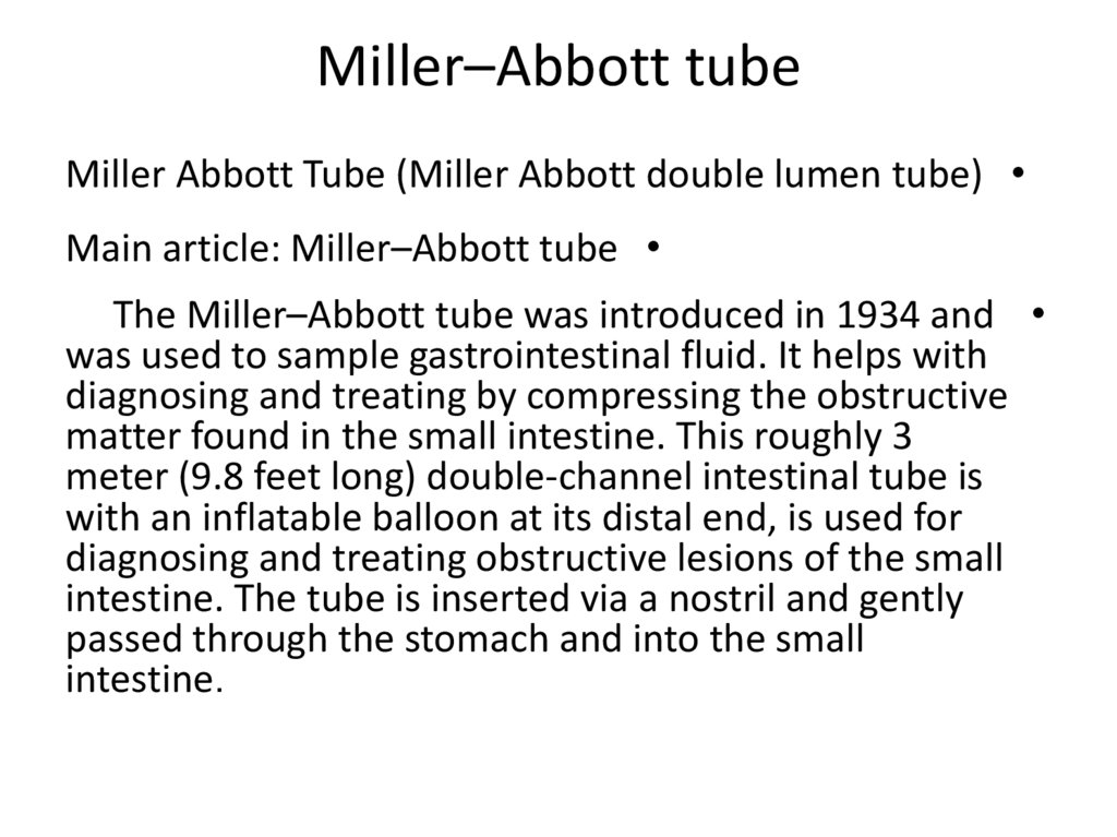
Miller–Abbott tubeMiller Abbott Tube (Miller Abbott double lumen tube)
Main article: Miller–Abbott tube
The Miller–Abbott tube was introduced in 1934 and
was used to sample gastrointestinal fluid. It helps with
diagnosing and treating by compressing the obstructive
matter found in the small intestine. This roughly 3
meter (9.8 feet long) double-channel intestinal tube is
with an inflatable balloon at its distal end, is used for
diagnosing and treating obstructive lesions of the small
intestine. The tube is inserted via a nostril and gently
passed through the stomach and into the small
intestine.
9.
[5] Still largely unchanged in 2011, once this instrument weavesdown the esophagus and into the stomach, the tube is capable
of a handful of jobs at this point, from suctioning gastric juices
for testing and irrigation to ballooning open the entryway to the
small intestine, called the duodenum, for clearer radiology
testing and easier removal of many intestinal blockages.
.[5] The Miller-Abbott tube is named after American
gastroenterologists William Osler Abbott and Thomas Grier
Miller. These doctors also pioneered the surgical procedures that
set the stage for easier diagnosis and removal of stomach and
intestinal lesions, blockages and ulcers.
10.
[6] With the instrument having its double-barreled design,one of the pipes, called a lumen, is responsible for pumping
up a thin balloon at the tip for easy exploration into the
intestines at the duodenum. The other lumen tube can then
suction fluids out or pump fluids in, depending on the
procedure. For radiology, a barium solution can be pumped
into the duodenum to isolate potential damage and produce
clear images of it. Allowing the tube to proceed into the
intestines also might help dislodge identified blockages
causing pain or digestive disorders.
11.
. In 2011, the Miller-Abbott tube might beaccompanied by another, called a laparoscope. This
latter tube combines a light and camera to give
physicians a three-dimensional, colored view of
whatever blockage is occurring. It can also help the
doctor know exactly when the Miller-Abbott tube's
balloon is at the perfect location in the duodenum —
a process that depends on the slow and steady
peristaltic contractions of the digestive tract.[6]
12.
Abbott–Rawson TubeIn 1937 Abbott helped create a new instrument for
gastrointestinal surgery called the Abbott–Rawson tube.[4]
The tube may be used for Jejunal feedings and for
administrating potassium, antibiotics or vitamins.[7] Although
many uses beyond that for which it was first employed have
been developed, it remains a much neglected surgical
adjunct. For a time after it was described in 1937, the device
was widely used for the emptying of the gastrointestinal tract
at times of surgical stress. Now it is described in only one
textbook on surgery.[7]
13.
Thomas Grier MillerWilliam Osler Abbott did not create the Miller Abbott tube all
alone, he had significant help from his coworker and good
friend Thomas Grier Miller, who was an American internist.
Miller was born on September 18, 1886 in Satesville, North
Carolina. He received an A.B. from the University of North
Carolina in 1906 and graduated in medicine from the
University of Pennsylvania in 1911 and then commenced
clinical investigation in the department of medicine, but this
was interrupted by the 1st World War where he served in the
army as a captain. Just like Abbott, Miller also accomplished
many achievements throughout his career. In 1926, Miller
founded the Gastro Intestinal Section of the Medical Clinic at
the Hospital of the University of Pennsylvania and was chief of
the section from 1928 until his retirement in 1952.
14.
From 1913 to 1952, he also held posts in the School ofMedicine at the University, becoming professor of clinical
medicine in 1934. Miller published on many areas of
medicine, but concentrated mainly on gastroenterology and in
1934 commenced a series of papers with William Osler
Abbott and W. G. Carr on intubation and studies of the small
intestine which became classics and were made possible by
the invention of the double lumen tube. This arose when
Abbott was unable to keep a tube with one distended balloon
at a fixed point of the duodenum and Miller suggested that a
second open tube be tied to the bag to see if this would make
sampling easier. Miller later went on to die on November 15,
1981, Hospital of the University of Pennsylvania.[8]
15.
DeathIn September of May 1942, Abbott was honorably discharged
from the army because of a physical disability and died of
myelogenous leukemia.[3] Abbott spent the remaining
months before he died doing leukemia research.[9] It is
believed this happened following the excess X-ray exposure
he received in screening the position of the tube in volunteers
and patients he was investigating.[3
16.
Years after deathA small 5 series collection about William Osler Abbott was
assembled by Catharine G. Leeke, his secretary at the Gastro
Intestinal Clinic at the Hospital of the University of
Pennsylvania. It contains personal information, a summary of
his professional work, and information on his final years. On
June 7, 1972, the collection was donated by Thomas A.
Urbine, Jr. on behalf of Catharine Leeke to the Historical
Society of Pennsylvania and then transferred to the College of
Physicians.[2]