
 Химия
ХимияПохожие презентации:


Proteins. Functions, structure, classification
1.
Proteins. Functions,Structure, classification
2.
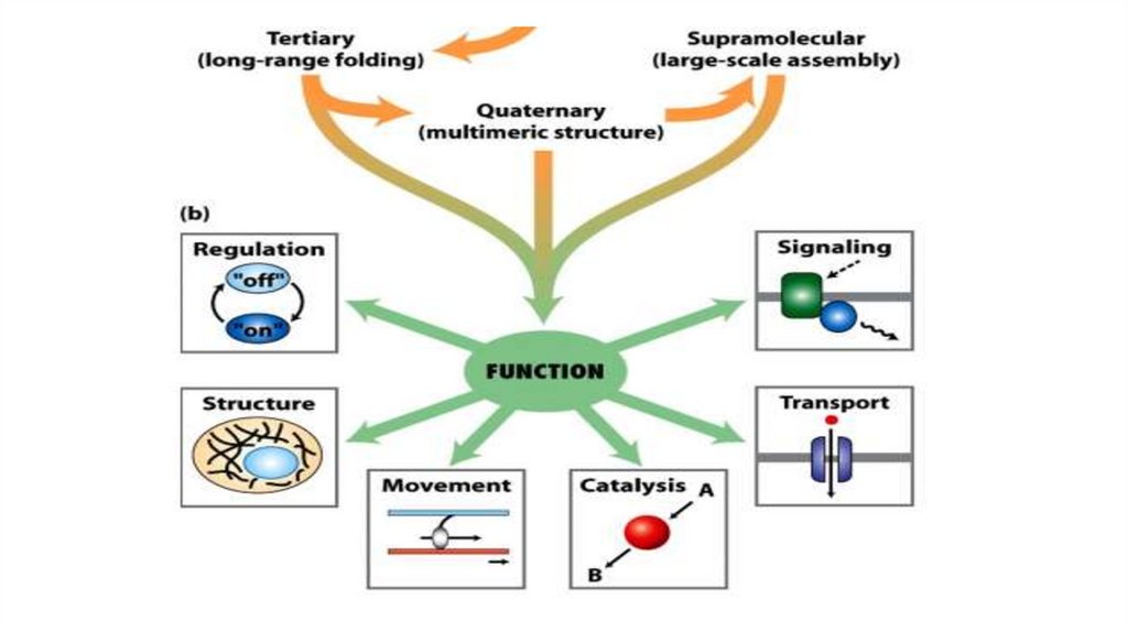
Agenda1. Functions of Proteins
2. Overview of Protein Structure - levels of organization of
protein molecules
Primary Structure
Secondary Structure
Tertiary and Quaternary Structures
3. Classification of proteins
3.
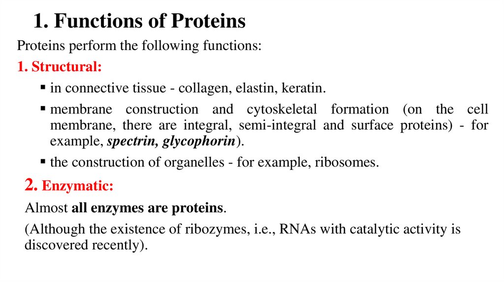
1. Functions of ProteinsProteins perform the following functions:
1. Structural:
in connective tissue - collagen, elastin, keratin.
membrane construction and cytoskeletal formation (on the cell
membrane, there are integral, semi-integral and surface proteins) - for
example, spectrin, glycophorin).
the construction of organelles - for example, ribosomes.
2. Enzymatic:
Almost all enzymes are proteins.
(Although the existence of ribozymes, i.e., RNAs with catalytic activity is
discovered recently).
4.
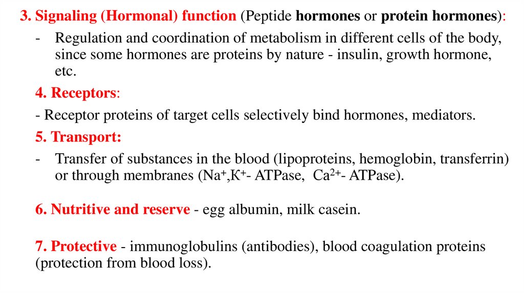
3. Signaling (Hormonal) function (Peptide hormones or protein hormones):- Regulation and coordination of metabolism in different cells of the body,
since some hormones are proteins by nature - insulin, growth hormone,
etc.
4. Receptors:
- Receptor proteins of target cells selectively bind hormones, mediators.
5. Transport:
- Transfer of substances in the blood (lipoproteins, hemoglobin, transferrin)
or through membranes (Na+,К+- ATPase, Са2+- ATPase).
6. Nutritive and reserve - egg albumin, milk casein.
7. Protective - immunoglobulins (antibodies), blood coagulation proteins
(protection from blood loss).
5.
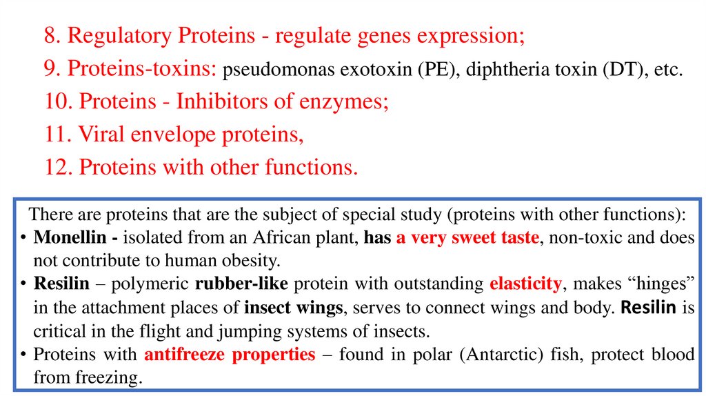
8. Regulatory Proteins - regulate genes expression;9. Proteins-toxins: pseudomonas exotoxin (PE), diphtheria toxin (DT), etc.
10. Proteins - Inhibitors of enzymes;
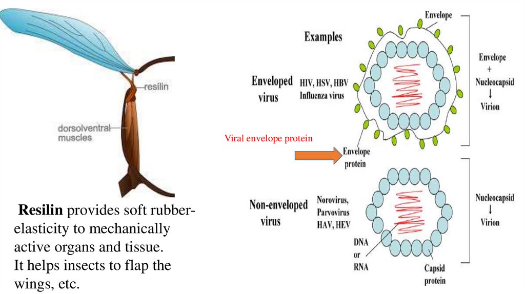
11. Viral envelope proteins,
12. Proteins with other functions.
There are proteins that are the subject of special study (proteins with other functions):
• Monellin - isolated from an African plant, has a very sweet taste, non-toxic and does
not contribute to human obesity.
• Resilin – polymeric rubber-like protein with outstanding elasticity, makes “hinges”
in the attachment places of insect wings, serves to connect wings and body. Resilin is
critical in the flight and jumping systems of insects.
• Proteins with antifreeze properties – found in polar (Antarctic) fish, protect blood
from freezing.
6.
Viral envelope proteinResilin provides soft rubberelasticity to mechanically
active organs and tissue.
It helps insects to flap the
wings, etc.
7.
8.
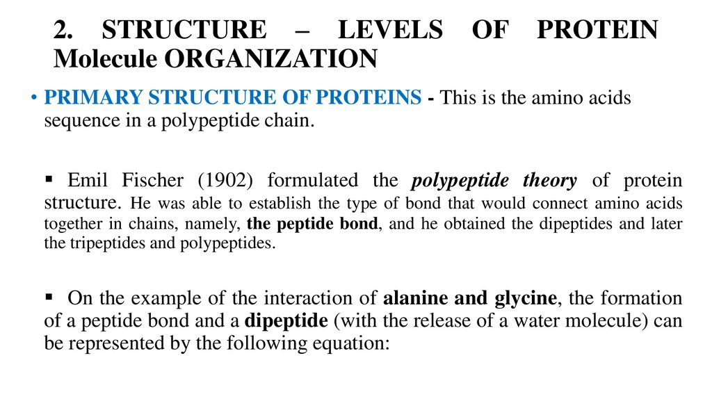
2. STRUCTURE – LEVELSMolecule ORGANIZATION
OF
PROTEIN
• PRIMARY STRUCTURE OF PROTEINS - This is the amino acids
sequence in a polypeptide chain.
Emil Fischer (1902) formulated the polypeptide theory of protein
structure. He was able to establish the type of bond that would connect amino acids
together in chains, namely, the peptide bond, and he obtained the dipeptides and later
the tripeptides and polypeptides.
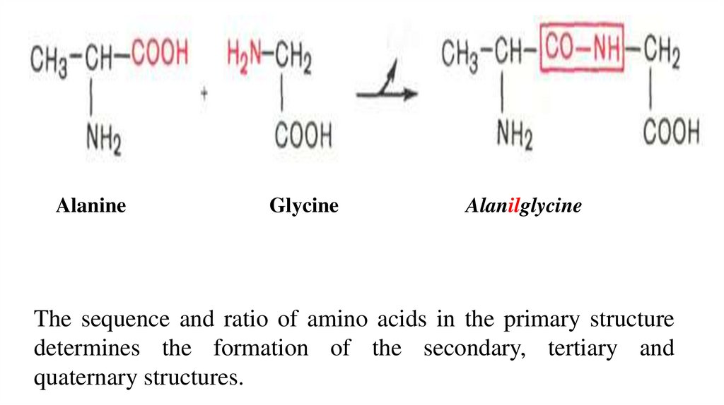
On the example of the interaction of alanine and glycine, the formation
of a peptide bond and a dipeptide (with the release of a water molecule) can
be represented by the following equation:
9.
AlanineGlycine
Alanilglycine
The sequence and ratio of amino acids in the primary structure
determines the formation of the secondary, tertiary and
quaternary structures.
10.
11.
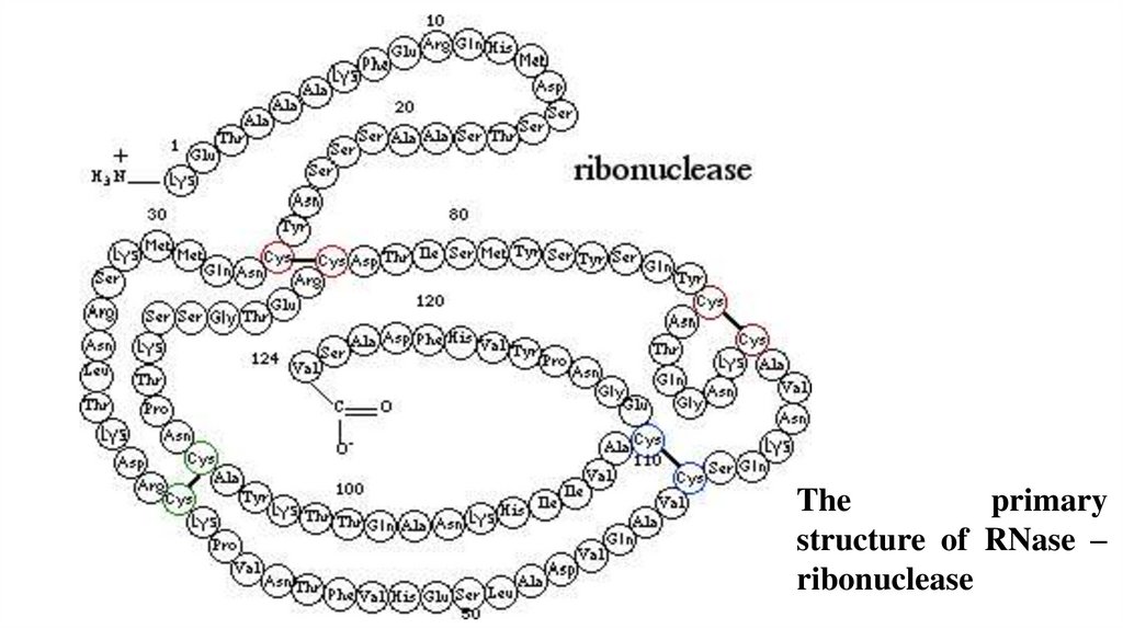
Theprimary
structure of RNase –
ribonuclease
12.
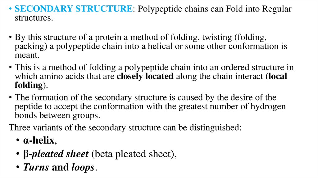
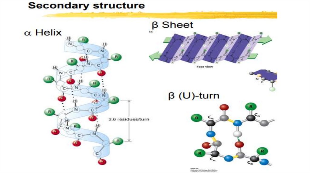
• SECONDARY STRUCTURE: Polypeptide chains can Fold into Regularstructures.
• By this structure of a protein a method of folding, twisting (folding,
packing) a polypeptide chain into a helical or some other conformation is
meant.
• This is a method of folding a polypeptide chain into an ordered structure in
which amino acids that are closely located along the chain interact (local
folding).
• The formation of the secondary structure is caused by the desire of the
peptide to accept the conformation with the greatest number of hydrogen
bonds between groups.
Three variants of the secondary structure can be distinguished:
• α-helix,
• β-pleated sheet (beta pleated sheet),
• Turns and loops.
13.
14.
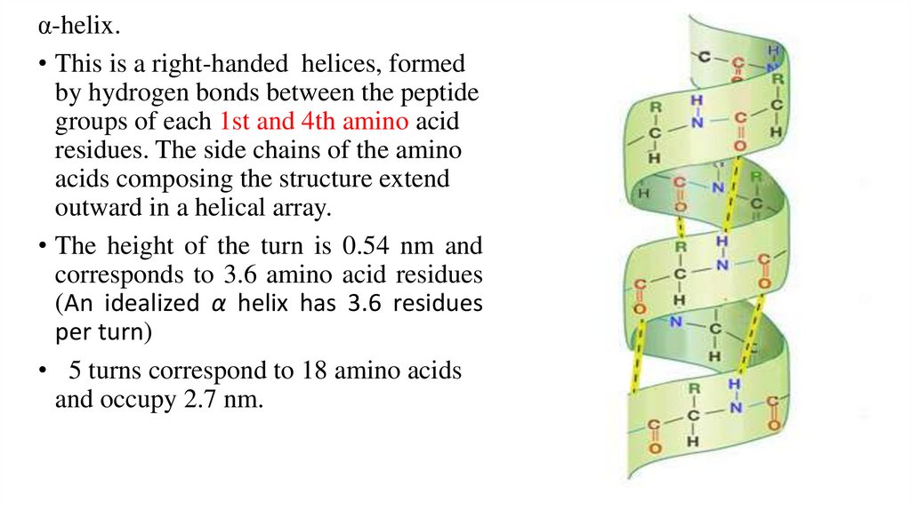
α-helix.• This is a right-handed helices, formed
by hydrogen bonds between the peptide
groups of each 1st and 4th amino acid
residues. The side chains of the amino
acids composing the structure extend
outward in a helical array.
• The height of the turn is 0.54 nm and
corresponds to 3.6 amino acid residues
(An idealized α helix has 3.6 residues
per turn)
• 5 turns correspond to 18 amino acids
and occupy 2.7 nm.
15.
16.
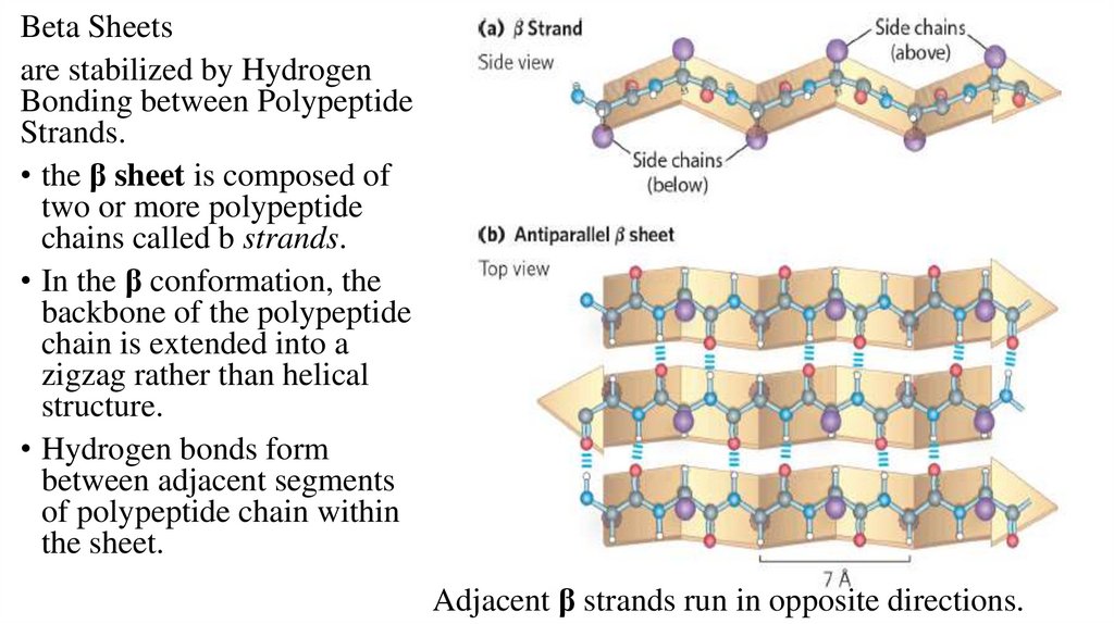
Beta Sheetsare stabilized by Hydrogen
Bonding between Polypeptide
Strands.
• the β sheet is composed of
two or more polypeptide
chains called b strands.
• In the β conformation, the
backbone of the polypeptide
chain is extended into a
zigzag rather than helical
structure.
• Hydrogen bonds form
between adjacent segments
of polypeptide chain within
the sheet.
Adjacent β strands run in opposite directions.
17.
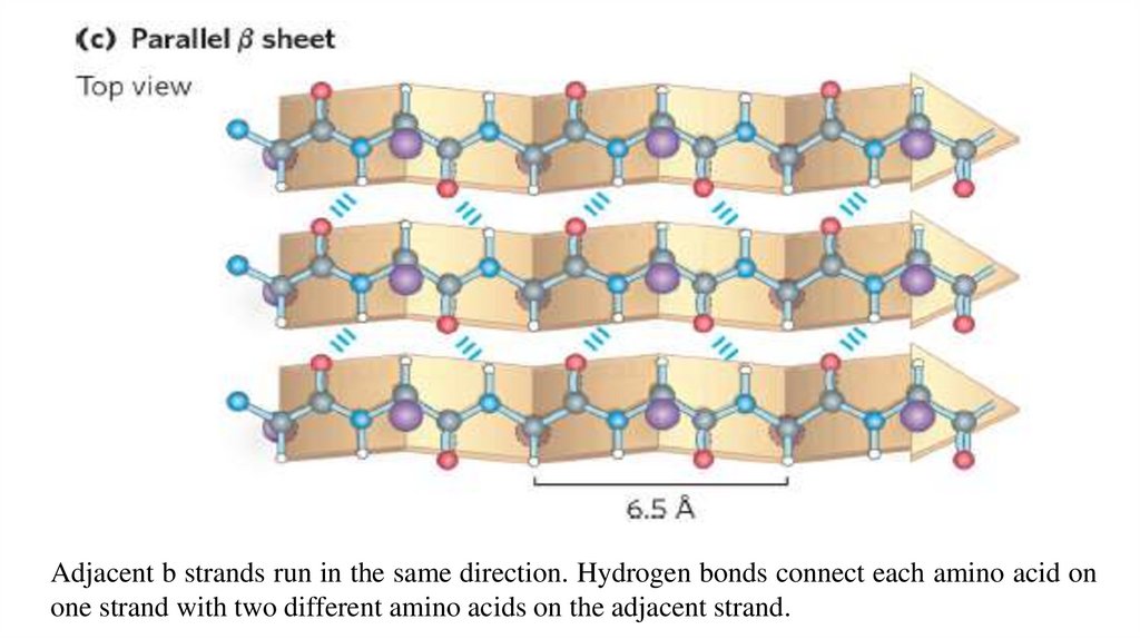
Adjacent b strands run in the same direction. Hydrogen bonds connect each amino acid onone strand with two different amino acids on the adjacent strand.
18.
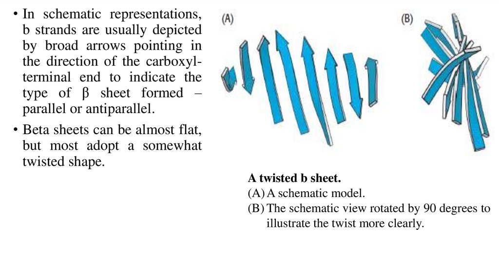
• In schematic representations,b strands are usually depicted
by broad arrows pointing in
the direction of the carboxylterminal end to indicate the
type of β sheet formed –
parallel or antiparallel.
• Beta sheets can be almost flat,
but most adopt a somewhat
twisted shape.
A twisted b sheet.
(A) A schematic model.
(B) The schematic view rotated by 90 degrees to
illustrate the twist more clearly.
19.
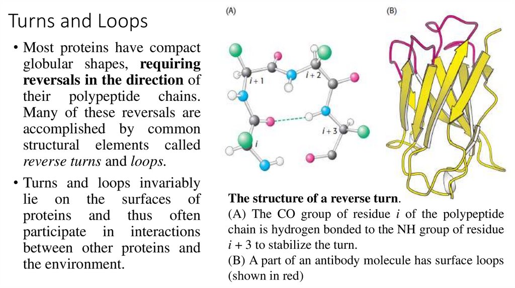
Turns and Loops• Most proteins have compact
globular shapes, requiring
reversals in the direction of
their polypeptide chains.
Many of these reversals are
accomplished by common
structural elements called
reverse turns and loops.
• Turns and loops invariably
lie on the surfaces of
proteins and thus often
participate in interactions
between other proteins and
the environment.
The structure of a reverse turn.
(A) The CO group of residue i of the polypeptide
chain is hydrogen bonded to the NH group of residue
i + 3 to stabilize the turn.
(B) A part of an antibody molecule has surface loops
(shown in red)
20.
Tertiary Structure: water-soluble Proteinsfold into Compact structures
• The tertiary structure, refers to the spatial arrangement of amino acid residues
that are far apart in the sequence and to the pattern of disulfide bonds.
• This level of structure is the result of interactions between the R groups of the
peptide chain.
• Thus, the overall three-dimensional arrangement of all atoms in a protein is
referred to as the protein’s tertiary structure.
• The tertiary structure includes longer-range aspects of amino acid sequence.
21.
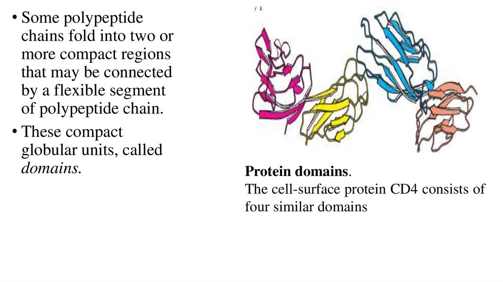
• Some polypeptidechains fold into two or
more compact regions
that may be connected
by a flexible segment
of polypeptide chain.
• These compact
globular units, called
domains.
Protein domains.
The cell-surface protein CD4 consists of
four similar domains
22.
Bonds involved in the formation of the tertiarystructure
Various bonds are involved in the formation of the tertiary structure:
1. Mainly:
• hydrogen
• Van der Waals communications.
2. Additional, but ve hano less significant:
• disulfide
• pseudopeptide
• Ionic bonds.
23.
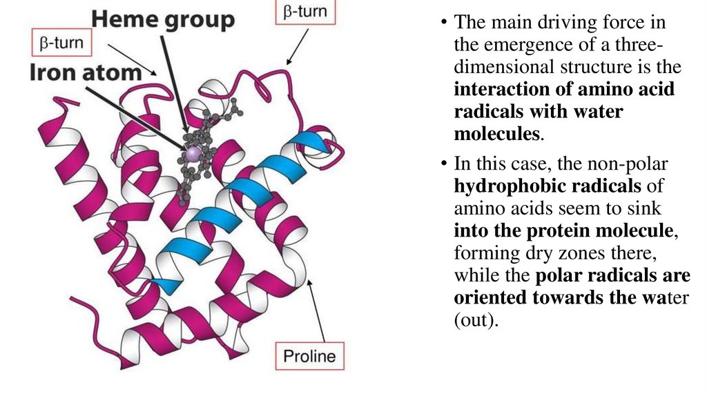
• The main driving force inthe emergence of a threedimensional structure is the
interaction of amino acid
radicals with water
molecules.
• In this case, the non-polar
hydrophobic radicals of
amino acids seem to sink
into the protein molecule,
forming dry zones there,
while the polar radicals are
oriented towards the water
(out).
24.
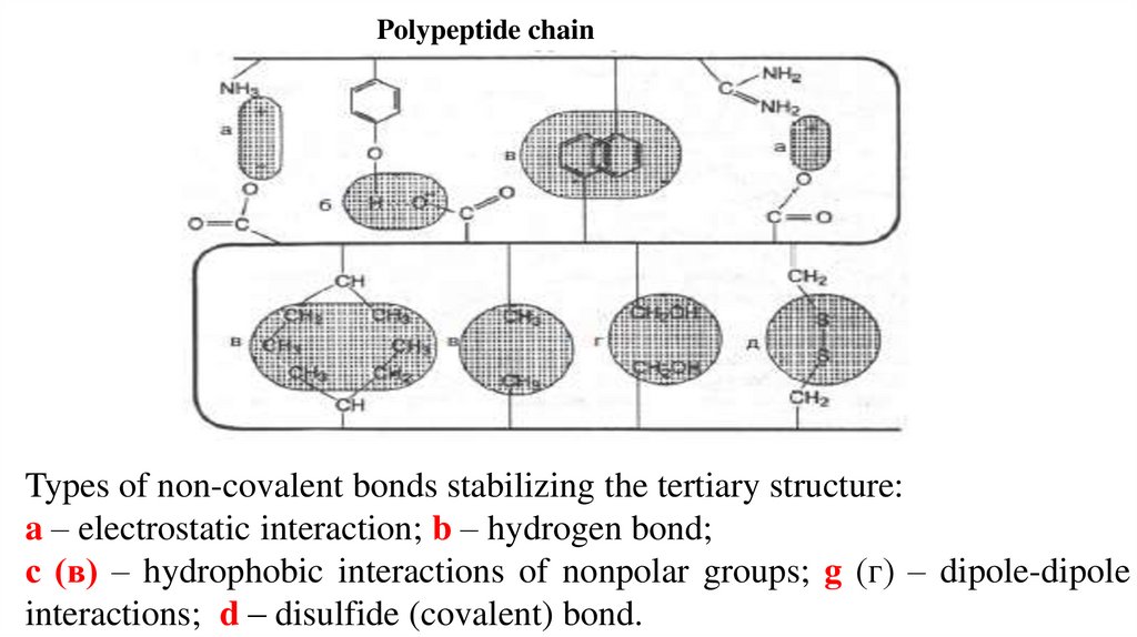
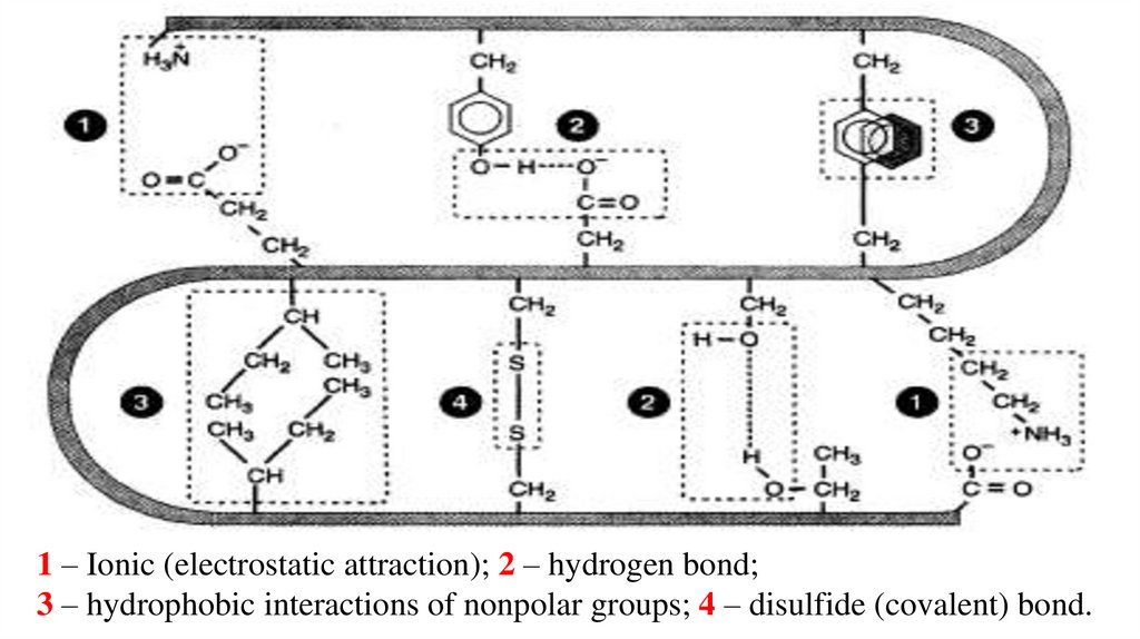
Polypeptide chainTypes of non-covalent bonds stabilizing the tertiary structure:
a – electrostatic interaction; b – hydrogen bond;
с (в) – hydrophobic interactions of nonpolar groups; g (г) – dipole-dipole
interactions; d – disulfide (covalent) bond.
25.
1 – Ionic (electrostatic attraction); 2 – hydrogen bond;3 – hydrophobic interactions of nonpolar groups; 4 – disulfide (covalent) bond.
26.
1 – Ionic (electrostaticattraction);
2 – hydrogen bond;
3 – disulfide bond
4 – hydrophobic interactions,
5 – hydrated groups.
27.
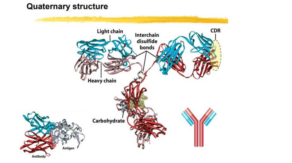
Quaternary Structure: Multiple Polypeptide Chains Can Assemble into aSingle Protein
• Proteins consisting of more than one polypeptide chain display
quaternary structure; each individual polypeptide chain is called a
subunit.
• Quaternary structure can be as simple as two identical subunits or as
complex as dozens of different subunits. In most cases, the subunits are
held together by noncovalent bonds.
28.
29.
SUMMARY on Protein Tertiary and Quaternary Structures• Tertiary structure is the complete three-dimensional structure of a polypeptide chain.
Many proteins fall into one of two general classes of proteins based on tertiary
structure: fibrous and globular.
• Fibrous proteins, which serve mainly structural roles, have simple repeating elements
of secondary structure.
• Globular proteins have more complicated tertiary structures, often containing several
types of secondary structure in the same polypeptide chain. The first globular protein
structure to be determined, by x-ray diffraction methods, was the structure of
myoglobin.
• The complex structures of globular proteins can be analyzed by examining folding
patterns called motifs (also called folds or supersecondary structures). Domains are
regions of a polypeptide chain that can fold stably and independently.
• Quaternary structure results from interactions between the subunits of multisubunit
(multimeric) proteins or large protein assemblies. Some multimeric proteins have a
repeated unit consisting of a single subunit or a group of subunits, each unit called a
protomer.
30.
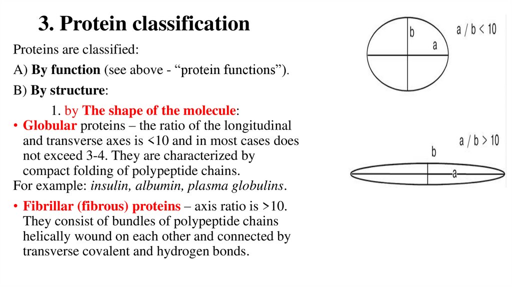
3. Protein classificationProteins are classified:
A) By function (see above - “protein functions”).
B) By structure:
1. by The shape of the molecule:
• Globular proteins – the ratio of the longitudinal
and transverse axes is <10 and in most cases does
not exceed 3-4. They are characterized by
compact folding of polypeptide chains.
For example: insulin, albumin, plasma globulins.
• Fibrillar (fibrous) proteins – axis ratio is >10.
They consist of bundles of polypeptide chains
helically wound on each other and connected by
transverse covalent and hydrogen bonds.
31.
2. By the number of protein chains in one molecule:monomeric protein - have one subunit (protomer),
polymer protein– have several subunits.
For example: hemoglobin (4 subunits), lactate dehydrogenase (4 subunits),
creatine phosphokinase (2 subunits), E. coli RNA polymerase (5 chains),
3. By the chemical composition:
Simple proteins – contain only amino acids (albumins, histones,
protamines, collagen, elastin).
Complex proteins – in addition to amino acids, have non-protein
components. A non-protein group is called a ligand (in phosphoproteins,
lipoproteins, chromoproteins, glycoproteins, nucleoproteins).
32.
As a ligand can be :• molecules that perform a structural function in a protein:
- lipids, carbohydrates, nucleic acids, mineral elements,
• any other organic and inorganic compounds: heme in hemoglobin:
- copper (Cu) in ceruloplasmin;
- molecules transferred by proteins: iron in transferrin,
hemoglobin residue in haptoglobin, heme in hemopexin.