


")
 occur in reproductive life and can be confused with neoplastic lesions.")


 – can occur but are rare. They are low differentiated tumours, have very malignant course, the tumour quickly grows, burgeons a capsule, yields metastasises in retroperitoneal lymphonoduses, hematogenic - in a liver,")


 Медицина
Похожие презентации:


Benign tumors of the female genital organs
1. .
Benign tumorsof the female genital
organs
.
2.
3.
Benign ovarian cysts are commonly encounteredproblem in gynecological practice and often
asymptomatic the clinical course and tend to
regress. Their presence is one of the most common
causes of hospitalization of patients in gynecological
hospitals. So at the age of 65 years, according to
some authors, 4% of all women have ever been
hospitalized with this diagnosis.
4.
90% of ovarian tumors are benign,although this index varies with age.
Among the tumors that require surgical
treatment in the premenopausal period,
13% are malignant, and after the
menopause, this index reaches 45%.
5.
The main objective in the management ofpatients with benign ovarian tumors avoidance the possibility risk of
malignant growth and prevention of
complications. While at younger women
should avoid unnecessary interventions
that violate fertility.
6.
Ovarian tumors are physiological or pathological andformed with any tissue, which is included in the
composition of the ovary. Most benign ovarian tumors
are cysts, but the presence of solid components
increases the risk malignization. However, benign
tumors such as fibroma, Corpus Luteal cysts, mature
teratoma (Dermoid Cyst) and Brener’s tumor, usually
contain solid components.
The management of each of ovarian tumors is
different. However, only when a specimen is analysed
in the pathology laboratory can we know for sure what
the diagnosis is. A clinician has to utilise his clinical
acumen and the results of investigations to help
determine management and make a clinical diagnosis.
7.
Tumours of ovaries are dividedinto two basic groups:
• blastomatous (proliferating) tumours of an
ovary, or a cystoma;
• non blastomatous (not proliferating)
tumours of an ovary, or a cyst.
• Blastomatous tumours (cystoma) are the
true tumours having unlimited growth.
• Not blastomatous tumours (cysts) have
limited growth and reach small size.
8. The classification of benign ovarian tumors (for the histological conclusion)
Functional cysts* Follicular cyst
* Corpus Luteal cyst
Germinogenic benign ovarian tumors
* Mature teratoma (Dermoid Cyst)
* Immature teratomas
Epithelial ovarian tumors
* Serous cystadenoma
* Mucinous cystadenoma
* Endometrioid cystadenoma
* Brener’s tumor
* Clear Cell tumor
Benign sex cord / stromal ovarian tumors
* Granulosa tumors
* Theca cell tumors
* Fibroma
* Sertoli/Leydig tumors
9. Follicular and Corpus Luteal cysts (lutein cysts) occur in reproductive life and can be confused with neoplastic lesions.
follicular cysts - tumorous formations which educe on a backgroundof inflammatory process owing to accumulation of fluid in a cystatretic follicle, or can be caused by infringement of hypothalamichypophysial regulation of function of ovaries; cavitary, thin-walled
unicameral formation, unilateral, 2-7 cm in diameter; sometimes
hormonactivity as contain estrogens.
lutein cysts - are surveyed as anatomical variant of normal
constitution of corpus luteum; can be consequence of inflammatory
diseases of ovaries or hyperproduction of gonadotrophic Hormones
by adenohypophysis; at girls appearance of cyst is connected to
hypersecretion of Prolactinum, arise in the period of sexual maturity,
is more often at biphase menstrual cycle.
The management is usually by observation alone. These cysts can
also be treated by suppressing ovarian activity with the
contraceptive pill. Sometimes for failure of conservative therapy it is
necessary perform an ovarian cystectomy.
10.
11.
Germinogenic benign ovarian tumorscan be benign mature teratoma (Dermoid
Cysts, Struma of an ovary) or immature
teratomas. They contain elements from all
three embryonic germ lines (mesoderm,
ectoderm, and endoderm).
12.
Mature teratomas account for a quarter of all benign ovariancysts (50% in women under 20). Mature teratomas are dermoid
cysts arise from all three rudimentary lists, is more often
onesided, mobile, a dense or irregular consistence, with a smooth
surface, growth is sluggish, have tendency to peduncle torsion,
one or multichamber, lumens are filled with lard, a hair, jelly-like
cloudy mass; sometimes the tumour is proved pains of different
intensity and duration; signs of premature puberty which do not
regress after an oncotomy are sometimes observed; a malignancy
infrequent in a teratoblastoma. Clinically is shown by peduncle
torsion or breakage of capsule. Treatment: an oncotomy together
with the damaged ovary. Diagnostics of dermoid cyst does not
represent difficulties: a firm consistence of a tumour, motility, a
locating ahead from a uterus, very sluggish growth.
Struma of an ovary - a tumour which on histological structure
is very similar to a thyroid gland. This tumour concerns to a
mature teratoma. It is routinely onesided, grows quickly, but
preserves benign character. Occurs early as the increasing
phenomena of thyrotoxicosis combined to presence of tumour of
ovary (a fast-growing, dense consistence, with pulled surface,
concerning the small dimensions, on a peduncle). Treatment is
surgical - removal of cystoma together with an ovary.
13.
14. Ummature teratomas (teratoblastoma) – can occur but are rare. They are low differentiated tumours, have very malignant course, the tumour quickly grows, burgeons a capsule, yields metastasises in retroperitoneal lymphonoduses, hematogenic - in a liver,
Ummature teratomas (teratoblastoma) – can occur butare rare. They are low differentiated tumours, have very
malignant course, the tumour quickly grows, burgeons a
capsule, yields metastasises in retroperitoneal
lymphonoduses, hematogenic - in a liver, lungs, a brain. At
gynecologic survey the tumour that leaves an ovary is
defined tuberous, an irregular consistence. In a blood a
high level of α-fetoprotein. Treatment: a hysterectomy with
bilateral removal of ovaries, an omentum, an appendix, in
the postoperative period - a chemotherapy, as at embrional
to a carcinoma.
15.
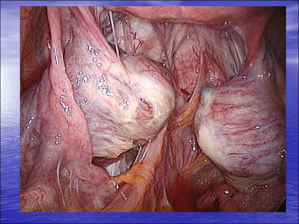
Serous cystadenomas are the most common epithelial tumorsand account for about 50% of malignant and 20% of benign cysts.
20% of benign cysts are bilateral and classically they are unilocular,
can reach the huge dimensions with tendency to torsion, and
contain a straw coloured fluid (light serous contents). Serous tumors
may be borderline. Borderline tumors with serous papillary
components are the worst type of borderline tumors with the
highest predisposition to dedifferentiate.
• The simple serous cystoma - is more often unicameral formation
with serous contents. A capsule wall is smooth. Treatment of
cystoma is only surgical.
• The papillary cystoma (proliferating) more often bilateral,
multichamber, has a tachyauxesis, is inclined to a malignant
degeneration (up to 80% of cases).
On an internal surface of capsule an abundant papillary growths are
formed. At malignant degeneration the growth pass on visceral
peritoneum and a peritoneum of the next organs. The papillary
cystoma of routinely small dimensions (its maximal size - about a
neonatal head), is frequently accompanying by an ascites. Contents
of a cyst is serous or serobloody.
• Treatment is surgical, as well as at a simple serous cystoma.
16.
17.
• Mucinous tumours (mucinous cystadenoma). Mucinoustumors can be borderline. Benign tumors are classically
multiloculated. Mucinous cystadenoma - multichamber with a
plenty of septum, filled gel-similar contents with a finely divided
suspension, are more often onesided, are characterized by a fast
growth. These form about 20% of ovarian cysts and are benign
90% of the time. When benign they are usually unilateral but 20%
of malignant mucinous tumors occur on both sides. Contents of
cystoma cavity of - a pseudomucin - heavy-bodied jelly-like fluid of
various colour (yellowish, brown, pale or dark green, dark red).
• The pseudomucinous cystoma educes asymptomatically, the
abdomen yet will not start to be enlarged. The tumour, even the big
dimensions, sometimes is not accompanied by any clinical
exhibiting. However at peduncle torsion or a necrosis of a capsule
early there is a pain in the abdominal low or loin. Signs of
compression of the next organs (urinary bladder, rectum, sacral
neuroplex, lymphatic and venous vessels) occur late.
• Treatment: at cystadenomas (serous and mucinous cystoma)
removal of an ovary even is carried out when the ovary is submitted
by a thin membrane around cystoma. At young age the tumour is
removed together with an ovary. Bilateral removal of ovaries are
after 48 years.
18.
19.
• Pseudomyxoma - rarely meeting tumour of an ovary(type of pseudomucinous tumour). A cystoma is
multichamber. The breakage of a capsule descends
spontaneously or at gynecologic research. Contents of
capsule get in abdominal cavity. The pseudomucin is not
soaked up by a peritoneum, and incapsulating, there is
dissemination of pseudomucin over all abdominal cavity.
Clinically the pseudomyxoma is accompanied by an
abdominal distention, a pain at palpation of abdomen.
Schotkin’s sign is weak positive in the inferior
departments of an abdomen. At a breakage of a capsule
of pseudomyxoma the signs of an acute irritation of a
peritoneum can appear.
Treatment surgical. It is impossible to remove jelly-like
masses from an abdominal cavity considerably. After
operation it is necessary to administrate cytostatics.
20.
21.
Endometriotic cysts are sometimescalled chocolate cysts. They are cysts of
endometriosis that occur on the ovary and
contain chocolate appearing material
formed from old blood. We'll talk about
endometriotic cysts the second part of this
lecture.
22.
23.
24.
• Brenner’s tumour - it is epithelial–connective tissue. Itmeets rarely, mainly at women than 50 years are more
senior. They are normally benign but can be malignant.
They often occur in association with serous tumors.
Clinically is shown disturbance of a menstrual cycle, can
have hormoneproduce character - hyperoestrogenic
(glandular-cystic hyperplasia endometrium at
postclimacteric bleedings) or a masculanization. Clinically
under the form, size and consistence a Brenner tumour
is similar to a fibroma. It is benign, onesided, a dense
consistence, ovoid form. Treatment is surgical - an
oncotomy together with the damaged ovary.
• Clear Cell tumor. These are always malignant and
carry a poor prognosis.
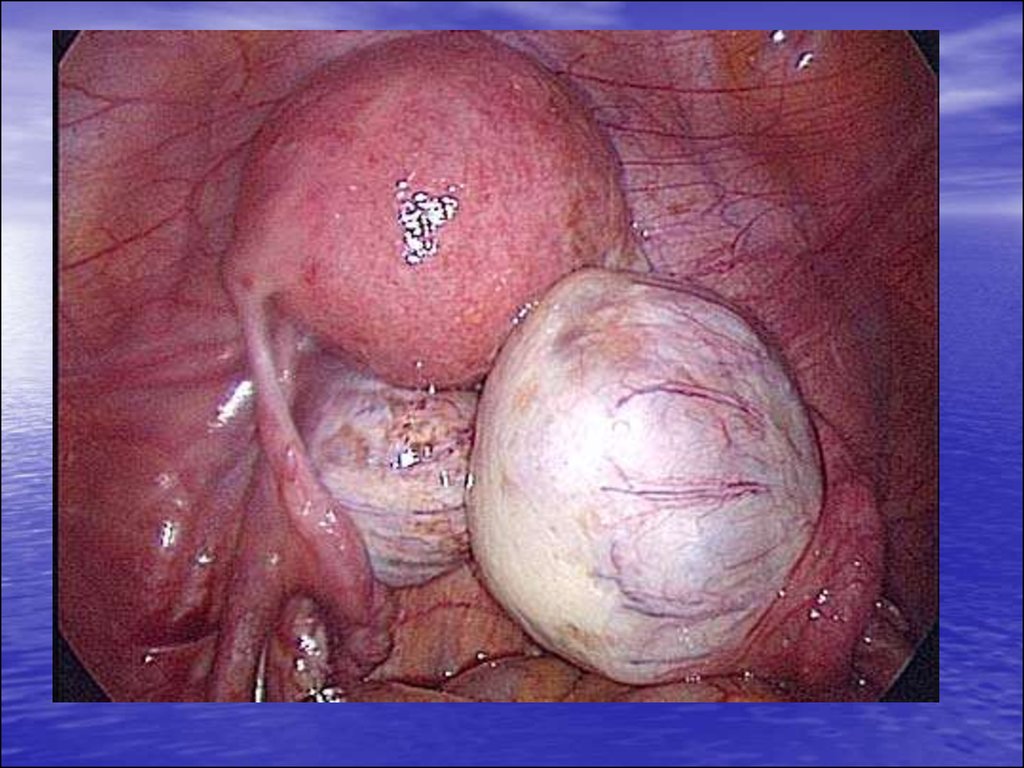
25. The right ovary is replaced by a Brenner tumor, the left ovary by a mucinous cystadenoma. The cervical tumor is a non-keratinizing squamous carcinoma.
26.
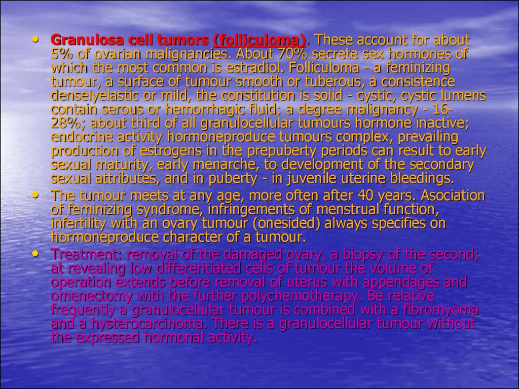
• Granulosa cell tumors (folliculoma). These account for about5% of ovarian malignancies. About 70% secrete sex hormones of
which the most common is estradiol. Folliculoma - a feminizing
tumour, a surface of tumour smooth or tuberous, a consistence
denselyelastic or mild, the constitution is solid - cystic, cystic lumens
contain serous or hemorrhagic fluid; a degree malignancy - 1628%; about third of all granulocellular tumours hormone inactive;
endocrine activity hormoneproduce tumours complex, prevailing
production of estrogens in the prepuberty periods can result to early
sexual maturity, early menarche, to development of the secondary
sexual attributes, and in puberty - in juvenile uterine bleedings.
• The tumour meets at any age, more often after 40 years. Asociation
of feminizing syndrome, infringements of menstrual function,
infertility with an ovary tumour (onesided) always specifies on
hormoneproduce character of a tumour.
• Treatment: removal of the damaged ovary, a biopsy of the second;
at revealing low differentiated cells of tumour the volume of
operation extends before removal of uterus with appendages and
omenectomy with the further polychemotherapy. Be relative
frequently a granulocellular tumour is combined with a fibromyoma
and a hysterocarcinoma. There is a granulocellular tumour without
the expressed hormonal activity.
27.
28.
Arrenoblastoma (an androblastoma, masculinizing tumour) - arisefrom embryonal germ of a man's component of female sexual gland,
localization is more often onesided, reach the big dimensions which
can be caused by absence of the expressed symptomatology at
early stages of tumoral growth; a solid constitution; frequently are
hormoneproduce, androgens define clinical sings of virilization,
disturbance of a menstrual cycle, an amenorrhea; frequency
malignancy 10-30%. Development of a tumour is show also a
masculanization (dfeminization) women (an involution - a
hypoplasia of uterus and the second ovary, an endometrium
atrophy, an amenorrhea, an atrophy of mammas, loss of sexual
sense, growth of hair on the face, breasts and legs, voice
roughening, a hypertrophy of clitoris).
• The association of virilization and hypooestrogenization signs is
possible.
• Treatment: at a hemilesion of gonad and the favourable urgent
cytologic diagnosis it is possible to confine an adnexectomy on the
one hand; after operation the menstrual cycle is restored, the
hirsutism decreases.
Gynandroblastoma – is very infrequent tumour, blended type, secretes
estrogens and androgens, with the conforming clinical pattern of a
feminization up to an endometrium hyperplasia and uterine bleedings and
virilization with a hirsutism, enlargement of clitoris.
• Treatment: an adnexectomy of uterus from the side of lesion.
29.
• Fibroma and a thecoma (thecablastoma, thecacellular, feminizinigtumour) are usually benign tumors secretes estrogens, invokes signs of a
feminization (a premature puberty in girls of prepuberty age) or later fading
of menstrual function (55-60 years) (early or late a feminizing syndrome).
At children's age prematurely there are the secondary sexual attributes
(mammas educe, occur cyclic or acyclic uterine bleeding, growth of a hair
on a pubis). At women of genital age the menstrual cycle is broken (the
menorrhagia, a metrorrhagia, amenorrhea), is marked infertility or
predilection to abortions.
In the period of a menopause are occurring acyclic bleeding, the uterus is
enlarged due to a hypertrophy and a hyperplasia of cells of a myometrium,
mammas are enlarged, the hyperplasia of a mucosa of a vagina and cervix
of a uterus, and also are developed the sexual drive strengthens. The
thecoma educes at women after 40 years, a tumour onesided of dense
consistences or dense elastic consistences more often, can reach the big
dimensions. The thecoma is quite often accompanied by disturbance of a
menstrual cycle such as a menometrorrhagia, hemorrhagic metropathias,
and also infertility.
The fibroma of an ovary exceeds the dimensions of medium man's fist,
educes more often at young women on the one hand. The tumour mobile,
is on a peduncle, grows sluggishly. Clinical signs are shown at hemorrhages
and a necrobiosis, torsion peduncle of tumour. In these cases there are
signs of a irritation of a peritoneum. On occassion (at a bilateral lesion) the
fibroma of an ovary is accompanied by Meigs triad (an ascites - a
polyserositis, an anemia, a cachexia), that specifies a malignant
degeneration of a tumour. In senior children can cause an anemia, an
ascites. Treatment: removal of the damaged ovary.
30. Fibroma ovary
31.
Sertoli/Leydig tumors. These accountfor less than 1% of ovarian tumors and
are usually benign. They occur in younger
women (teens and early 20s) and may
produce androgens.
32. Sertoli/Leydig tumors
33.
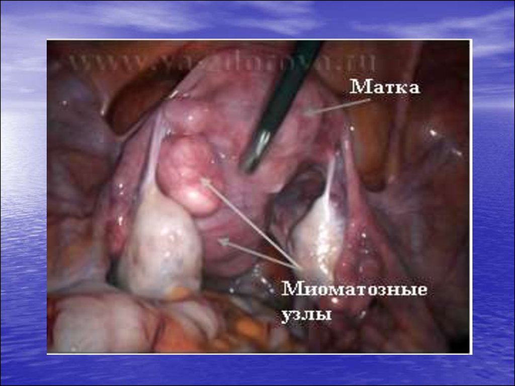
LEUOMYOMA OF THE UTERUS• Leuomyoma of uterus (fibromyoma,
leuomyoma) - the limited benign tumour
consisting from smoothmuscular cells and
fibrous elements of connective tissue.
34.
Epidemiology.• Leuomyoma is the most wide-spread
gynecologic disease: occurs approximately
from 10 to 27% of gynecologic patients.
Should pay attention that it meets at 20%
of the women who have achieved 30years age.
35.
ETIOLOGY.
The leuomyoma occurs as a result of local proliferation smoothmuscular
cells. On a measure of growth in structure of leuomyoma fibrous elements
start to dominate. It is established that a tumour can develop from a
smooth-muscle cells and grow under the influence of estrogens. The
amount and activity of the total progesterone receptors on a cell at uterine
myoma is lower than for healthy women, and the estrogens receptors – is
higher. Depending on the sizes of the myoma the level of progesterone
receptions in a tumour of the myometrium changes.
With age, and also due to accompanying ovarian dysfunction, the role of
absolute or relative (against a background of hyperoestrogenism) the
deficiency of progesterone increases. These hormonal disorders can result
to hyperplastic processes of the endometrium, cystic changes in the
ovaries, frequently developing at patients with uterine myoma.
The term “fibroma”, or “fibromyoma”, is not precise, because the initial
element of this tumour is the smooth muscle cells.
Under the influence of hormonal stimulation during pregnancy the myoma
can enlarge, become more soft consistency, which complicates its diagnosis
during palpation. After delivery, the sizes of the tumour, as a rule,
decrease.
Contributing factors of development of the myoma are preanemic states
and an iron deficiency anemia, an idiopathic hypertension, IHD (ischemic
heart disease), the chronic locuses of an infection contamination (a
tonsillitis, a genyartritis, an otitis), a thyrotoxicosis, a diabetes, chronic
diseases GIT (a gastritis, a cholecystitis, a colitis).
36.
Classification• Approximately in 95% of cases the
myoma develops in the corpus uteri, in
5% - in the cervix. 80% of the women
have multiple nodes of leuomyoma.
• Depending on the site concerning the
uterine wall the leuomyoma are
differentiated: subserous,
intraligamentous, intramural, submucousal
or cervical myoma (leiomyoma).
37.
• Subserous leiomyoma is located under the peritoneal (serous) surface ofthe uterus, it can be small or large, and in some cases has a pedicle. The
subserous myoma can receive additional blood supply from the omentum
due to a fusion formed with it (parasitic tumour).
Intraligamentous leiomyoma is characterized by a lateral growth or
primary development between the leaves of the broad ligament of the
uterus.
Intramural (interstinal) leiomyoma develops in the uterine wall. With
the small sizes it can not cause changes in the contours of the uterus.
Increasing, such a uterine myoma gets a nodular asymmetric form. With
the large sizes the myoma is distributed up to the serous and mucous
membrance of the uterus.
The subserous and intramural uterine myoma before reaching large
sizes, as a rule, is asymptomatic.
The submucous myoma is rare (5-10% of cases), but a dangerous type
of benign uterine tumour (strong bleedings can be observed, infected nodes
with distribution of the infection onto the uterus).
The cervical myoma occurs most frequently on the posterior surface of
the cervical leiomyoma is accompanied by symptoms of compression of the
bladder.
Leuomyosarcoma is found in 0,1-0,5% of the patients with leuomyoma;
however its development from leuomyoma is not established.
38.
39.
40.
41.
CLINICAL SYMPTOMATOLOGY OF THE LEIOMYOMA
Signs considerably vary in dependence on the dimensions, amounts
and localizations of nodes.
Pathological menstrual bleedings (routinely a hypermenorrhea) - the
most typical attribute of a leiomyoma. Intensity of bleedings
gradually increases, that can result to the expressed anemia.
Pain. Uncomplicated leuomyoma of the uterus is routinely painless.
There can be constant whining pains in the lower part of an
abdomen, loin-sacrul range - are connected to a distention of a
peritoneum at growth subserous nodes, pressure of myomatous
nodes upon neuroplexes of a small pelvis or separate nerves.
The compression of organs of the small pelvis routinely arises, if the
myomatous uterus or node achieves the dimensions conforming to
12-14 weeks of pregnancy and more.
a. Urinary retention arises at a uterus retroversio owing to
myomatous growth; thus the cervix uterus is moved anterior and
presses a urethra to a pubic articulation.
b. Constipations and difficulties of defecation can be caused by large
myomas of a back wall of a uterus.
Infertility - at women with submucous or intramural myomas
abortions and premature births are more often.
42.
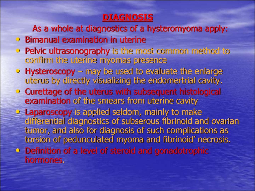
DIAGNOSIS
As a whole at diagnostics of a hysteromyoma apply:
Bimanual examination in uterine
Pelvic ultrasonography is the most common method to
confirm the uterine myomas presence
Hysteroscopy – may be used to evaluate the enlarge
uterus by directly visualizing the endomertrial cavity.
Curettage of the uterus with subsequent histological
examination of the smears from uterine cavity
Laparoscopy is applied seldom, mainly to make
differential diagnostics of subserous fibrinoid and ovarian
tumor, and also for diagnosis of such complications as
torsion of pedunculated myoma and fibrinoid’ necrosis.
Definition of a level of steroid and gonadotrophic
hormones.
43.
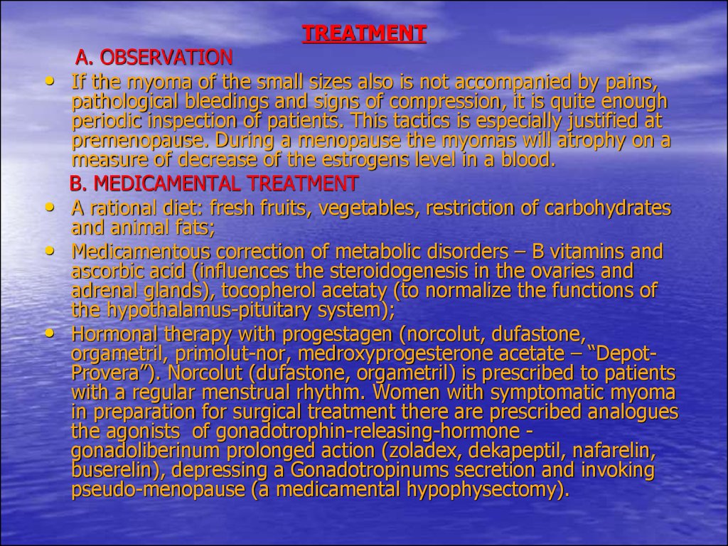
TREATMENTА. OBSERVATION
If the myoma of the small sizes also is not accompanied by pains,
pathological bleedings and signs of compression, it is quite enough
periodic inspection of patients. This tactics is especially justified at
premenopause. During a menopause the myomas will atrophy on a
measure of decrease of the estrogens level in a blood.
B. MEDICAMENTAL TREATMENT
A rational diet: fresh fruits, vegetables, restriction of carbohydrates
and animal fats;
Medicamentous correction of metabolic disorders – B vitamins and
ascorbic acid (influences the steroidogenesis in the ovaries and
adrenal glands), tocopherol acetatу (to normalize the functions of
the hypothalamus-pituitary system);
Hormonal therapy with progestagen (norcolut, dufastone,
orgametril, primolut-nor, medroxyprogesterone acetate – “DepotProvera”). Norcolut (dufastone, orgametril) is prescribed to patients
with a regular menstrual rhythm. Women with symptomatic myoma
in preparation for surgical treatment there are prescribed analogues
the agonists of gonadotrophin-releasing-hormone gonadoliberinum prolonged action (zoladex, dekapeptil, nafarelin,
buserelin), depressing a Gonadotropinums secretion and invoking
pseudo-menopause (a medicamental hypophysectomy).
44.
C. SURGICAL TREATMENT– Indications:
• а. The bleeding invoked by myomas, is especial in case of the expressed
anemia. The hypermenorrhea routinely happens at submucous or
intramuscular myomas.
b. The strong pains supposing of necrosis or peduncle torsion of
myomatous node.
c. Enlargement of a myomatous uterus up to the dimensions conforming to
12-weeks duration of gestation.
d. At grow of myomatous nodes (4-5 weeks and more in a year) it is
necessary to carry out immediate inspection for exception of their malignant
degeneration. Presence of the big subserous node on the thin peduncle also
is the indication to operative treatment because of high risk of torsion node.
e. Submucose leuomyoma – it is reason of significant DUB, metrorrhagia
and other disorders of menstrual function, conservative treatment without
effect.
f. Disorder of nutrition of myomatous node – necrosis of node, when “acute
abdomen” develops.
g. Suspicion on malignant changes in the myomatous node (fast grow,
softening of node).
h. The Hydronephrosis and other expressed signs of urinary bladder
compression, an intestine or the urethras, revealed at ultrasonic or an
intravenous pyelography.
i. A myoma in a combination to precancerous endometrium pathology,
ovaries.
k. Infertility as a result of a hysteromyoma.
45.
The kind of a surgical intervention depends on age of
the woman, signs, and also desire to have children
in the future.
А. The myomectomy - erasion of single or plural
myomas with conservation of uterus; this operation
routinely carry out to the women, wishing to become
pregnant and not having contraindications.
The main complications - a bleeding during and after
operation, and also the early and late intestinal
obstruction caused by adhesions between an intestine
and a uterus after a myomectomy.
The probability of repeated originating myomas after a
myomectomy depends on age of the woman, and also
volume of previous carried out myomectomy; at 30%
of cases repeated originating myomas is observed
within 10 years after operation.
Probability of offensive of pregnancy after a
myomectomy - 40%.
46.
B. Hysterectomy. If there are surgical indications, and the woman
does not plan to have more children, operation of a choice complete removal of the uterus.
It is necessary to conserve ovaries at women more youngly 40-45
years.
Before hysterectomy or other medical procedures, especially elderly
woman, it is necessary to carry out a diagnostic currettage of a
cavity of the uterus for exact definition of the cause of bleeding
(myoma or endometrium cancer).
The hysterectomy completely eliminates risk of repeated originating
of a leiomyoma.
There are no convincing data about rising risk of development in a
cancer in a ovary, to women more youngly 40-45 years ovaries are
necessary for conserving.
C. Semiradical operative treatment applies to conservation of
menstrual function at women in premenopause.
The defundation is carried out when the locating of myomatous
node allows to keep a corpus uterus without its fundus.
High supravaginal amputation of the uterus differs from routine that
cut a corpus uterus much above internal os.
Selective embolisation uterine arterias by Seldinger.
47.
PROPHYLACTIC MEDICAL EXAMINATION:
Patients with the increased risk of development of
leiomyoma (often relapses of chronic salpingooophoritis, endomyometritis, accompanying with
metabolic-endocrine infringements);
With infringement of menstrual cycle from the menarche
and a late menopause;
With numerous abortions and diagnostic curettages of a
uterus;
With disorders of menstrual cycle on background of long
course of extragenital diseases;
With presence of a leiomyoma and oncologic diseases at
close relatives;
With a leiomyoma at incipient state of development;
After the carried out operative and conservative
treatment;
With contraindications to operative treatment.
Control surveys 1 time in 6 months.
48.
Endometriosis• The endometriosis is a pathological benign
process which is formed on a background
of the broken hormonal and immune
homeostasises and is characterized by
presence epithelial or stromal the
elements similar to endometrioid structure
in myometrium or in other organs of
sexual system.
49.
The etiology of an endometriosis is completely unknown.
At the moment there are following the theory of development of endometriosis.
Embrional theory (dysontogenetic), suggested by Recklingaysen (1896): endometrioid
heterotopies educe from paramezonephral ducts or from a germinal material of which
generative organs including an endometrial tissue are formed.
Metaplastic theory (Ivanov N.S., Ulezko-Stroganova K.P., 1887; R.Meyer, 1909): the
endometriosis educes as a result of a metaplasia embrional peritoneums or a coelomic
epithelium.
Implantation theory (or the theory retrograde menses) (for the first time it was offered by
J.A.Sampson, 1925) - now the most recognized: formation of the locuses of an endometriosis
descends in result retrograde runaway in a abdominal cavity of endometrium cells which were
tore away during a menses and their further implantation on surrounding organs and a
peritoneum.
The theory of an ratrogenic dissimination: transmission or a translocation of endometrioid
particles in a small pelvic cavity can take place as a result of surgical manipulations, including
a diagnostic currettage, at obstetric and gynecologic operations.
The lymphogenous and hematogenous theory (transport hypothesis): endometrioid cells are
transported on lymphatic and to blood vessels.
Genetical theory: family forms of an endometriosis, high their frequency among patients with
developmental anomalies of genitalias.
The hormonal theory: development of all forms of an endometriosis is spoken changes of
hormonal function of ovaries and hypothalamo-pituitary system. As processes of a
proliferation and secretory transformation of endometrium are controlled by steroid
Hormonums, infringement of a secretion of gonadotrophic Hormonums and a steroidogenesis
in ovaries (chaotic peak emissions of FSH, LH, decrease of a basal level of Progesteronum,
hypoostrogenia) frame necessary conditions for development of endometrioid implants and
support of their awake state.
The immune theory (M.V.Jonesco et G.Popesco, 1975): development of an endometriosis is
probably only in conditions of the broken local immunodefence.
50.
Risk factors of development of an endometriosis:
а) Hereditary predisposition;
b) Reproductive age;
c) Disorders of menstrual function;
d) Absence of labor or one labor in an anamnesis;
e) Frequent abortions and diagnostic currettages of a
uterus;
f) Long use of endometrial contraceptives;
g) Retrograde wave contractions of a uterus from
uterine cervix to the fundus during a menses;
h) Anovulation.
51.
TOPICAL CLASSIFICATION OF THE ENDOMETRIOSISA genital endometriosis
• I.
• 1. An internal endometriosis
• 1.1. An endometriosis of a uterine body (I, II, III (adenomyosis) stages in
dependence on depth of a lesion of a myometrium):
- Glandular, cystic, the diffuse form;
- Focal, nodal, diffuse forms.
1.2. Endometriosis of the cervical canal.
1.3. Endometriosis intramural parts of uterine tubes.
2. An external endometriosis.
2.1. Peritoneal endometriosis:
- Endometriosis of ovaries (infiltrative, tumoral forms);
- Endometriosis of uterine tubes;
- Endometriosis of a pelvic peritoneum (red, black, white forms).
2.2. Extraperitoneal endometriosis:
- Endometriosis of vaginal part of the uterine cervix;
- Endometriosis of a vagina, a vulva;
- Retrocervical endometriosis;
- Endometriosis of uterine ligaments;
- Endometriosis parametral, paravesical, paravaginal fats (without and with
germination in urinary bladder, a rectum).
• 3. An external-internal endometriosis.
• 4. Associated forms of a genital endometriosis (a genital endometriosis in an
association to other genital or extragenital pathology).
II.
An extragenital endometriosis (an endometriosis of a gastrointestinal
path, urinary organs, skin, a umbilicus, postoperative wounds, lungs, pleuras, etc.).
52.
Internal endometriosis of uterine corpus
(adenomyosis) (Adamjan L.V., 1998):
I degree - the pathological process
circumscribed to a submucosa of a body of the
uterus;
II degree - passes pathological process to a
muscle layer;
III degree - diffusion of pathological process on
all depth of a muscular wall of a uterus up to its
serous coat;
IV degree - recruitment phenomenon in
pathological process, except for a uterus,
parietal peritoneums of a small pelvis and the
next organs.
53.
CLINIC• Clinical exhibitings and anatomic-morphological changes of many respects
depend on localization, the form, diffusion of the given pathological
process.
COMPLAINTS
• A pain sign - nagging pains in the low part of abdomen or in lumbosacral
range, strengthen at the eve and during time menses, sometimes they
imitate a pattern of an acute abdomen (algodysmenorea; a pain in the low
part of the abdomen, at range of a pelvis and a loin, untied with a
menstrual cycle; dyspareunia);
• Disorders of the menstrual cycle - the sign of an endometriosis second on
frequency: a long, profuse menses (hyperpolymenorrhea), before menses
and after frequently scanty dark–bleeding discharge;
• Infertility (primary, secondary) is caused by anovulation, failure of a corpus
luteum, adhesive process in a small pelvis, a lesion of uterine tubes,
inferiority of endometrium function;
• Long unefficient treatment of chronic adnexites, metrites;
• Psychoneurological distresses;
• Infringement of function of next organs (dysuria; painful defecation).
The basic signs of a genital endometriosis: a dysmenorrhea, pelvic pains,
painful coitus (dyspareunia), a meteorism, infringement of a defecation
(dyschesia), infertility, dysuria. More infrequent signs: a proctorrhagia, an
intestinal obstruction, etc.
Sometimes patients do not show complaints that can depend on features of
topical localization endometrioid heterotopias.
54.
Internal endometriosis (adenomyosis). Pathognomonic clinical criteria
of an internal endometriosis: painfull and long and-or profuse menses which
result in development of the secondary anemia, pains in the low parts of
the abdomen on the eve and in the first days of a menses, enlargement of
the uterine dimensions, especially expressed before menses.
Diffuse, nodal, focal forms of an internal endometriosis are distinguished.
Focal and nodal forms are observed a little bit less often diffuse. At these
forms of disease at women at reproductive age and at premenopause the
hyperplasia of a muscle tissue which surrounds the locuses heterotopic
endometrium is always defined. Clinical exhibiting of the nodulose form of
an endometriosis, except for the signs described above, it is characterized
by more appreciable pain reaction on a menses with the expressed
vegetative infringements - a nausea, a vomiting, a headache, a
fervescence, a loss of consciousness. Development of typical exhibiting of
an endometriosis is preceded quite often with infertility.
Infrequent forms of an endometriosis: an endometriosis isthmic part of a
uterus, isthmic-cervical part.
The internal endometriosis is frequently combined with a leomyoma, less
often - with tumours of ovaries, a chronic inflammation of appendages of a
uterus, an endometriosis of other organs and tissues (endometrioid cysts of
ovaries and a retrocervical endometriosis).
At differential diagnostics it is necessary to take into account an opportunity
of a combination of an internal endometriosis of a corpus uterus with an
endometrium adenocarcinoma.
55.
Endometriosis of the uterine cervix
On researches of last years, frequency of an endometriosis of the
uterine cervix has sharply increased.
The macroscopic locuses of an endometriosis of a vaginal part of
uterine cervix look like "eyes", “Nabothian follicles” more often. At
survey endometrioid heterotopias have light pink or reddish colour.
Most legiblly they are defined at the end of lutein phase: formations
of blue-crimson colour, boldly act above a surface of the cervix
uterus. Distinctive feature of an endometriosis - superficial its
locating on vaginal part of the uterus, distal part of a mucosa of the
cervical canal, pre-and postmenstrual scanty bloody discharge,
contact discharge. Pains at an endometriosis of the cervix uterus are
absent.
The endometriosis of the cervix uterus quite often arises after a
diathermy and other surgical interventions, labors which are
accompanied by a trauma.
Endometriosis of the cervix uterus it is necessary to differentiate
from endometrioid metaplasia of separate endocervix glands,
adenocarcinoma in situ, Nabothian follicles with hemorrhagic
contents - formations which too are accompanied pre-and
postmenstrual bloody discharge.
56.
The endometriosis of uterine tubes meetsrarely, much more often illness is observed in a
combination with endometrioid heterotopias of
other localizations (an endometriosis of a uterus
and ovaries). The clinical pattern of the given
disease practically does not differ from the
conforming clinical exhibiting the listed
localizations. The algomenorrhea remains as a
leading sign.
57.
58.
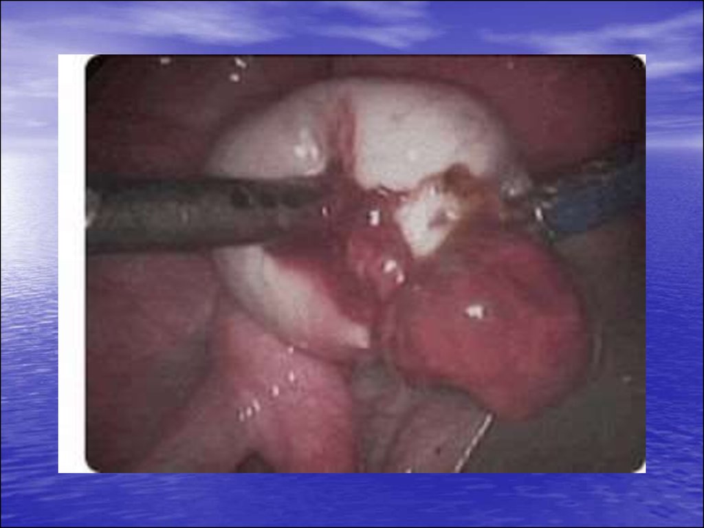
Endometriosis of ovaries. Among all localizations of an external
endometriosis the lesion of ovaries wins first place. Endometrioid cysts of
ovaries at long existence get a characteristic kind. The dimensions of them are
0,6-10 cm. Cysts more than 10 cm meet rarely. Macroscopicly: an endometrioid
cyst capsule is thick (0,2-1,5 sm), numerous dense comissuras on a external
surface, hemorrhagic contents of a chocolate kind, in past named them
“chocolate cysts”. The clinical pattern of an endometriosis of ovaries is very
various.
The basic complaint - a pain syndrome of different intensity: constant whining
pain, periodically strengthen, irradiate in a rectum, a loin, pains achieve a
maximum at the eve and during a menses. Sharp pains are observed when
there are microperforations of a wall of a cyst and its contents are poured out
in abdominal cavity. At an intensive pain syndrome patients will be frequently
hospitalized with diagnoses: an acute appendicitis, a salpingocuesis, torsion
peduncle of an ovarian tumour, an acute pelviperitonitis.
The "acute abdomen" syndrome educes at 26% of patients. At patients the
progressing algomenorrhea is marked, is accompanied by a vomiting, a loss of
consciousness and the common weakness with decrease of a working capacity
more often. Endometrioid ovarian cysts are always accompanied by
development of adhesive process in a small pelvis that results in infringement
of function of an intestine and urinary bladder (constipations, the dysuric
phenomena). Very much frequently at patients there are marked scanty preand postmenstrual bloody discharge from sexual ways.
At presence endometrioid ovarian cysts there can be a subfebrile temperature,
rising of a blood sedimentation rate, a leukocytosis. Patients frequently are
unsuccessfully treated concerning “inflammatory process”.
At gynecologic inspection at range of appendages of a uterus a formations are
defined one or bilateral, inactive, tense elastic consistence.
59.
60.
The retrocervical endometriosis is defined at women of30-40 years more often.
* Clinic: very sharp pain, irradiate in rectum, the vagina, a
perineum, external generative organs and is more often
in a femur. Intensifying pains is marked at sexual
contacts, the act of defecation. A menses are
accompanied by a vomiting, a loss of consciousness, a
cold snap of extremities, general delicacy, irritability, a
unbalance, tearfulness, often headaches, infringement of
a rhythm of dream, a hypoactivity of a thyroid gland,
other endocrine glands, a gastrointestinal path
dyskinesia. Patients complain of constipations which
fractionally strengthen before menses. Constipations
gradually strengthen before development of a particulate
intestinal obstruction. Constipations can be alternated to
diarrheas with a mucifying and bloods from a rectum,
that is the indication to hospitalization in an infectious
diseases hospital with suspicion on dysentery.
61.
62.
Endometriosis of a vagina. Theprimary vaginal endometriosis can
sometimes be combined with
developmental anomaly of a organ
(padding in part vagina aplasia), with an
endometriosis of cervix uterus.
63.
DIAGNOSTICS:
1. Speculum and bimanual examination.
2. Ultrasonic.
3. A colposcopy, colpocervicscopy.
4. A hysteroscopy.
5. A laparoscopy.
6. Express methods of research of organs of
extragenital endometriosis localization.
7. Histological research of biopsian material and
a material of postoperative preparations (after
hysterectomy, adnexectomy, etc.).
Cytologic and histological research of a material
from a cavity of the uterus and cervix uterus
poorly informatively.
64.
DIFFERENTIAL DIAGNOSTICS with:• - Leuomyoma of a uterus; a chronic
salpingo-oophoritis (adnexitis);
• - Tumours of genitalias; tumours of an
intestine;
• - Hyperplastic processes of endometrium;
• - Extrauterine pregnancy;
• - Nephroptosis; a urolithiasis; an
appendicitis; a paraproctitis; a proctitis; a
colitis; an adhesive intestinal obstruction.
65.
Diagnostics in dependence on topical localization of anendometriosis:
Internal endometriosis (an endometriosis of uterine corpus, the
cervical canal, intramural parts of uterine tubes)
Bimanual examination: moderate enlargement of a uterus at anteriorposterior dimension; painful at a palpation;
Ultrasonic (transabdominal, vaginal, the rectal sensor): the moderate
enlargment of a uterus, is especial it anterior-posterior dimension; rotundity
of its form; dilating of an isthmus; a thickening of one of walls of a uterus;
roughness of contours of a uterus; deformation the M-echo (i.e. cavities of
the uterus with endometrium); enlargement of acoustic frame of a
myometrium (I, II, III stage in dependence on depth of a lesion of a
myometrium), deformation and dilating of region increased echogenic
around the M-echo, presence unechogenic incorporations with echogenic
contour, formation of regions increased echogenic irregular;
A hysteroscopy (the stage is not defined):
- Presence of dark-blue or crimson spots, cysts; albescent nodules,
roughnesses; a rigidity of endometrium relief;
- Endometrioid ductus foramens.
A hysterosalpingography (the stage is not defined):
- Presence after contours shades;
- A proximal tubal occlusion.
A laparoscopy (at III stage of an adenomyosis):
- Dark-blue, crimson, cystic or nodulose formations;
- Albescent or grayish brown nodules.
66.
Endometriosis of ovaries, uterine tubes, peritoneums• Bimanual research: enlargement, bracing at range of
appendages of a uterus; restriction in motility of organ of a
small pelvis; painfull at a palpation;
• Ultrasonic (transabdominal, vaginal, the rectal sensor):
enlargement of appendages of a uterus, their
inhomogeneous acoustic density, bracing in the posterior
fornix (Duglas’ pouch); presence of formation of the
spherical form with a legible capsule, weak echogenic
internal structure ("cloud"); attributes of perifocal adhesive
process.
• A laparoscopy:
- Dark-blue, crimson, cystic, spotted or nodulose structures;
- Albescent or grayish nodules;
- Presence of cicatrical changes, adhesive process;
- Bluish cystic structures - endometrioid cysts.
• A computer tomography, magneto-resonance tomography:
- Formations of the spherical form with enough dense
capsule;
- Adnations with other structures.
67.
Retrocervical endometriosis, endometriosisligaments, fats.
• Bimanual examination: a thickening, contraction
sacrouterine, cardinal ligaments; small-sizes
nodulose structures behind of cervix uterus at a
level of internal os, frequently sharply painful;
restriction of motility of organs of a small pelvis;
painfull at a palpation.
• A laparoscopy:
- Dark-blue, crimson, cystic, spotted or nodulose
structures;
- Albescent or grayish nodules;
- Presence of cicatrical changes, adhesive
process.
68.
Endometriosis of vaginal part of the cervixuterus (intracervical, subepithelial),
vagina, vulva.
• Bimanual research: without features or dense
painfull nodes, seams, thickenings in a wall of a
vagina, a vulva.
• Colpocervicoscopia: nodules, a stains or a points
of dark-blue, crimson colour on cervix uterus, a
vulva, a vagina.
External-internal endometriosis.
• Various combinations of diagnostic attributes
which are inherent at the topical forms of an
endometriosis listed earlier.
69.
TREATMENT OF THE ENDOMETRIOSIS:The basic directions of an endometriosis therapy:
hormonal, immunocorrecting, antioxidanting,
desensitizing, anti-inflammatory (inhibitors of
Prostaglandinums), symptomatic therapy, surgical.
1. Conservative therapy.
2. Surgical treatment.
The choice of treatment tactics depends from:
- Age of the woman;
- Localizations and degrees of disease diffusion;
- Expressivenesses of signs and duration of disease;
- Presence of a fertility and necessity of regeneration of
reproductive function at infertility;
- Presence of concomitant gynecologic diseases;
- Efficacy of previous treatment;
- States of other organs and systems.
70.
Indications for surgical treatment of a genital
endometriosis:
1. An internal endometriosis in a combination to
hyperplastic processes of ovaries and-or a precancer
endometrium.
2. An adenomyosis (the diffuse or nodulose form) which
is accompanied by a hyperplasia endometrium.
3. Endometrioid ovarian cysts (there are dimensions
more than 5 cm which function is stable).
4. Absence of effect from medicamental treatment which
was carried out continuously during 6 months.
5. Recruitment phenomenon in pathological process of
other organs and systems with infringement of their
function.
6. An endometriosis of postoperative cicatrix.
7. A combination of an endometriosis to some anomalies
of generative organs.
8. Presence of a somatic pathology which excludes an
opportunity of carrying out of long hormonal therapy.
71.
Criteria of efficacy of treatment:• 1. Absence of relapses of disease.
• 2. Regeneration of genesial function (at
conservative treatment and organretaining
operations).
• 3. Positive dynamics of life quality.
72.
Internal endometriosis (a method of a choice - conservative therapy)Hormonal therapy:
1.
а) Datum level FSH, eu- or slight hyperoestrogenia, deficiency of Progesteronum and
excess LH:
At childbearing age: an oestrogen-gestagen drugs (non-ovlonum, ovidonum,
Rigevidonum, marvelonum, femodenum, diane-35, logest, janinum, etc.). The
preference is given the monophasic combined oral contraceptives with strong
progestagen effect. A method of administration: for 1 tabl/day in a continuous
regimen during 6-9-12 months, enlarging a dose up to 2-3 tab. at broken through
bleedings.
At perimenopause: gestagen drugs:
Progesteronum (utrogestanum) - 200-300 mg/day in 2 receptions from 14 to 26 day
or from 5-th to 26 day of a cycle, are peroral or vaginaly, 6-9 months;
Didrogesteronum (dufastonum) - 10 mg*1-3 time/day from 14 to 26 day or from 5th to 26 day of a cycle, perorally, 6-9 months;
Medroxyprogesteronum acetas (provera) - 10 mg*3 time/day, perorally,
continuously, within 3 months; depot-provera - 50 mg*1 in a week or 100 mg*1
time/in 2 weeks or 150 mg at 14 day of a menstrual cycle intramuscularly, 6 months;
17-pregnenoldione capronat - 12,5% 1 ml at 7, 14, 21 day of a menstrual cycle,
during 3-6 months;
Norethisteronum (Norcolutum, Primolutums-nor) - 5-10 mg/day from 14 to 26 day or
from 5-th to 26 day of a cycle, perorally, 6-9 months;
gestonoronum capronat (Depostatum) - 200 mg 1 time/week, intramuscularly, during
3 months;
linoestrenol (orgametril) - 5-10 mg/day from 14 to 26 day or in a continuous
regimen, 6-9-12 months.
73.
b) Datum level FSH, LH, hyperproduction of oestrogens.Antigonadotrophic drugs (with the count of material opportunities
and wishes pacients): danasolum (danoval, danol, danogen) on 200 mg*14 once a day after meal from 5-th till 26-th day of a menstrual cycle; at
perimenopause - in a continuous regimen of 3-6 months.
c) Datum level FSH, LH, Progesteronum, expressed hyperoestrogenia.
Antioestrogenic drugs: Tamoxifenum (zitazonium, Nolvadexum) 20-40
mg/day of 6-9 months; toremifen (fareston) 10-20 мг*2-3 time/day of 6-9
months.
d) Hyperproduction FSH, LH, oestrogens.
Agonists Gonadotropinum-releasing Hormonums (with the count of
material opportunities and wishes of the patient): triptorelinum
(diferelinum, a decka-peptil) 3,75 mg subcutaneously, in a anterior
abdominal wall, in any of the first 5 days of a menstrual cycle; a repeated
injection - in 28 days; course of treatment - 3-6 months.
hoserelinum acetas (zoladex) 3,6 mg (under the similar schema);
buserelinum (suprefact-depot) of 900-1200 mg/day intranasal or
200-400 mg/day, 3-6 months;
nafarelinum acetas (synarel) 0,4-0,8 g/day intranasal in 2 receptions,
3-6 months;
leyprolid (lupronum) 3,75 mg subcutaneously, at anterior abdominal
wall, in any of the first 5 days of a menstrual cycle; a repeated injection - in
28 days; course of treatment - 3-6 months.
74.
• 1 Nonspecific anti-inflammatory therapy:- Not steroid anti-inflammatory drugs (diclophenak (voltaren) to 1 suppository it is
rectal, or 25-50 mg*2-3 time/day after meal; Indomethacinum 25-50 mg*2-3
time/day after meal; nimesulidum (mesulidum, nimegesik) 100 mg*2 time/day or it is
rectal on 1 suppository during 10-15 days, etc.);
- kontrikalum 10000 Units on 200 ml of a Sodium chloridum, intravenously, are
trickling, during 10-15 days;
2
Agents which influence the central nervous system (sedative drugs,
small tranquilizers, a psychotherapy).
3
Resorptional therapy (systemic ensimotherapy - vobensim, flagensim: 3-5
tabl*3 time/day, 1-2 months).
4.
Immunomodulating factors, antioxidants, a vitamin therapy (redoxon,
vitamin A, reproduction vitamin 1 caps. 1-3 time/day, the Т-activin 1 ml
subcutaneously; an interferon (laferon) 1 million Units intramuscular during 10 days,
etc.).
• 5.
Agents which sustain function of a gastrointestinal path and
hepatobilian systems (hepatoprotectors (hepabene 1 caps*3 once a day;
Essentiale 2 caps*2-3 time/day; chophitolum 2 tabl /2-3 time/day during meal during
20-30 days)).
6.
Physiotherapeutic methods (at presence of adhesive process):
electrophoresis of copper and Zincum; electrophoresis with Lydasum, Trypsinum;
radon baths; acupuncture; low intensive laser radiance; a magnetotherapy in a
pulsed operation) 15-20 sessions.
• 7.
• 8.
Treatment of concomitant genital and extragenital diseases.
A diet according to concomitant diseases.
75.
The control of efficacy of treatment of an intrinsicendometriosis:
• 1. At a positive effect - a dispensary observation once
in 3-6 months, periodic courses of therapy.
• 2. At an inefficiency of hormonal therapy, infertility,
tumorous forms of an internal endometriosis, suspicion
on a malignancy - surgical treatment:
• а) At reproductive age - organretaining surgical
treatment by laparotomy or lapascopy access. In the
subsequent, conservative treatment, treatment of
infertility.
• б) At perimenopause - surgical treatment in volume of a
hysterectomy; hysterectomia with tubes.
At absence of treatment a progressing disease with
development of wide-spread, tumorous and malignant
forms are possible.
76.
Treatment of an ovarian endometriosis.
1. The tumorous form - surgical treatment with the subsequent
control of efficacy and conservative treatment (similar with
treatment of an internal endometriosis). At positive effect - a
dispensary observation once in 3-6 months, periodic courses of
therapy.
2. Infiltrative the form - conservative treatment (it is similar with
treatment of an intrinsic endometriosis). At contraindications to
hormonal therapy - surgical treatment.
At an inefficiency of conservative treatment of infiltrative forms,
presence of infertility, development of tumorous forms - surgical
treatment:
а) At reproductive age - organretaining volume of operation by
laparotomy or laparoscopy access: a cystectomy, a resection of an
ovary, adnexectomia, a laser vaporization, an electrocoagulation,
use of a ultrasonic scalpel, argonum coagulator, presacral
neurotomy. Further, conservative treatment (it is similar to
treatment of an internal endometriosis), treatment of infertility.
б) At perimenopause - a hysterectomy.
77.
Endometriosis of uterine tubes.
Conservative treatment (it is similar to treatment of an
internal endometriosis) - the control of efficacyy:
а) At an inefficiency of conservative treatment, presence
of infertility - surgical treatment (as it mentioned above):
at reproductive age (a laser vaporization of the locuses,
electro-, a thermocoagulation; use of a ultrasonic
scalpel); at perimenopause - tubeectomy by laparotomy
or laparoscopy access.
After surgical treatment at reproductive age padding
complex of conservative therapy are carried out
(similarly therapy of an internal endometriosis).
At absence of treatment progressing disease with
development of wide-spread and tumorous forms are
possible.
78.
Endometriosis of a pelvic peritoneum.• Surgical treatment by laparotomy or
laparoscopy access (erasion of the locuses
of an endometriosis with the help of the
carbonic laser, electro-, a
thermocoagulation).
• Further, conservative treatment (it is
similar to treatment of an internal
endometriosis).
• At absence of treatment progressing
disease with development of wide-spread
forms are possible.
79.
Endometriosis vaginal part of an uterine
cervix, a vagina, a vulva endometriosis.
Conservative treatment (it is similar to treatment
of an internal endometriosis) - the control of
efficacy:
а) At an inefficiency of conservative treatment cryosurgical treatment, removal of the locuses of
an endometriosis. Further, conservative
treatment (it is similar to treatment of an
internal endometriosis).
б) At a positive effect - dispensary observation
once 3-6 months, periodic courses of treatment.
At absence of treatment a progressing disease is
possible with development of wide-spread and
tumorous forms which demand surgical
treatment in volume of a trachelectomy,
resections of a vagina, vulvectomy.
80.
Retrocervical endometriosis, ligaments, fat
endometriosis.
Surgical treatment by laparotomy or laparoscopy access:
а) At reproductive age - removal of the locuses of an
endometriosis with the help of a laser vaporization,
electric-, thermocoagulations. At diffusion on interfacing
organs - with participation of the conforming experts.
Further, conservative treatment (it is similar to treatment
of an intrinsic endometriosis).
б) at perimenopause - removal of an endometriosis
locuses with a hysterectomy with appendages; at a
germination in a rectum or urinary bladder, urethras with participation of the conforming experts. Further,
conservative treatment (it is similar to treatment of an
internal endometriosis).
At absence of treatment progressing disease with
development of wide-spread and tumorous forms,
infringement of function of interfacing organs is possible.
81.
External-internal endometriosis
Conservative treatment (it is similar to treatment of an
internal endometriosis) - the control of efficacy:
а) At an inefficiency of conservative treatment, presence
of infertility, tumorous forms - surgical treatment at
reproductive age with performance organo-conserved
operations (as it mentioned above). After surgical
treatment at reproductive age conservative therapy is
carried out (similarly therapy of an internal
endometriosis).
At perimenopause - a hysterectomy.
б) At a positive effect - a dispensary observation once in
3-6 months, periodic courses of therapy.
At absence of treatment a progressing disease is possible
with development of wide-spread and tumorous forms, a
malignant degeneration, infringement of function of
interfacing organs.
82.
The prognosis• It is relatively favourable also depends on
the form of an endometriosis, a degree of
a lesion of a organ, an expressiveness of
adhesive process and a pain syndrome,
infringement of function of interfacing
organs. Efficiency of treatment of infertility
depends on a degree of a lesion of female
generative organs, an expressiveness of
adhesive process, features of infringement
of function hypothalamo-pituitary-ovarian
system and immunological distress.