


 It is appearance of different clones of tumor cells, which form the small cellular association. Part of these cells perishes under action of immune cells, and the other part slips out and is di")


 Медицина
Похожие презентации:


Introduction into morphology of tumors tumors from epithelium
1. Introduction into morphology of tumors Tumors from epithelium
ZAPOROZHZHIAN STATE MEDICAL UNIVERSITYThe department of pathological anatomy and forensic
medicine with basis of low
Introduction into morphology
of tumors
Tumors from epithelium
Lecture on pathological anatomy for the
3-rd year students
2.
Neoplasia (tumor, neoplasm) - it is theprocess of new uncontrolled growth,
characterized by violation of cellular
genome
with
the
un-regulation
reproduction of new cell populations and
losing a capability for differentiation.
In modern usage, a tumor/neoplasm
develops in the wrong shape, in the wrong
place, and persists after the initiating
stimulus is removed.
3.
There are 3 types of tumors accordingto prognosis:
1. benign tumors – slowly growing, not
threatening to patient life;
2.
malignant
–
quick
progress,
threatening to patient life without
medical treatment;
3. tumors of transitional type – they
have
an
behavior
unforeseeable
biological
4. All tumors, benign and malignant, have two basic components:
1) proliferating neoplastic cells thatconstitute their parenchyma
2) supportive stroma - connective
tissue and blood vessels
The nomenclature of tumors is based
on the parenchymal component
5. Tumor Nomenclature
I. In general, benign tumors are designated byattaching the suffix -oma to the cell type from
which the tumor arises. A benign tumor
arising in fibrous tissue is a fibroma.
II. Malignant neoplasms arising in mesenchymal
tissue or its derivatives are called sarcomas
(sarcos = fleshy). A malignant tumors of
fibrous tissue origin is a fibrosarcoma.
Malignant neoplasms of epithelial cell origin
are called carcinomas.
6. Tumor Nomenclature
III. If the tumor originated in glandularepithelium, use the root adeno- (adenoma or
adenocarcinoma).
If the tumor originated in squamous or
transitional epithelium, is benign, and
protrudes above the epithelial surface, use the
root papillo- (papiloma).
If the tumor originated in non-glandular
epithelium and is malignant, name it for the
cell of origin.
Examples: Squamous cell carcinoma (skin)
Basal cell carcinoma (skin)
7. Tumor Nomenclature
IV. You can add adjectives as appropriate.- papillary
- well-differentiated
- keratinizing
V. A handful of tumors that are thoroughly
malignant have "benign" names:
- lymphoma
- mesothelioma
- myeloma ("multiple", plasma cell)
- melanoma
VI. Some tumors arise in "totipotential cells" and
contain a variety of different mature and/or
immature tissues from different germ layers, and
they are called teratomas ("monster").
8. Tumor Nomenclature
VII. A hamartoma is "not a tumor, but is adevelopmental anomaly" which contains the
same tissues as the organ in which it is found,
but in the wrong proportions.
VIII. A tumor which ends in blastoma is
composed of cells that resemble those seen in a
developing organ. Most blastomas are
malignant.
9. Benign Tumors
The suffix “-oma” is added to denotebenign tumors.
For example, a benign tumor arising from
adipose cells is called a lipoma,
- a cartilaginous tumor is a chondroma
- a tumor of osteoblasts is an osteoma.
10. Characteristics of benign tumors:
1. Cells resemble normal cells and tumorarchitecture resembles that of the mature
organ - homological by appearance, to
architectonics, color, consistence.
2. Usually are spherical and compress the
surrounding tissues (giving rise to the
appearance of a "capsule") - expansive type
of growth.
3. Grow slowly and have few mitotic figures –
only tissue atypism.
4. Never give metastasis and relapses.
11. Characteristics of malignant tumors
1. Malignant tumors generally grow more rapidlythan benign tumors.
2. Cells differ morphologically and functionally from
normal cells, and tumor architecture is less
organized than that of parent tissue heterological.
3. Tumor cells are locally invasive; the tumor
grows into the surrounding tissues and destroys
them - infiltrative type of growth .
4. It is characterized by cellular and by tissue
atypism.
5. The tumor will eventually metastasize, spreading
to another site remote from the original tumor.
6. Secondary changes in tissue – necrosis and
haemorrhage – are seen.
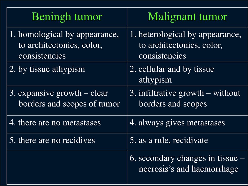
12.
Beningh tumor1. homological by appearance,
to architectonics, color,
consistencies
2. by tissue athypism
Malignant tumor
3. expansive growth – clear
borders and scopes of tumor
1. heterological by appearance,
to architectonics, color,
consistencies
2. cellular and by tissue
athypism
3. infiltrative growth – without
borders and scopes
4. there are no metastases
4. always gives metastases
5. there are no recidives
5. as a rule, recidivate
6. secondary changes in tissue –
necrosis’s and haemorrhage
13. Stages of malignant tumor
1. The before tumor changes of tissue –is dysplasia (duration of this period is
from a few months to about 2-3 years)
2. Formation of tumor cells clone – uninvasion stage (the complete recovery
is possible).
3. The Invasive stage – penetration of
tumor in the neighbor tissues.
4. Stage of metastasis formation.
14.
DYSPLASIA ("atypia", "pre-cancer"): It isabnormal epithelium with "loss of uniformity
of the individual cells, as well as a loss of their
architectural orientation".
This includes "atypical hyperplasia" and
"atypical metaplasia".
“Hyperplasia" and "metaplasia" imply the
tissue cells look normal. In dysplasia, they
look distinctly abnormal, and the changes
resemble those seen in cancer cells. These
weird changes are called ANAPLASIA.
15. Dysplasia
This stage can be recognized only bymicroscopy of tissue or by the zoned
authentication of DNA. This is rapid
proliferation and slow differentiation.
Microscopically: great number
of cells, without stratification
of layers, polyploidy, hyperchromasia
of
cytoplasm
(cellular atypia).
16.
Dysplasia really can reflect:- initial stage of development of cancer
- the process of reparation of tissue
through dysplasia
- dyshormonal alteration of cellular pool
(generations).
Tactic of doctor:
medical treatment > repeated biopsy
through month > final conclusion.
17. Dysplasia – it is facultative pre-cancer in:
- Breаst- cervix of uterus
- endometrium
- bronchioles
- gastric ulcer
- mucous membrane of stomach
at the chronic gastritis.
18. The stage of formation of tumor rudiment (non-invasive stage) It is appearance of different clones of tumor cells, which form the small cellular association. Part of these cells perishes under action of immune cells, and the other part slips out and is di
The stage of formation of tumorrudiment
(non-invasive stage)
It is appearance of different clones of
tumor cells, which form the small
cellular association. Part of these cells
perishes under action of immune cells,
and the other part slips out and is
divided further. This stage is
recognized by chance – during biopsy.
19.
Microscopically:1. intensive proliferation of cells with the
presence of pathological mitosis’s
2. structural atypism (cellular and tissue):
a) cellular – difference of daughter's cells from
maternal (cellular and nuclear pleomorphism,
hyperchromatic nuclei, and tumor giant cells)
b) tissue – violation of normal correlation between
parenchyma and stroma
20.
The Invasive stage – migration of tumorcells into surrounding tissues. Tumor cells
connects with fibronectin and leave its
tumor association. Tumor cells synthesize
the molecules of adhesion and enzymes
which destroy surrounding tissues, that
allows to migrate up to the eventual points
of migration – lymphatic and blood vessels
(it is revealed at microscopy).
21.
Metastatical stage– tumor cells must grow
up to the vessels. Cancer cells have the tropism
to the lymphatic vessels (nodules).
Cells, that are growing up to the vessels, must
penetrate into the vessel, where, the part of cells
are destroyed by immune cells of the organism.
Аnd a part of tumor cells becomes enveloped by
fibrin and migrates by blood or lymph. Surviving
tumor cells get into the organ, where a lot of
macrophages that possess the macrophagical
activity.
22. Metastatic spread:
There are four routes:1) Spreading by serous surfaces
2) Mechanical transplantation (rare, typically
iatrogenic)
3) Via lymphatic vessels (carcinomas). Tumors
spread first to regional lymph nodes, then to
any lymph nodes or organs
4) Via blood vessels (sarcomas, because the
tumor cells are in direct contact with blood
vessels from the beginning)
The common sites for metastatic spread for many
common cancers include:
lymph nodes, lung, liver, bone, and brain.
23. Features of metastatic stage
1. the first tumor cells are destroyed bymacrophages
2. there is the program of the repeated
migration through the vascular wall, further
in parenchyma of organ.
In parenchyma of organ a process can go as:
- tumor cells are destroyed in parenchyma of
organ by tissue macrophages;
tumor
cells
slip
out
from
the
immunological supervision and secure the
appearance of new tumor cells.
24. The Anatomy-histological classification
According to the appearance:- cellular infiltrate in tissue
- nodules
- polypus
- ulcer
- cyst
25.
According to the type of growth:- unicentrical – from one tumor rudiment
- policentrical (in stomach)
- expansive growth – without destroying
surrounding tissues
- infiltrating (invasive) - tumor cells
invade an organ diffusely without
changing its shape growth, destroying
surrounding tissues
- exophytic ("fungating") growth – tumor
grows as a lump
- endophytic - tumor grows as an ulcer
26.
According to the degree ofmaturity tumors are devided:
- differentiate – characterized
by the slow growth and late
metastases (I-II grade)
- undifferentiated – have not
functions,
except
for
the
division, hasty growth and early
metastases (III-IV grade)
27. Grade and Stage:
Tumor grade: assigned by thepathologist to reflect the cancer's
degree of differentiation.
Grade 0: benign tumor
Grade I: Well-differentiated, cells look like
normal organ
Grade II: Not so well-differentiated
Grade III: Worse than that
Grade IV: Even worse
Grade V: Worst of all (most tumor types
are graded I-III)
28. Grade and Stage:
Tumor stage: assigned by the clinician onthe basis of all available information on
the extent of tumor spread.
Stage I might mean the tumor is smaller
than 1 cm diameter, without metastases
Stage II might mean the tumor is larger
than 1 cm and/or is symptomatic and/or
there are metastases to the regional
lymph nodes
Stage III might mean the tumor has
infiltrated a non-resectable structure
and/or there are distant metastases
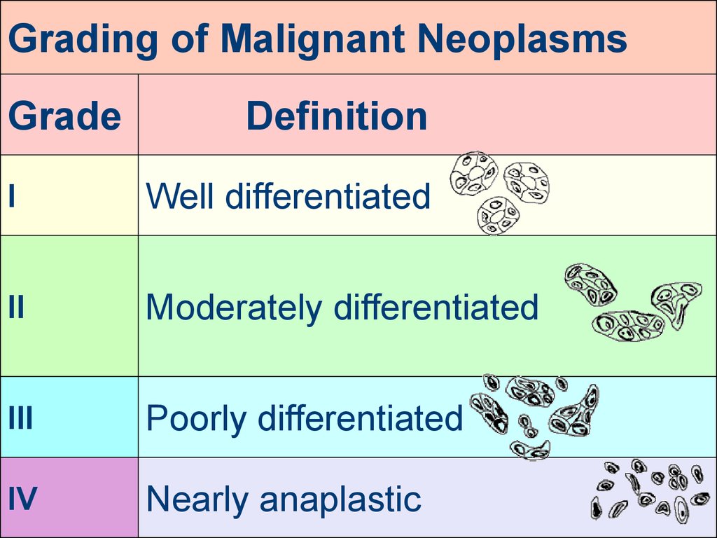
29.
Grading of Malignant NeoplasmsGrade
Definition
I
Well differentiated
II
Moderately differentiated
III
Poorly differentiated
IV
Nearly anaplastic
30.
Alternative system: TNM"T" for tumor:
T1 might mean primary tumor is smaller than 1 cm in
diameter
T2 might mean primary tumor is larger than 1 cm in
diameter
T3 might mean primary tumor is invading something
non-resectable
Tx might mean primary tumor can’t be found
"N" for regional lymph nodes:
N0 would mean no tumor in regional lymph nodes
N1 might mean tumor in a few nearby lymph nodes
N2 might mean many nodes, or some nodes farther
downstream, are involved
Nx might mean unable founding tumor in lymph nodes
"M" for metastases:
M0 would mean no distant metastases
M1 would imply distant metastases, etc.
Mx might mean unable founding of tumor metastases
31. Generally, tumors of high grade present at high stage, while tumors of low grade present at low stage
32. Benign epithelial tumors
Benign epithelial tumors aresubdivided according to their origin
from different types of epithelium
into the tumors of integumentary
epithelium (papillomas), tumors of
glandular epithelium (adenomas).
33. Papillomas
It is broad-based superficial tumor ofbranching villous vascular stroma covered
by neoplastic epithelium.
Papilloma has following features:
• Bening tumor.
• Origin from the skin and mucous
membranes.
• It looks like a ledge or a bush of
branching papillae.
• Exophytic tumor.
• Slow growth.
34. Adenomas
Benign epithelial tumor from the epitheliumof the glands and glandular organs.
More often they can be found in the breast,
thyroid gland, liver, ovaries, prostatic gland,
gastrointestinal tract.
According to the histological composition
adenoma may be tubular and alveolar.
35. Squamous cell carcinomas
These arise anywhere there is a stratifiedsquamous epithelium, either healthy
(skin, esophagus, mouth, many others)
or metaplastic (endocervix, bronchi).
Look for any (or even all) of the following:
• keratin (will stain pink-red on H&E)
• pearls (i.e., whorls that mimic little hairs)
• desmosomes ("intercellular bridges",
"prickles")
• tonofilaments (electron microscopy)
• single-cell apoptosis (they're at the top of the
epidermis)
36. Adenocarcinomas
These arise anywhere there are glands, evensingle-celled glands (i.e., goblet cells)
Look for any (or even all) of the following:
• lumens (intercellular, intracellular)
• especially, glands-within-glands ("Swiss cheese")
• or even "inside-out" glands, with the malignant cells
growing around a fibrous stalk ("papillary growth")
• mucin (intercellular "lakes", intracellular)
• other secretory products, depending on the gland of
origin (immunostain may be required)
• cells forming cohesive nests, or at least sticking to
one another
• signet-ring cells containing mucin, alone or in
clusters
• microvilli
37.
Nomenclature of NeoplasiaBased upon origin:
I. Maligant neoplasms arising from tissue
embryologically derived from ectoderm or
endoderm are usually carcinomas. Examples
include:
Squamous cell carcinoma of cervix
Adenocarcinoma of stomach
Hepatocellular carcinoma
Renal cell carcinoma
II. Malignancies arising from mesoderm are
usually sarcomas. Examples include:
Leiomyosarcoma
Chondrosarcoma
Osteosarcoma
Liposarcoma
38.
Nomenclature of NeoplasiaBased upon origin:
III. Neoplasms with more than one cell
type but arising from only one germ layer
are called "mixed tumors".
The best example is the benign mixed
tumor of salivary gland.
IV. Neoplasms with more than one cell
type and arising from more than one germ
layer are called teratomas (in ovary)
39.
Nomenclature of NeoplasiaBased upon origin:
V. Neoplasms ending in "-blastoma"
resemble primitive embryonic tissues.
Examples include:
Retinoblastoma
Neuroblastoma
Hepatoblastoma
Medulloblastoma
40.
Nomenclature of NeoplasiaBased upon origin:
VI. Not all malignant neoplasms have benign
counterparts:
Hematopoietic and lymphoid cells (as
in bone marrow and lymph node) give rise
to leukemias and lymphomas.
They have no benign counterpart.
Gliomas (astrocytomas,
oligodengrogliomas, glioblastoma
multiforme, etc) arise from glial cells in the
CNS. They have no benign counterpart.