


 Медицина
Похожие презентации:


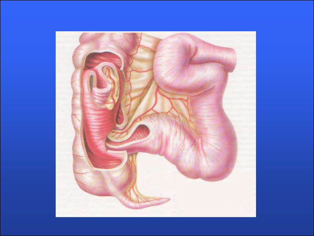
Intussusception definition
1.
INTUSSUSCEPTIONLection
2. INTUSSUSCEPTION DEFINITION
Telescopingof a proximal segment of
the intestine (intussusceptum) into a
distal segment (intussuscipiens)
3.
4. INTUSSUSCEPTION ANATOMIC LOCATIONS
ILEOCOLIC– MOST COMMON IN CHILDREN
ILEO-ILEOCOLIC
– SECOND MOST COMMON
ENTEROENTERIC
– ILEO-ILEAL, JEJUNO-JEJUNAL
– MORE COMMON IN ADULTS
– MAY NOT BE SEEN ON BARIUM ENEMA
CAECOCOLIC, COLOCOLIC
– MORE COMMON IN ASIAN CHILDREN
5.
6. PATHOPHYSIOLOGY
Precipitating mechanism unknownObstruction of intussusceptum mesentery
Venous and lymphatic obstruction
Ischemic necrosis occurs in both
intussusceptum and intussuscipiens
Pathologic bacterial translocation
7. PATHOPHYSIOLOGY
Majority occur in the region of the ileocecalvalve (80%)
– DISPROPORTIONATE DIAMETERS OF ILEUM AND
CECUM
– LYMPHOID AGGREGATES MORE NUMEROUS IN
TERMINAL ILEUM
– ILEOCECAL REGION ANATOMIC NEURAL
TRANSITION ZONE
8.
9. ETIOLOGIES
Majority of pediatric intussusceptionsidiopathic (85-90%)
– LYMPHOID HYPERPLASIA POSSIBLE ETIOLOGY
Mechanical abnormalities may act as “lead
points”
– CONGENITAL MALFORMATIONS
(MECKEL’S DIVERTICULUM, DUPLICATIONS)
– NEOPLASMS (LYMPHOMA, LYMPHOSARCOMA)
– POLYPOSIS
– TRAUMA (POST-SURGICAL, HEMATOMA)
– MISCELLANEOUS (APPENDICITIS, PARASITES)
10. EPIDEMIOLOGY
Incidence 2 - 4 / 1000 live birthsUsual age group 3 months - 3 years
Greatest incidence 6-12 months
No clear hereditary association
No seasonal distribution
Frequently preceded by viral infection
– ADENOVIRUS
11. INTUSSUSCEPTION CLINICAL CHARACTERISTICS
Early Symptoms– PAROXYSMAL ABDOMINAL PAIN
– SEPARATED BY PERIODS OF APATHY
– POOR FEEDING AND VOMITING
Late Symptoms
– WORSENING VOMITING, BECOMING BILIOUS
– ABDOMINAL DISTENTION
– HEME POSITIVE STOOLS
– FOLLOWED BY “RASPBERRY JELLY” STOOL
– DEHYDRATION (PROGRESSIVE)
Unusual Symptoms
– DIARRHEA
12. CLINICAL SYMPTOMS BY AGE
INTERMITTENTPAIN (85%)
VOMITING (78%)
BLOOD IN STOOL (36%)
PATIENTS < 1 YR
INTERMITTENT
PAIN (95%)
VOMITING (55%)
BLOOD IN STOOL (5%)
PATIENTS > 1 YR
13. CLINICAL SYMPTOMS BY DURATION
INTERMITTENTPAIN (85%)
VOMITING (78%)
BLOOD IN STOOL (36%)
SYMPTOMS 0-6 HRS
INTERMITTENT
PAIN (95%)
VOMITING (55%)
BLOOD IN STOOL (5%)
SYMPTOMS > 6 HRS
14. PHYSICAL EVALUATION
Moderately to severely illIrritable, limited movement
Most are at least 5-10% dehydrated
80% have palpable abdominal masses
Paucity of bowel sounds
Rectal examination (blood, mass)
Abdominal rigidity
“Knocked Out” syndrome
15. INTUSSUSCEPTION STAGES
I.Bright clinical manifestation
II.
Pseudodysenteric stage
III.
Peritonitis
16. Ultrasonic diagnostics
17.
18.
19.
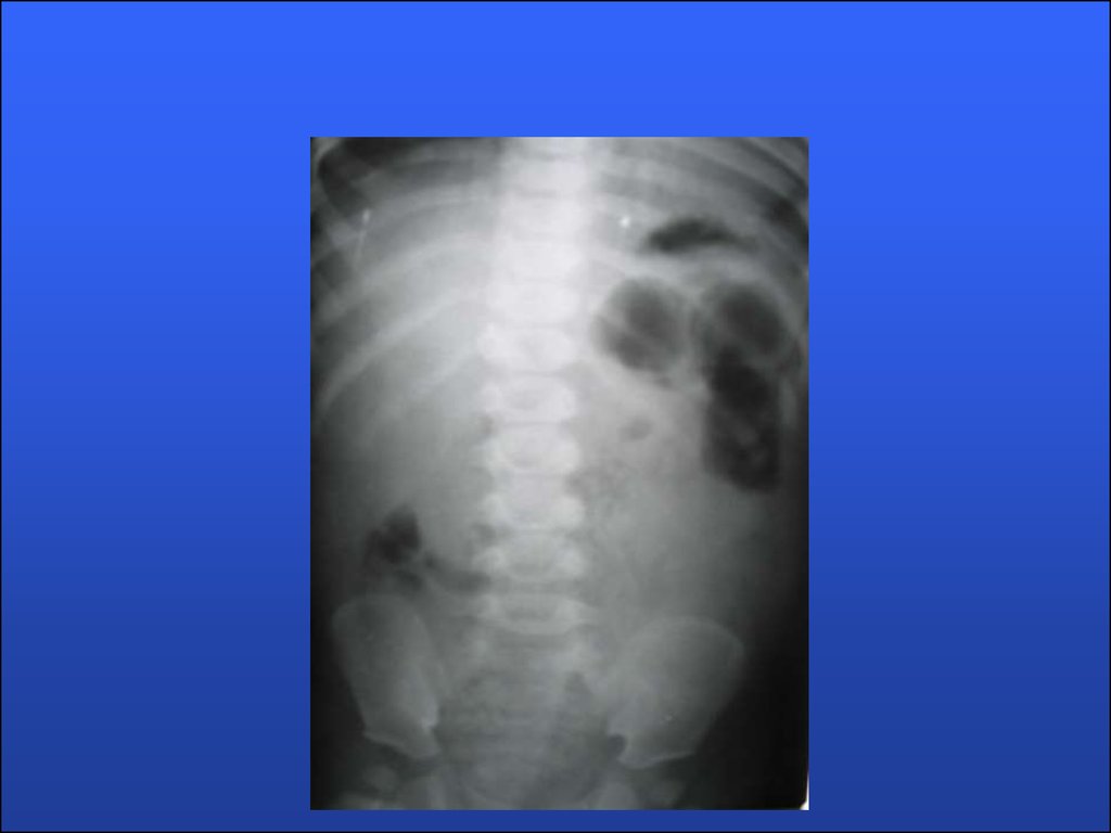
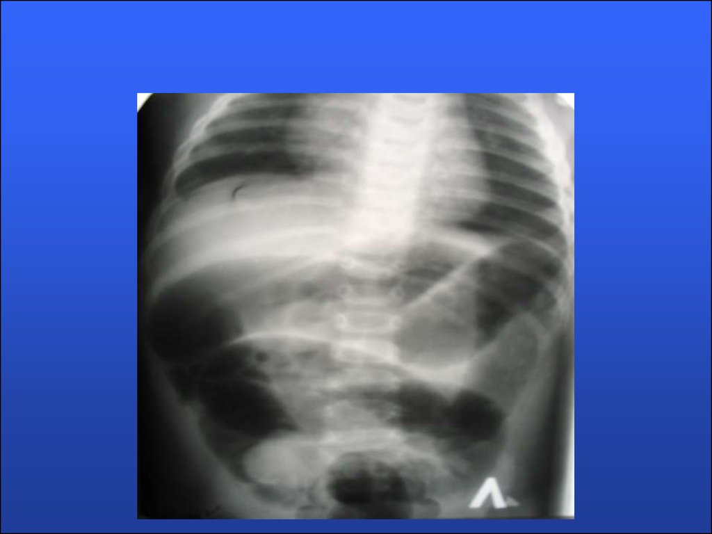
20. RADIOGRAPHIC EVALUATION
Plain radiographs (acute abdominal series)Plain films suggestive in majority, but
cannot rule out diagnosis
– PAUCITY OF LUMINAL AIR IN INTESTINAL
– SMALL BOWEL DISTENTION, AIR FLUID LEVELS
– LUMINAL AIR CUTOFFS (CECUM, TRANSVERSE
COLON)
Suggestive clinical symptoms and
compatible or nonspecific plain films
should undergo evaluation with air or
barium enema
21.
22.
23.
24.
25. TREATMENT
Obstructive surgical emergencyPediatric surgeon notified immediately
Supportive Therapy
– AGGRESSIVE FLUID RESUSCITATION
– ELECTROLYTES
– NASOGASTRIC TUBE PLACEMENT AND DRAINAGE
– ANTIBIOTICS IF ISCHEMIC BOWEL SUSPECTED
Arrange radiographic evaluation
Physician should accompany patient
– FREQUENT MONITORING OF FLUID STATUS
26.
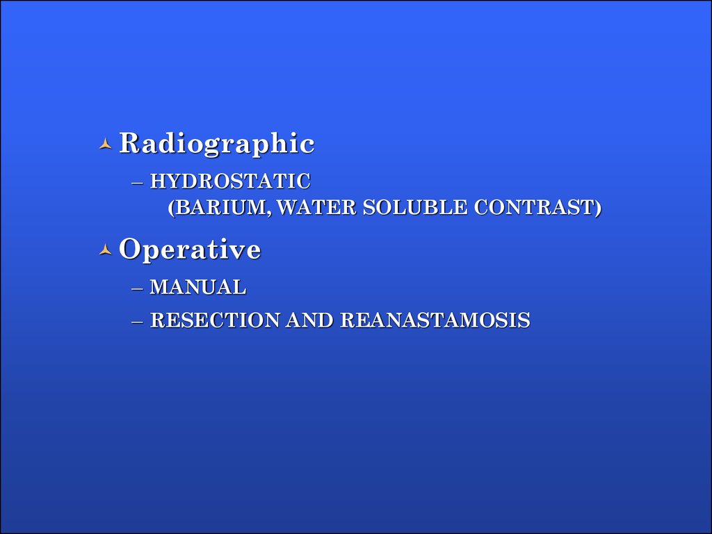
Radiographic– HYDROSTATIC
(BARIUM, WATER SOLUBLE CONTRAST)
Operative
– MANUAL
– RESECTION AND REANASTAMOSIS
27. INTUSSUSCEPTION PNEUMATIC REDUCTION
Theoretical Advantages– LESS INFLAMMATION IF PERFORATION OCCURS
Method
– AIR INSUFFLATION LIMITED TO MAXIMUM
“RESTING “ PRESSURE OF 120 mmHg
– MAXIMUM PRESSURE MAINTAINED FOR 3 MIN
– USUALLY 3 ATTEMPTS AT REDUCTION
Success Rate (75-90%)
– MUST OBSERVE AIR IN THE TERMINAL ILEUM
– LESS RECURRENCES (5-10%)
– LOW PERFORATION RATE (1%)
28. INTUSSUSCEPTION NON-OPERATIVE REDUCTION CONTRAINDICATIONS
Absolute Contraindications– PERITONEAL SIGNS
– SUSPECTED PERFORATION
Relative Contraindications
– SYMPTOMS > 24-48 HRS
– RECTAL BLEEDING
– POOR PROGNOSTIC INDICATORS
29. INTUSSUSCEPTION FAILURE OF NON-OPERATIVE REDUCTION
Factors associated with failure– SYMPTOMS > 48 HRS
– RECTAL BLEEDING
– SMALL BOWEL OBSTRUCTION
RADIOGRAPHICALLY
– ILEOILEOCOLIC OR SMALL BOWEL TYPES
– PRESENCE OF MECHANICAL LEAD POINT
– AGE < 3 MONTHS
Operative Reduction
30. INTUSSUSCEPTION POST-REDUCTION TREATMENT
Admit patient for 24 hoursMay attempt feeding within 12 hrs
Return to fluoroscopy for suspected
recurrence (occurs in ~ 4%)
– CONSIDER PATHOLOGIC LEAD POINT
– SCHEDULE MECKEL’S SCAN, ? ABDOMINAL CT
May also recur up to one year
Need to follow as outpatient
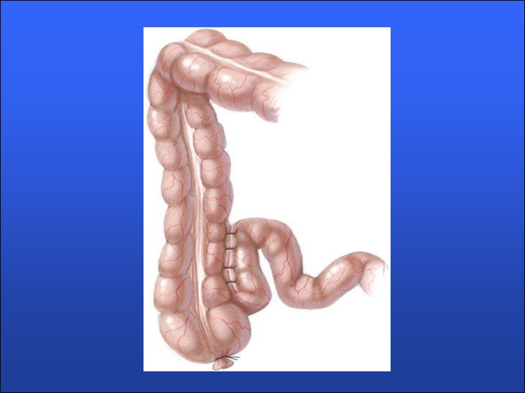
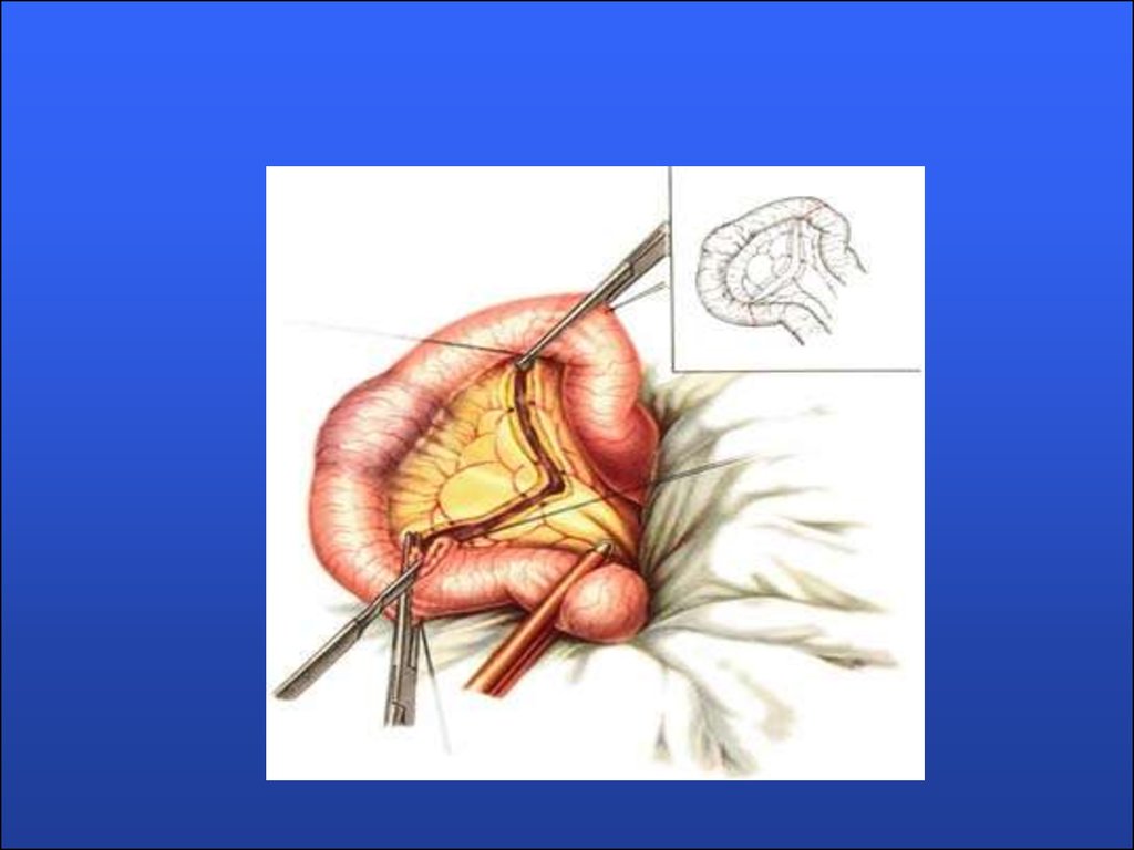
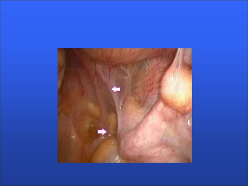
31. Surgical treatment
32.
33. Acquired intestinal obstruction
Acquired intestinal obstructionsare a partial or complete
blockage of the small or large
intestine, resulting in failure of
the contents of the intestine to
pass through the bowel
normally.
34.
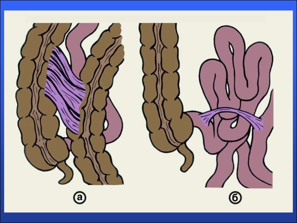
Intestinal obstructions can be mechanicalor nonmechanical.
Mechanical obstruction is caused by the
bowel twisting on itself (volvulus) or
telescoping into itself (intussusception).
Mechanical obstruction can also result
from hernias, fecal impaction, abnormal
tissue growth, the presence of foreign
bodies in the intestines, or inflammatory
bowel disease (Crohn's disease).
35.
36.
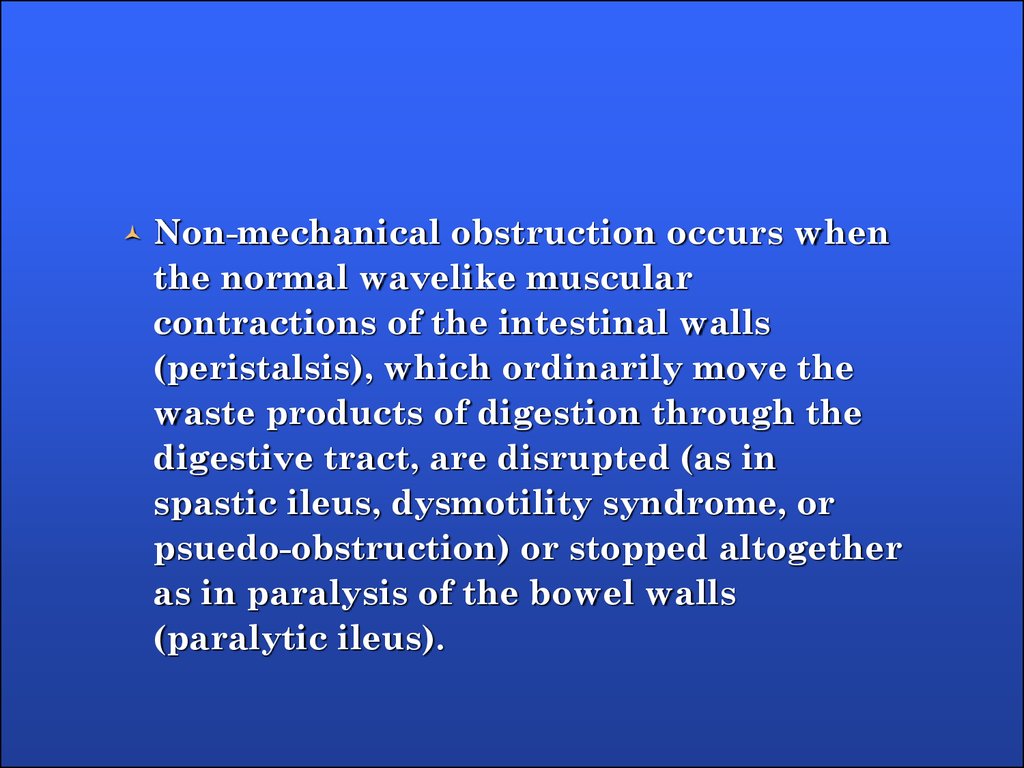
Non-mechanical obstruction occurs whenthe normal wavelike muscular
contractions of the intestinal walls
(peristalsis), which ordinarily move the
waste products of digestion through the
digestive tract, are disrupted (as in
spastic ileus, dysmotility syndrome, or
psuedo-obstruction) or stopped altogether
as in paralysis of the bowel walls
(paralytic ileus).
37. Clinic
1. Abdominal2.
pain
Vomiting
3. Constipation
4.
Intoxication syndrome
38. Diagnosis
1.X-ray examination
2.
Ultrasonic diagnostics
3.
Computed tomography
4.
Diagnostic testing will include a
complete blood count (CBC), electrolytes
(sodium, potassium, chloride) and other
blood chemistries, blood urea nitrogen
(BUN), and urinalysis. Coagulation tests
may be performed if the child requires
surgery.
39.
40.
41. Treatment
1.Preoperative preparation:
a. inserting a nasogastric tube to suction
out the contents of the stomach and
intestines
b. Intravenous fluids will be infused to
prevent dehydration and to correct
electrolyte imbalances that may have
already occurre
42. Surgical treatment
43.
44.
45.
46.
Thank you forattention!