


")


 Медицина
Похожие презентации:


Surgical Emergencies in the Newborn
1. Surgical Emergencies in the Newborn
2. Emergencies
TypesAirway/Respiratory
Intestinal Obstruction
Intestinal Perforation
Signs
Respiratory distress
Abdominal distension
Peritonitis
Pneumoperitoneum
3. Airway/Respiratory
Neck MassesCystic Hygromas
Tracheal anomalies
Thoracic masses/pulmonary lesions
Congenital lobar emphysema
• Overdistension of one or more lobes (nl histological lung)
Congenital cystic adenomatous malformation
• Multicystic mass of lung tissue, proliferation of bronchial structures
at the expense of alveoli
Pulmonary agenesis
• Absence of lung
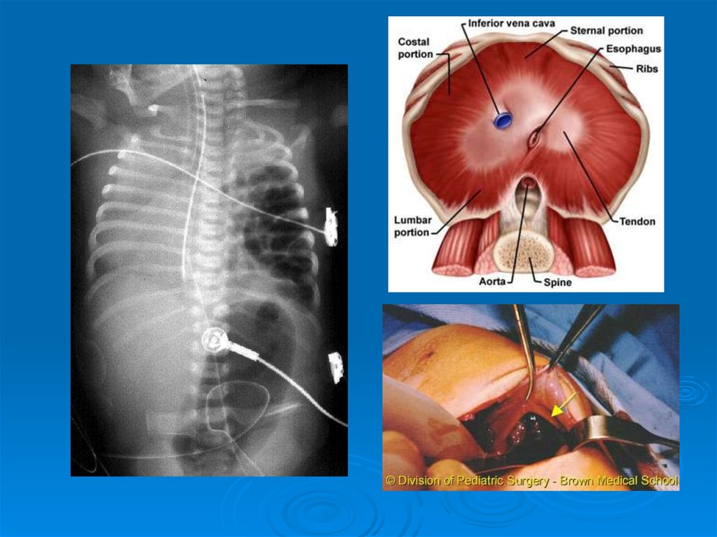
Congenital diaphragmatic hernia
Tracheoesophageal fistula
4. Cystic Hygroma
Multiloculated cystic spaces lined by endothelialcells
Separated by fine walls containing numerous smooth muscle
cells
Result of maldevelopment of lymphatic spaces
Incidence about 1 in 12,000 births
50-65% appear at birth, 85-90% appear by age 2
Neck-75%, Axilla 20%; can be seen in mediastinum,
retroperitoneum, pelvis, groin
Nuchal/post cervical CH’s have been associated with
chromosomal abnormalities—high mortality rate
5. Cystic Hygroma
ComplicationsRespiratory—large hygromas can extend into oropharynx and
trachea
Inflammation/Infection
Hemorrhage
Treatment
Dependent on size, location, symptoms/complications
Some pts require emergent surgery due to airway compromise
Best treatment is complete excision
Aspiration typically not effective due to rapid refilling of fluid
Sclerotherapy—Bleomycin, OK-432 (no longer available in US),
doxycycline, fibrin glue
6. Cystic Hygroma
7. Cystic Hygroma
8. Congenital Lobar Emphysema
Postnatal overdistension of one or more lobes ofhistologically normal lung
Probably due to cartilaginous deficiency in the tracheobronchial
tree
Obstruction causing the overdistension may be due to
• 1—chondromalacia of bronchi
• 2—extrinsic pressure on bronchus by anomalous pulmonary vein or
abnormally large PDA
• 3—idiopathic
Location
LUL 47%, RML 28%, RUL 20%; lower lobes <5%; Bilat rare
9. Congenital Lobar Emphysema
DiagnosisUsually can be made by plain CXR; Chest CT and V/P scans
may be helpful
Treatment
May require urgent surgical decompression with lobectomy
Selective bronchial intubation
Sometimes see spontaneous resolution—need close
observation
10. Congenital Lobar Emphysema
11. Congenital Cystic Adenomatous Malformation (CCAM)
Mass of cysts lined by ciliated cuboidal orcolumnar pseudostratified epithelium
Three types
I—few large cysts >2cm; thick walls, normal alveoli between the
cysts; ciliated pseudostratified columnar epithelium
II—numerous small cysts <1cm, thin muscular coat, large
alveolar-like structures between the cysts; ciliated cuboidal to
columnar epithelium; assoc w/other congenital anomalies
III—bulky firm masses of folded ciliated and non-ciliated cuboidal
epithelium and thick layer of smooth muscle; often occupy the
entire lobe or lobes of lung
More common on the left side, 2% bilateral
12. CCAM
DiagnosisCT scan allows differentiation of types
Some can be diagnosed on prenatal US
Treatment
Surgical excision, typically anatomical lobe resection, due to risk
of infection, malignant transformation
Some are performing fetal aspiration
13. CCAM
14. Congenital Diaphragmatic Hernia
IntroDX
1 in 200-5000 live births, females >males
Etiology unknown
Large percentage of fetuses are stillborn
Still high mortality of those that make it to birth
Frequently made prenatally
CXR
Treatment
Respiratory support
ECMO
Primary closure or patch closure when pt stable
15.
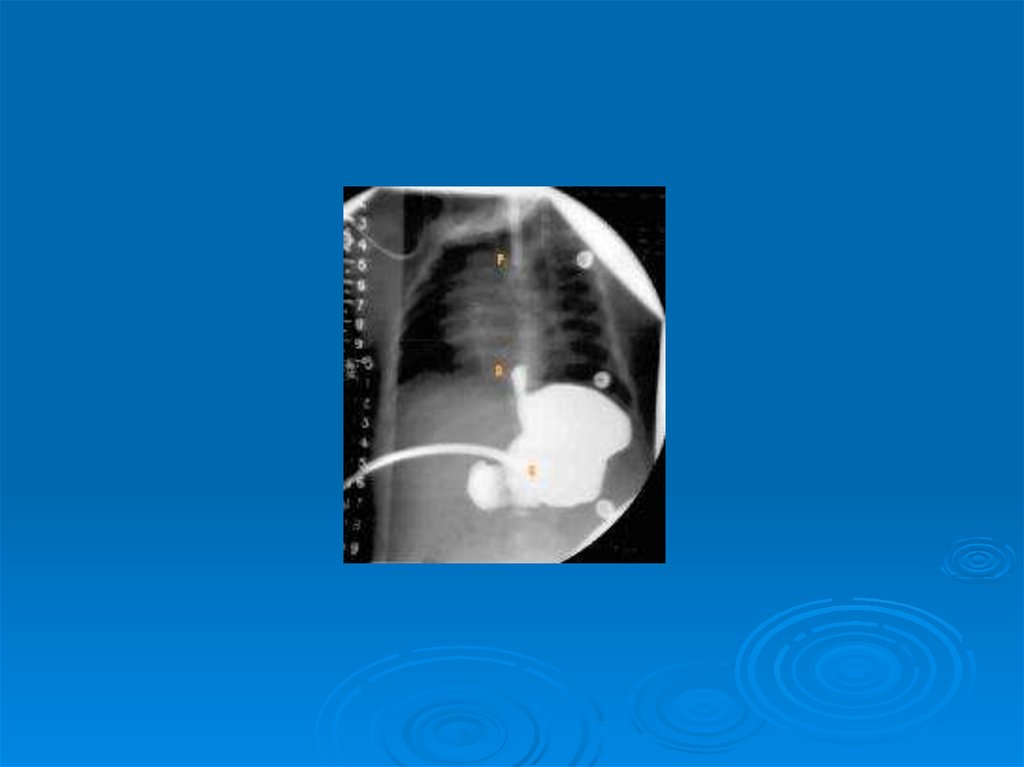
16. Tracheoesophageal Fistula and Esophageal Atresia
17.
18.
19.
Intestinal ObstructionIncidence
approx 1 per 500-1000 live
births
Approx 50% due to atresia or stenosis
Majority of neonates present shortly
after birth
20.
Anatomic DifferentiationUpper
GI
Duodenal atresias/webs
small bowel atresias
malrotation/midgut volvulus
GERD
Meconium ileus
pyloric stenosis
Inguinal hernia
NEC
21.
Anatomic DifferentiationLower
GI
Colonic atresia
Meconium plug
Hirschsprung’s
Small Left Colon Syndrome
Magalocystis-Microcolon-Intestinal
Hypoperistalsis Syndrome
Imperforate anus
22.
Urgency to TreatEmergencies
Free air on KUB
Peritonitis
Acute increase in abd distension
Clinical deterioration (incr pressors, dec
platelets, worsening acidosis)
Abd wall cellulitis/discoloration
23.
Urgency to TreatFurther
workup
Contrast enemas for distal obstructions
KUB/Cross-table lateral
Milk Scans for GERD
UGI for malrotation/proximal atresias
24.
Common DisordersNEC
Duodenal Atresia
Small
Bowel Atresia
Malrotation/Volvulus
Hirschsprung’s
25.
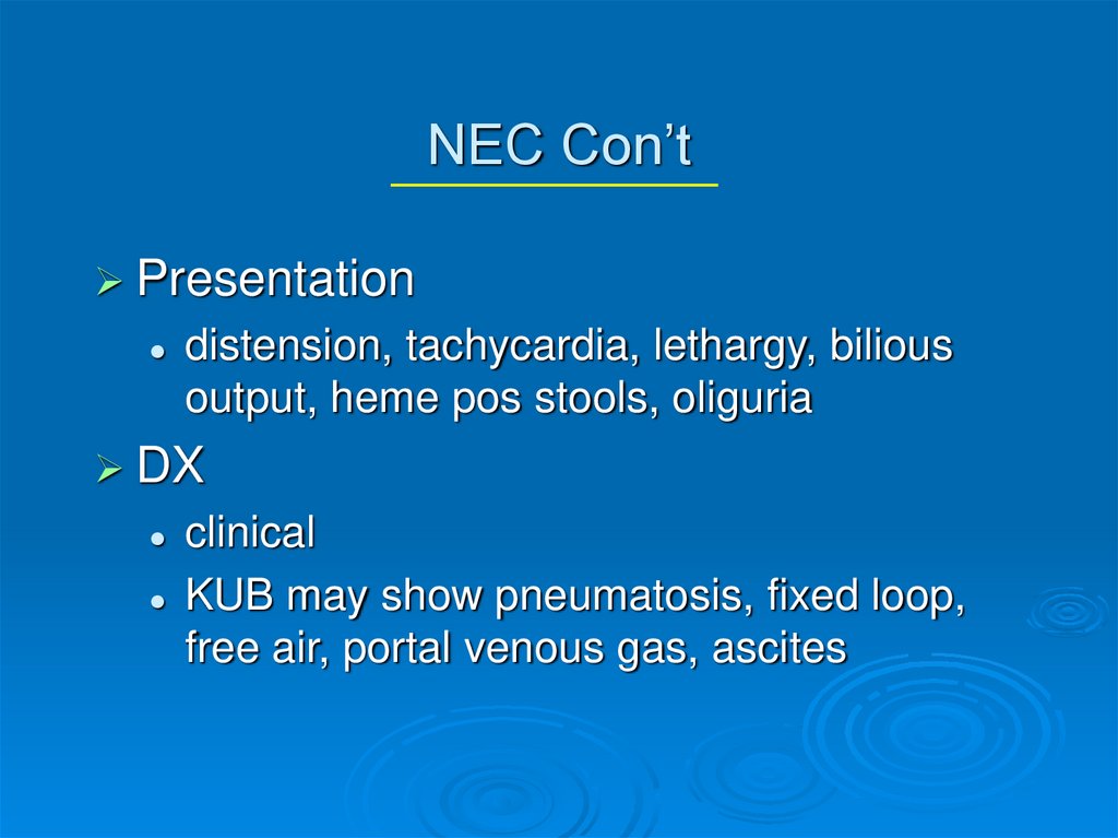
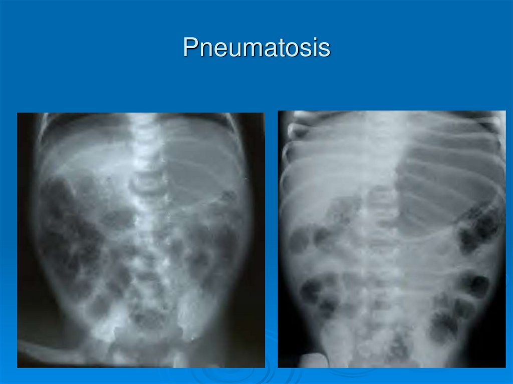
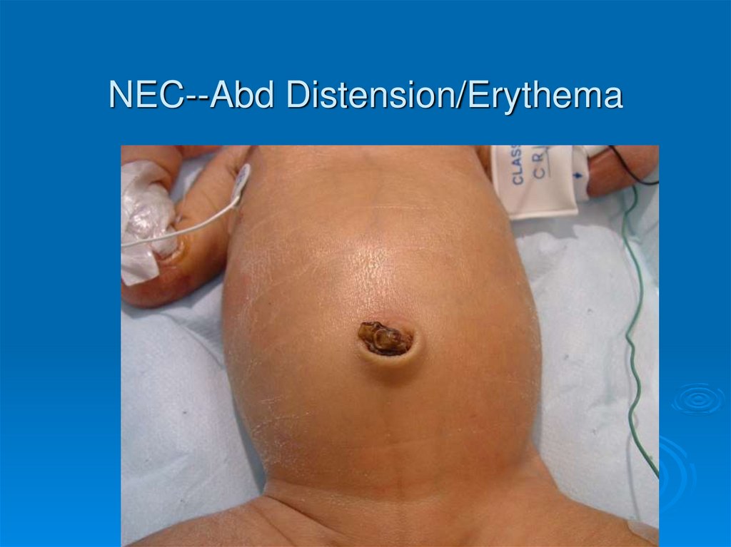
NEC Con’tPresentation
distension, tachycardia, lethargy, bilious
output, heme pos stools, oliguria
DX
clinical
KUB may show pneumatosis, fixed loop,
free air, portal venous gas, ascites
26.
NEC TreatmentMedical
NPO, sump tube, Broad Abx after cx’s
drawn, serial KUB/lateral x-rays, frequent
abd exams
Surgical
indications
Free air
Abd wall Cellulitis
Fixed loop on KUB
Clinical deterioration
27.
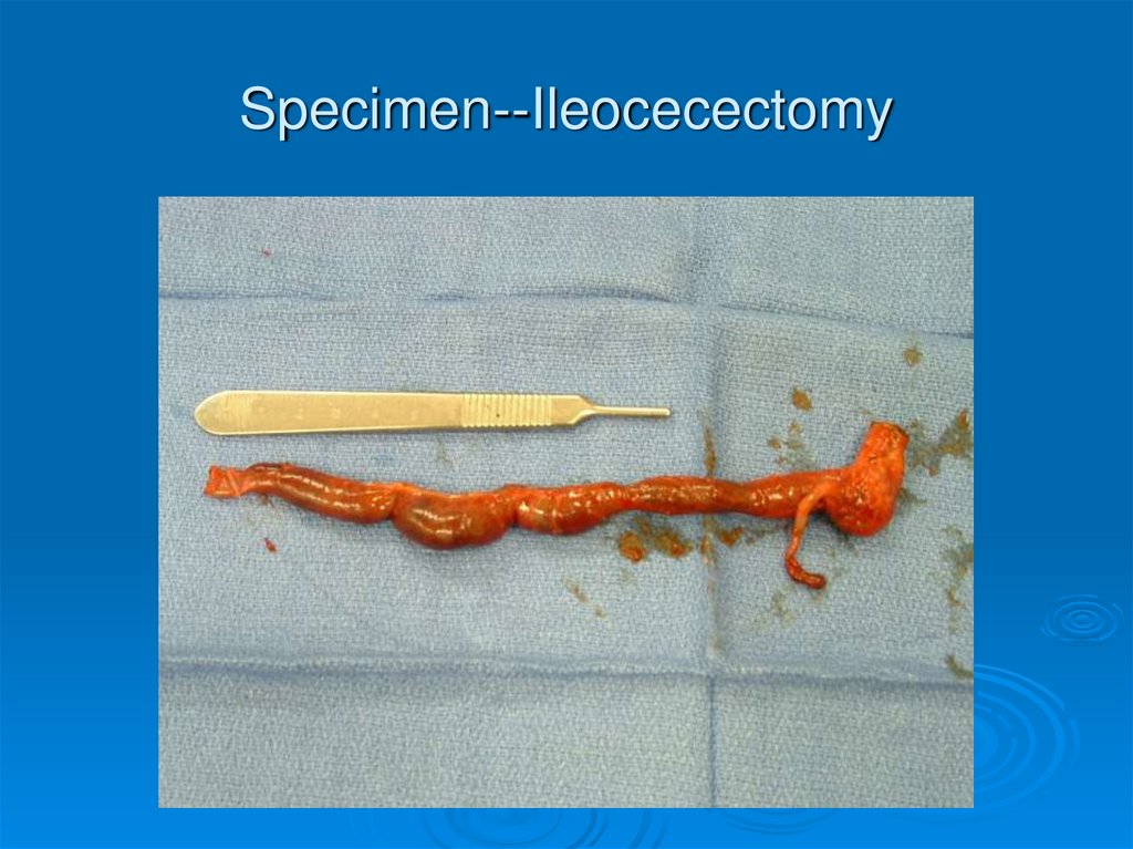
NEC OutcomesOverall
survival ~ 80%, improving in
LBW
In pts w/perforation, 65% perioperative
mortality, no perf--30% mortality
25% of Survivors develop stricture
6% pts have recurrent NEC
Postop NEC--Myelomeningocele,
Gastroschisis--45-65% mortality
28.
Pneumatosis29.
Pneumoperitoneum30.
NEC--Abd Distension/Erythema31.
Necrotic Segment Ileum32.
Resection33.
Specimen--Ileocecectomy34.
Ileostomy35.
Common DisordersNEC
Duodenal Atresia
Small
Bowel Atresia
Malrotation
Hirschsprung’s
36.
Duodenal AtresiaIncidence--1
in 5,000 to 10,000 live
births
75% of stenoses and 40% of atresias are
found in Duodenum
Multiple atresias in 15% of cases
50% pts are LBW and premature
Polyhydramnios in 75%
Bilious emesis usually present
37.
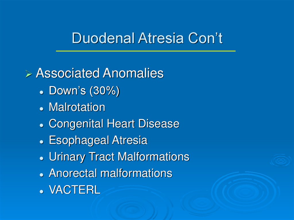
Duodenal Atresia Con’tAssociated Anomalies
Down’s (30%)
Malrotation
Congenital Heart Disease
Esophageal Atresia
Urinary Tract Malformations
Anorectal malformations
VACTERL
38.
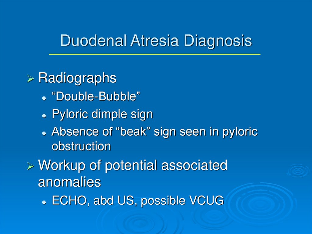
Duodenal Atresia DiagnosisRadiographs
“Double-Bubble”
Pyloric dimple sign
Absence of “beak” sign seen in pyloric
obstruction
Workup
of potential associated
anomalies
ECHO, abd US, possible VCUG
39.
“Double Bubble”40.
Duodenal Atresia TreatmentNasogastric
decompression, hydration
Surgery
Double diamond duodenoduodenostomy
Con’t prolonged NG decompression,
sometimes more than 2 weeks needed
41.
Common DisordersNEC
Duodenal Atresia
Small
Bowel Atresia
Malrotation
Hirschsprung’s
42.
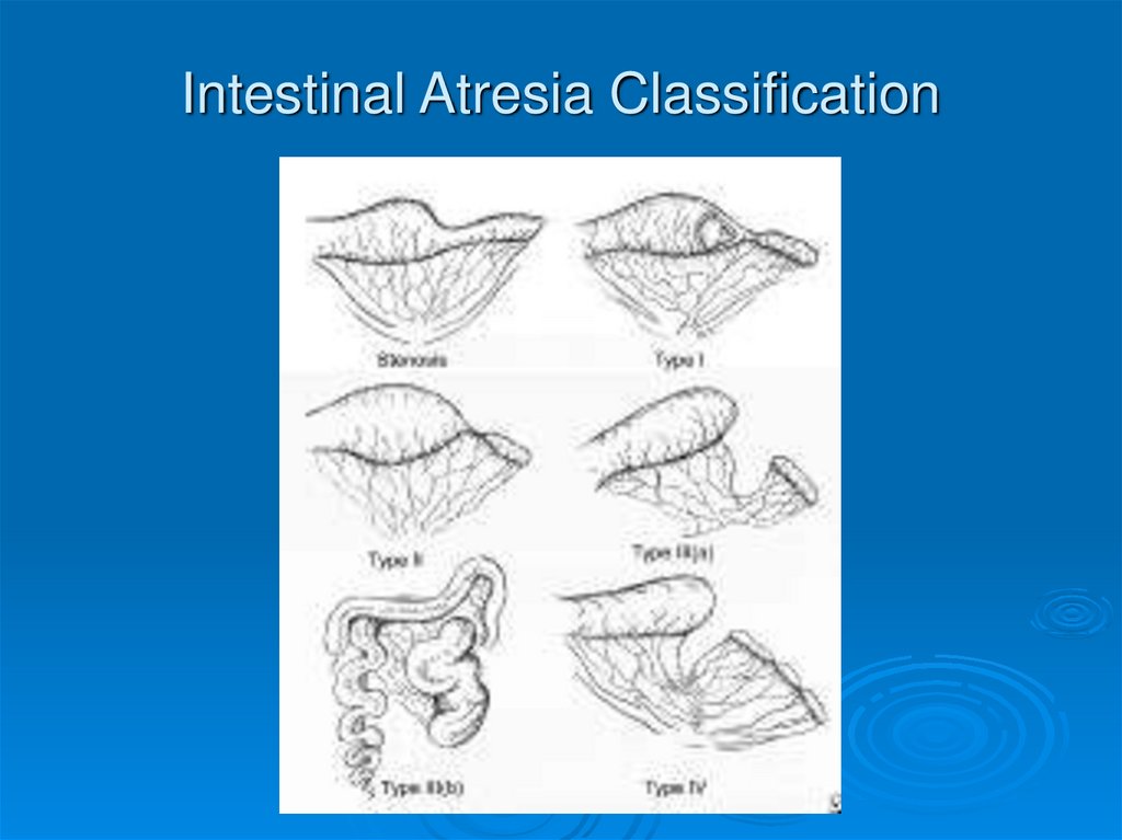
Small Bowel AtresiaJejunal
is most common, about 1 per
2,000 live births
Atresia due to in-utero occlusion of all or
part of the blood supply to the bowel
Classification--Types I-IV
Presents w/bilious emesis, abd
distension, failure to pass meconium
(70%)
43.
Intestinal Atresia Classification44.
Small Bowel Atresia Con’tAssociated Anomalies
other atresias
Hirschsprung’s
Biliary atresia
polysplenia syndrome (situs inversus,
cardiac anomalies, atresias)
CF (10%)
45.
Atresia--Diagnosis and TreatmentPlain films show dilated loops small bowel
Contrast enema shows small unused colon
UGI/SBFT shows failure of contrast to pass
beyond atretic point
Treatment is surgical
tapered primary anastamosis
check for other atresias/associated anomalies
46.
Common DisordersNEC
Duodenal Atresia
Small
Bowel Atresia
Malrotation/Volvulus
Hirschsprung’s
47.
Malrotation1 per 6,000 live births
can be asymptomatic throughout life
Usually presents in first 6 months of life
18% children w/short gut had malrotation with
volvulus
Etiology
physiologic umbilical hernia--4th wk gestation
Reduction of hernia 10th - 12th wks of gestation
48.
Normal Embryology49.
Malrotation ClassificationNonrotation
when neither duodenojejunal or cecocolic
limbs undergo correct rotation
Abn
causes Ladd’s bands to form across
duodenum
Abn
Rotation of Duodenojejunal limb
rotation of Cecocolic limb
cecum lies close to midline, narrow
mesenteric base
50.
Abnormal Rotation/Fixation51.
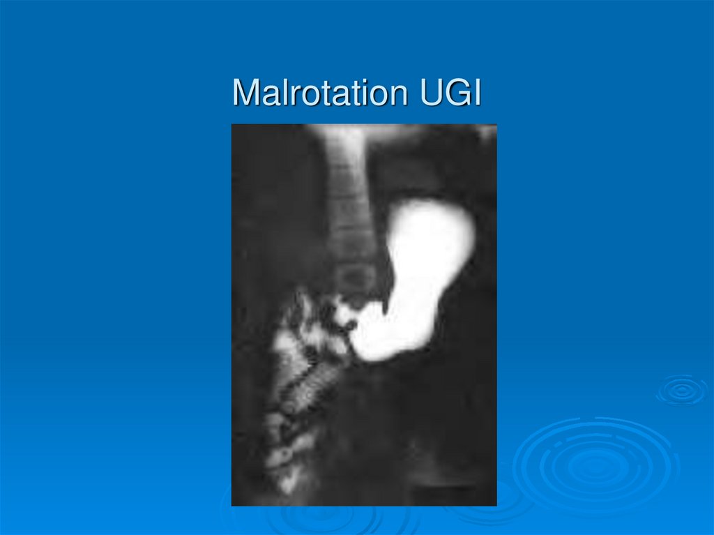
Malrotation DiagnosisVarying
symptoms from very mild to
catastrophic
**Bilious emesis is Volvulus until proven
otherwise**
Bilious emesis, bloody diarrhea, abd
distension, lethargy, shock
UGI shows abnormal position of
Duodenum
if Volvulus, see “bird’s beak” in duodenum
52.
Malrotation UGI53.
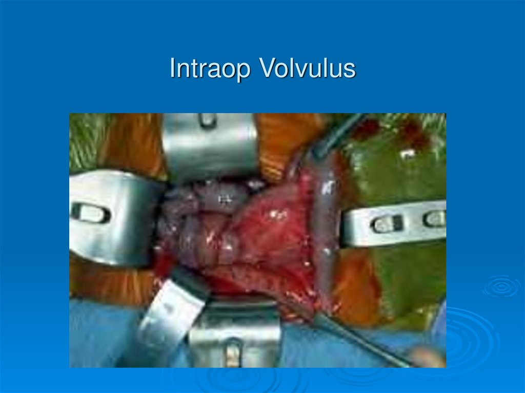
Intraop Volvulus54.
Bowel Necrosis--Volvulus55.
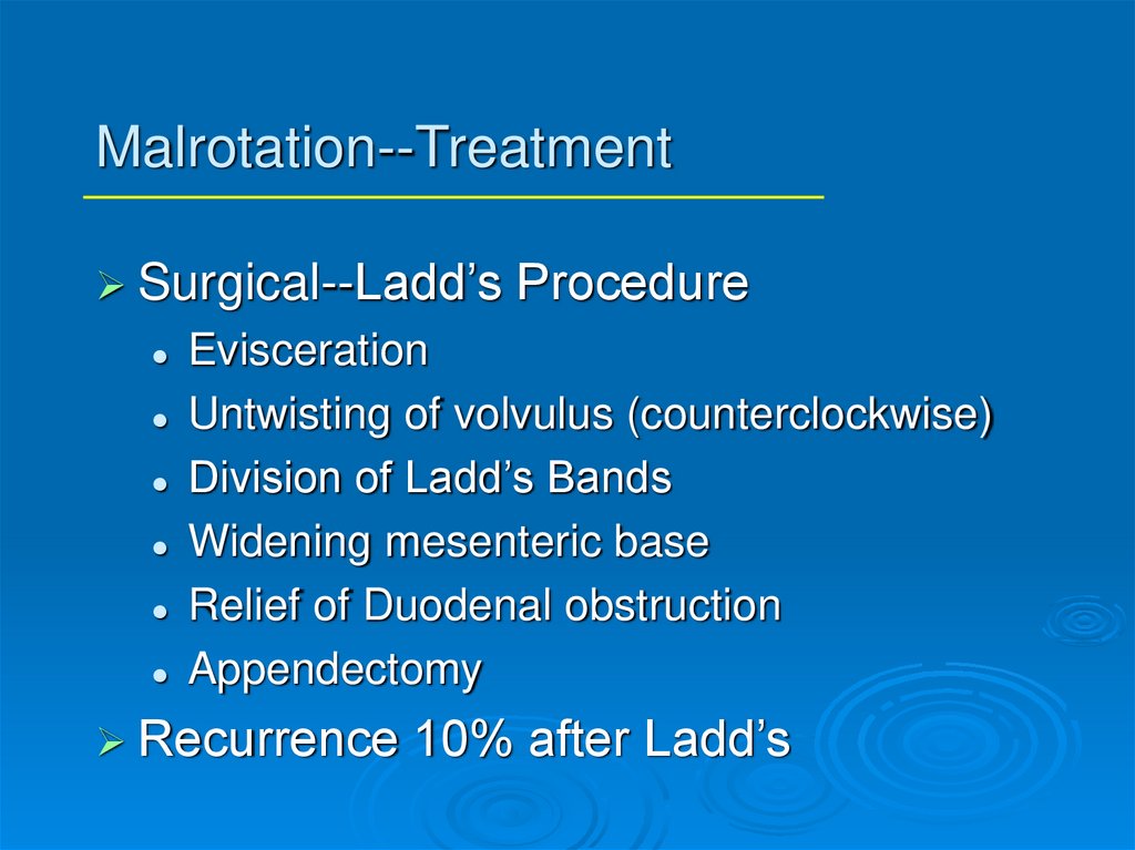
Malrotation--TreatmentSurgical--Ladd’s
Procedure
Evisceration
Untwisting of volvulus (counterclockwise)
Division of Ladd’s Bands
Widening mesenteric base
Relief of Duodenal obstruction
Appendectomy
Recurrence
10% after Ladd’s
56.
Common DisordersNEC
Duodenal Atresia
Small
Bowel Atresia
Malrotation
Hirschsprung’s
57.
Hirschsprung’s DiseaseMigratory
failure of neural crest cells
Incidence 1 in 5,000 live births, males
affected 4:1 over females
90% of pts w/H’sprung’s fail to pass
meconium in first 24-48 hrs
Abd distension, bilious emesis,
obstructive enterocolitis
58.
Hirschsprung’s DiagnosisBarium
Enema
Transition zone
Anorectal
shows failure of reflexive relaxation
not very helpful in infants, young children
Rectal
Manometry
Biopsy
Absence of Ganglion cells and hypertrophy
of nerves
59.
Transition Zone on BE60.
Hirschsprung’s TreatmentIn
neonates, can do primary pullthrough--bringing normal colon down to
anorectal junction
In older infants, may need diverting
colostomy first to decompress
May need prolonged dilatations and
irrigations
61.
Pull-Through Procedure62.
63.
SummaryBILIOUS
EMESIS IS VOLVULUS
UNTIL PROVEN OTHERWISE
Signs of surgical emergency
free air, abd wall cellulitis, fixed loop on
xray, rapid distension, peritonitis, clinical
deterioration
History
and plain films will guide
sequence of additional studies
Remember associated anomalies