


 Медицина
Похожие презентации:


")


Phylogenetic. Disorders of Human
1.
BIOLOGYPRESENTATION
SUBMITTED BY: Manas Yadav 194A
2.
Topic;Phylogenetic
Disorders of Human
Excretory System
Guided By:
Prof. Anna Zhukova
3.
1. INTRODUCTION2. Normal structure and Function of
excretory System.
3. Phylogenetic Disorders
4. Example and images
4.
INTRODUCTIONThe excretory system is a passive biological system that removes excess,
unnecessary materials from the body fluids of an organism, so as to help
maintain internal chemical homeostasis and prevent damage to the body. The
dual function of excretory systems is the elimination of the waste products of
metabolism and to drain the body of used up and broken down components in a
liquid and gaseous state. In humans and other amniotes (mammals, birds and
reptiles) most of these substances leave the body as urine and to some degree
exhalation, mammals also expel them through sweating.
It includes
Urinary system
Respiratory system
Integumentary system
Billary system
Gastrointestinal tract
5.
Normal structure and Function ofexcretory System
Urinary system
The kidneys are large, bean-shaped organs which are present on each side of the vertebral
column in the abdominal cavity. each kidney is supplied with blood from the renal artery. The
kidneys remove from the blood the nitrogenous wastes such as urea, as well as salts and excess
water, and excrete them in the form of urine. This is done with the help of millions of nephrons
present in the kidney. The filtrated blood is carried away from the kidneys by the renal vein (or
kidney vein). The urine from the kidney is collected by the ureter (or excretory tubes), one from
each kidney, and is passed to the urinary bladder. The urinary bladder collects and stores the
urine until urination. The urine collected in the bladder is passed into the external environment
from the body through an opening called the urethra.
Respiratory system
One of the main functions of the lungs is to diffuse gaseous wastes, such as carbon
dioxide, from the bloodstream as a normal part of respiration.
Gastrointestinal tract
The large intestine's main function is to transport food particles through the body and expel
the indigestible parts at the other end, but it also collects waste from throughout the body.
The typical brown colour of mammal waste is due to bilirubin, a breakdown product of
normal heme catabolism.[1] The lower part of the large intestine also extracts any remaining
usable water and then removes solid waste. At about 10 feet long in humans, it transports
the wastes through the tubes to be excreted.
6.
Biliary systemThe liver detoxifies and breaks down chemicals, poisons and other toxins that enter the body. For
example, the liver transforms ammonia (which is poisonous) into urea in fish, amphibians and mammals,
and into uric acid in birds and reptiles. Urea is filtered by the kidney into urine or through the gills in fish
and tadpoles. Uric acid is paste-like and expelled as a semi-solid waste (the "white" in bird excrements).
The liver also produces bile, and the body uses bile to break down fats into usable fats and unusable
waste.
Invertebrates lack a liver, but most terrestrial groups, like insects, possesses a number of blind guts that
serve the similar functions. Marine invertebrates do not need the ammonia conversion of the liver, as they
can usually expel ammonia directly by diffusion through the skin.
Integumentary system
The integumentary system comprises the skin and its
appendages acting to protect the body from various kinds
of damage, such as loss of water or damages from
outside. The integumentary system includes hair, scales,
feathers, hooves, and nails.
7.
1. Horseshoe Kidney.2. Polycystic Kidney.
3. Renal Agenesis.
4. Renal Hypoplasia.
5. Epispadias.
6. Renal Ectopia [ECTOPIC KIDNEY].
7. Doubling of Ureter.
8.
Horseshoe kidneyHorseshoe
kidney, also known as ren
arcuatus (in Latin), renal fusion or super
kidney, is a congenital disorder affecting
about 1 in 500 people that is more common
in men, often asymptomatic, and usually
diagnosed incidentally.In this disorder, the
patient's kidneys fuse together to form a
horseshoe-shape during development in the
womb. The fused part is the isthmus of the
horseshoe kidney. The abnormal anatomy
can affect kidney drainage resulting
increased frequency of kidney stones and
urinary tract infections as well as increase
risk of certain renal cancers.
9.
Signs andsymptoms
Although
often asymptomatic, the most
common presenting symptom of patients
with a horseshoe kidney is abdominal or
flank pain. However, presentation is often
non-specific.[Approximately a third of
patients with horseshoe kidneys remain
asymptomatic throughout their entire life
with over 50% of patients having no medical
issues related to their renal fusion when
followed for a 25 year period. As a result, it
is estimated that approximately 25% of
patients with horseshoe kidneys are
diagnosed incidentally with ultrasound or
CT imaging.[1
10.
Polycystic kidneydisease
Polycystic
kidney disease (PKD or PCKD,
also known as polycystic kidney syndrome) is a
genetic disorder in which the renal tubules
become structurally abnormal, resulting in the
development and growth of multiple cysts
within the kidney.These cysts may begin to
develop in utero, in infancy, in childhood, or in
adulthood.[6] Cysts are non-functioning tubules
filled with fluid pumped into them, which
range in size from microscopic to enormous,
crushing adjacent normal tubules and
eventually rendering them non-functional as
well.
PKD
is caused by abnormal genes that
produce a specific abnormal protein; this
protein has an adverse effect on tubule
development. PKD is a general term for two
types, each having their own pathology and
genetic cause: autosomal dominant polycystic
kidney disease (ADPKD) and autosomal
recessive polycystic kidney disease (ARPKD)
11.
SymptomsPolycystic kidney disease symptoms can include:
1. High blood pressure 2. Back or side pain 3. Headache 4. A feeling of
fullness in your abdomen 5. Increased size of your abdomen due to
enlarged kidneys 6. Blood in your urine 7. Kidney stones 8. Kidney
failure 9. Urinary tract or kidney infections
Causes
Abnormal genes cause polycystic kidney disease, which means that
in most cases, the disease runs in families. Rarely, a genetic mutation
occurs on its own (spontaneous), so that neither parent has a copy of
the mutated gene.The two main types of polycystic kidney disease,
caused by different genetic flaws, are: 1. Autosomal dominant
polycystic kidney disease (ADPKD). Signs and symptoms of ADPKD
often develop between the ages of 30 and 40.
12.
DiagnosisPolycystic kidney disease can be ascertained via a CT scan of
abdomen, as well as, an MRI and ultrasound of the same area. A
physical exam/test can reveal enlarged liver, heart murmurs and
elevated blood pressure
Treatment
There is no FDA-approved treatment. However, recent research indicates
that mild to moderate dietary restrictions slow the progression of
autosomal dominant polycystic kidney disease in mice. If and when the
disease progresses enough in a given case, the nephrologist or other
practitioner and the patient will have to decide what form of renal
replacement therapy will be used to treat end-stage kidney disease. That
will either be some form of dialysis, which can be done at least two
different way
13.
Renal AgenesisRenal
agenesis is a medical
condition in which one (unilateral)
or both (bilateral) fetal kidneys fail
to develop.
Unilateral
and bilateral renal
agenesis in humans, mice and zebra
fish has been linked to mutations in
the gene GREB1L.It has also been
associated with mutations in the
genes RET or UPK3A. in humans
14.
BilateralBilateral renal agenesis is a condition in which both kidneys of a fetus fail to develop during
gestation. It is incompatible with life.It is one causative agent of Potter sequence. This
absence of kidneys causes oligohydramnios, a deficiency of amniotic fluid in a pregnant
woman, which can place extra pressure on the developing baby and cause further
malformations. The condition is frequently, but not always the result of a genetic disorder,
and is more common in infants born to one or more parents with a malformed or absent
kidney.
Unilateral
This is much more common, but is not usually of any major health consequence, as long
as the single kidney is healthy. However, this kidney tends to be hypertrophied, ectopic
and prone to infection and damage.
It may be associated with an increased incidence of Müllerian duct abnormalities, which
are abnormalities of the development of the female reproductive tract and can be a
cause of infertility, blocked menstrual flow (hematocolpos), increased need for
Caesarean sections, or other problems. Herlyn-Werner-Wunderlich syndrome is one
such syndrome in which unilaterial renal agenesis is combined with a blind hemivagina
and uterus didelphys.[5] Up to 40% of women with a urogenital tract anomaly also have
an associated renal tract anomaly
15.
Renal hypoplasiaRenal
hypoplasia is relatively
common – it is estimated that one
baby in a few hundred is born with a
small kidney.It is not always possible
to know why renal hypoplasia
happens. In the majority of cases, it
is not caused by anything that the
mother does during her pregnancy,
and it is unlikely that a future
pregnancy will result in renal
hypoplasia or other problems with
the kidneys
16.
EpispadiasAn
epispadias is a rare type of
malformation in which the urethra
ends, in males, in an opening on the
upper aspect of the penis,[1] and in
females when the urethra develops
too far anteriorly. It occurs in
around 1 in 120,000 male and 1 in
500,000 female births.
17.
Signs and symptomsMost cases involve a small and bifid penis, which requires surgical closure
soon after birth, often including a reconstruction of the urethra. Where it is part
of a larger exstrophy, not only the urethra but also the bladder (bladder
exstrophy) or the entire perineum (cloacal exstrophy) are open and exposed
on birth, requiring closure. Many parts of this article are incorrect.
Causes
Epispadias is an uncommon and partial form of a spectrum of failures of
abdominal and pelvic fusion in the first months of embryogenesis known as
the exstrophy - epispadias complex. It occurs as a result of defective
migration of the genital tubercle primordii to the cloacal membrane, and so
malformation of the genital tubercle, at about the 5th week of gestation
18.
TreatmentThe main treatment for isolated epispadias is a comprehensive surgical repair
of the genito-urinary area usually during the first 7 years of life, including
reconstruction of the urethra, closure of the penile shaft and mobilisation of the
corpora. The most popular and successful technique is known as the modified
Cantwell-Ransley approach. In recent decades however increasing success
has been achieved with the complete penile disassembly technique despite its
association with greater and more serious risk of damage
Prognosis
Even with successful surgery, patients may have long-term problems
with:[citation needed]
•incontinence, where serious usually treated with some form of continent
urinary diversion such as the Mitrofanoff
•depression and psycho-social complications
•sexual dysfunction
19.
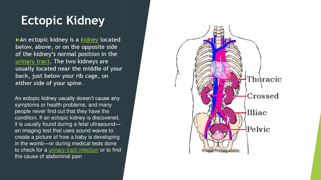
Ectopic KidneyAn
ectopic kidney is a kidney located
below, above, or on the opposite side
of the kidney’s normal position in the
urinary tract. The two kidneys are
usually located near the middle of your
back, just below your rib cage, on
either side of your spine.
An ectopic kidney usually doesn’t cause any
symptoms or health problems, and many
people never find out that they have the
condition. If an ectopic kidney is discovered,
it is usually found during a fetal ultrasound—
an imaging test that uses sound waves to
create a picture of how a baby is developing
in the womb—or during medical tests done
to check for a urinary tract infection or to find
the cause of abdominal pain
20.
An ectopic kidney may cross overand can become fused with the other
kidney (crossed renal ectopia).
An ectopic kidney may remain in
the pelvis, near the bladder (pelvic
kidney).
21.
What are the symptoms of an ectopic kidney?Most people with an ectopic kidney have no symptoms. If complications
occur, however, symptoms may include
•pain in your abdomen or back
•urinary frequency or urgency, or burning during urination
•fever
•hematuria, or blood in the urine
•lump or mass in the abdomen
•high blood pressure
How do health care professionals diagnose an ectopic
kidney?
Health care professionals use the following imaging tests to help
diagnose and manage an ectopic kidney.
•Ultrasounds
•Voiding cystourethrograms
• Radionuclide scans
•Magnetic resonance imaging (MRI)
22.
DuplicatedUreters
Ureters
are long, narrow tubes
that drain urine from the kidneys to
the urinary bladder. Normally one
ureter leads from each kidney to the
bladder. In the case of duplicated
ureters, two ureters drain a single
kidney. One ureter drains the upper
part of the kidney and the other
drains the lower part. This condition
may affect one or both kidneys.
23.
Duplicated ureters can take one of two forms:•Incomplete: Two separate ureters are attached to the same kidney but join together at some
distance away from the kidney to form a single ureter that enters the bladder.
•Complete: Two separate ureters lead away from the same kidney and remain separate.
How common are duplicated ureters?
About 0.7% of the healthy adult population and 2% to 4% of patients with urinary tract symptoms have
duplicated ureters. Incomplete duplication is three times more common than complete duplication,
which is estimated to appear in about one of every 500 people.
What causes duplicated ureters?
Duplicated ureters are a result of errors in cell division that occur during the development of a
fetus, the baby inside the mother’s womb. There is no proof that anything during pregnancy causes
the defect. However, there is evidence to show that the condition can be passed from parent to
child. If one parent has a duplicated ureter the child has a 50-50 chance of also being born with
this condition.
24.
symptomsA number of symptoms can also occur when one of the ureters is ectopic, which means it drains to
somewhere other than the bladder. Symptoms of an ectopic ureter include:
•Hydronephrosis: An ectopic ureter is usually narrower than it should be, leading to an obstruction in
the flow of urine. The urine gets backed up and causes the kidney and ureter to swell.
•Urinary tract infection (UTI): Poor drainage makes it easier for bacteria to enter urine and travel to
the bladder. UTIs result in painful urination.
•Vesicoureteral reflux: Urine backs up and flows in the wrong direction (up toward the kidney instead
of down toward the bladder). It is important for a doctor to grade the amount of reflux, as a child may
be able to outgrow a small amount of reflux but may need more extensive treatment if the reflux is
large. Kidney infections or other damage can result from reflux.
•Incontinence (inability to control urination):