


 Медицина
Похожие презентации:


Placenta accreta
1.
Reporter: Denis Larionov3 year student of Sechenov university
2021
2.
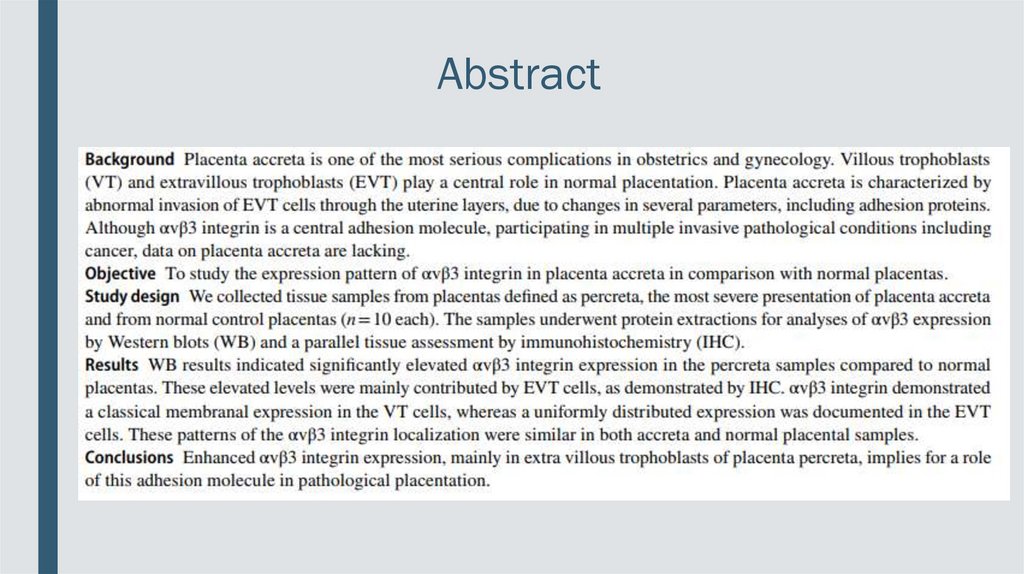
Abstract3.
Outlines■ Article and journal characteristics
■ Introduction
■ Materials and methods
■ Results
■ Discussion
■ Conclusion
■ Questions for discussion
4.
Article and journal characteristics■ Published in Archives of Gynecology and
Obstetrics (Q2, h-index of 68, IF =
2.344, Open access journal) on 28
October 2020.
■ Original research.
■ Retrospective study.
■ 20 female patients were devided into 2
groups based on their placental status
(n=10 of placenta accreta; n=10 normal
control placentas).
■ Tissue samples were collected from
January 2015 through April 2019.
■ References: 44 articles
5.
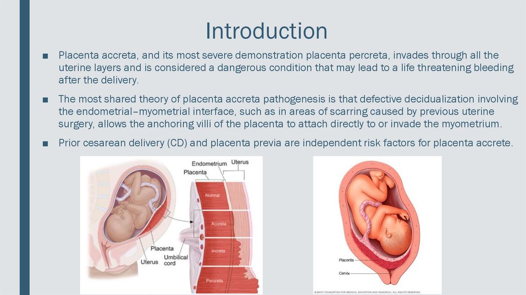
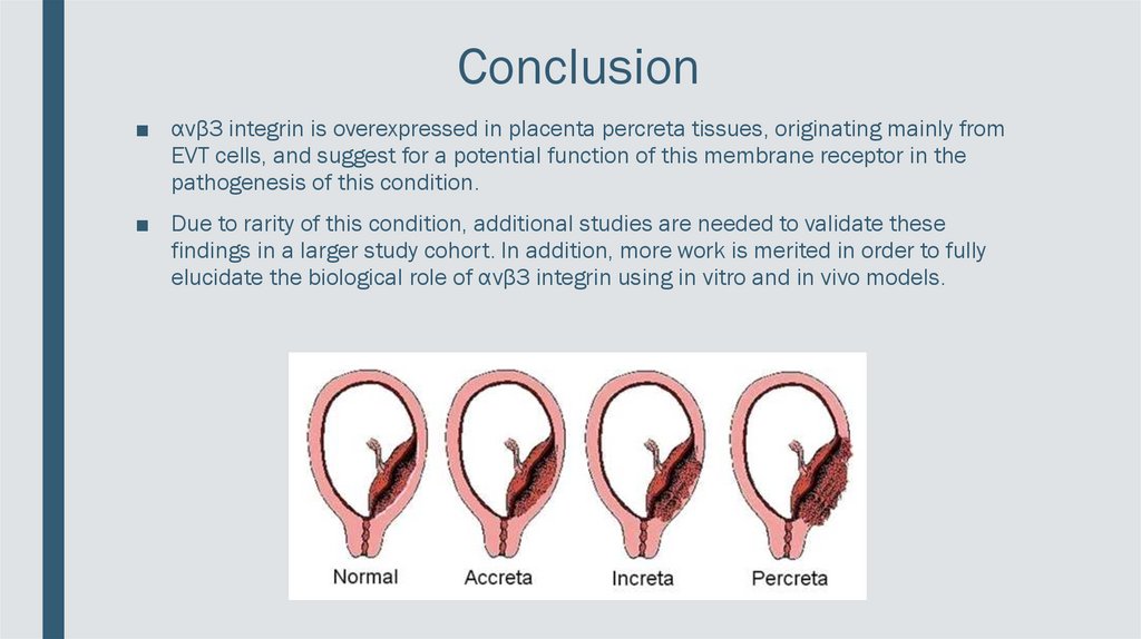
Introduction■ Placenta accreta, and its most severe demonstration placenta percreta, invades through all the
uterine layers and is considered a dangerous condition that may lead to a life threatening bleeding
after the delivery.
■ The most shared theory of placenta accreta pathogenesis is that defective decidualization involving
the endometrial–myometrial interface, such as in areas of scarring caused by previous uterine
surgery, allows the anchoring villi of the placenta to attach directly to or invade the myometrium.
■ Prior cesarean delivery (CD) and placenta previa are independent risk factors for placenta accrete.
6.
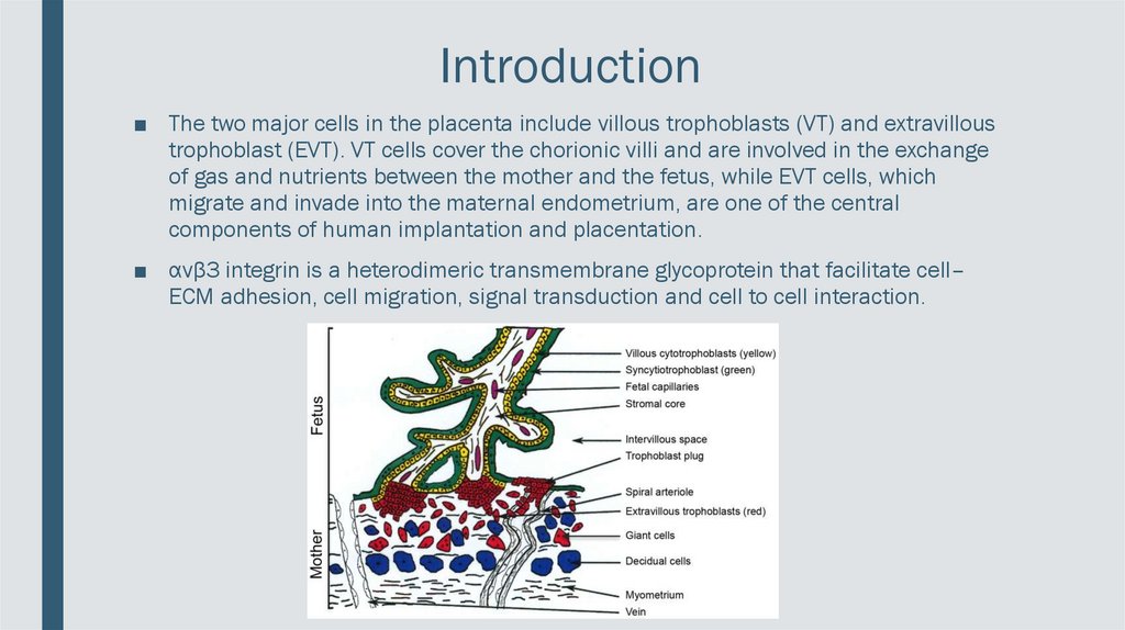
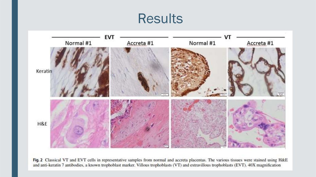
Introduction■ The two major cells in the placenta include villous trophoblasts (VT) and extravillous
trophoblast (EVT). VT cells cover the chorionic villi and are involved in the exchange
of gas and nutrients between the mother and the fetus, while EVT cells, which
migrate and invade into the maternal endometrium, are one of the central
components of human implantation and placentation.
■ αvβ3 integrin is a heterodimeric transmembrane glycoprotein that facilitate cell–
ECM adhesion, cell migration, signal transduction and cell to cell interaction.
7.
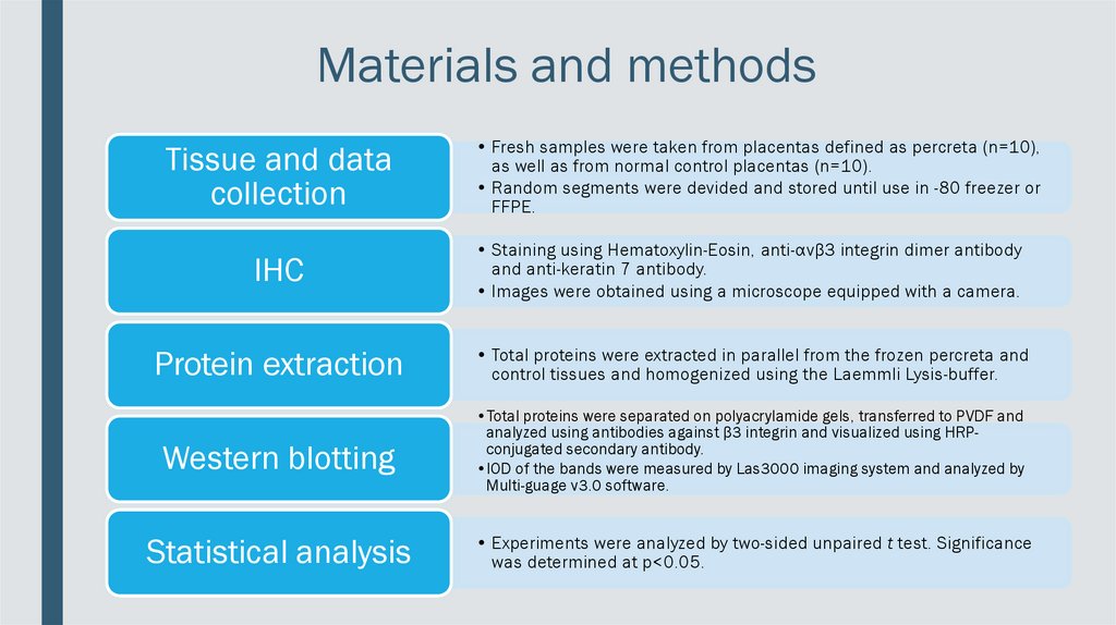
Materials and methodsTissue and data
collection
• Fresh samples were taken from placentas defined as percreta (n=10),
as well as from normal control placentas (n=10).
• Random segments were devided and stored until use in -80 freezer or
FFPE.
IHC
• Staining using Hematoxylin-Eosin, anti-αvβ3 integrin dimer antibody
and anti-keratin 7 antibody.
• Images were obtained using a microscope equipped with a camera.
Protein extraction
• Total proteins were extracted in parallel from the frozen percreta and
control tissues and homogenized using the Laemmli Lysis-buffer.
Western blotting
•Total proteins were separated on polyacrylamide gels, transferred to PVDF and
analyzed using antibodies against β3 integrin and visualized using HRPconjugated secondary antibody.
•IOD of the bands were measured by Las3000 imaging system and analyzed by
Multi-guage v3.0 software.
Statistical analysis
• Experiments were analyzed by two-sided unpaired t test. Significance
was determined at p<0.05.
8.
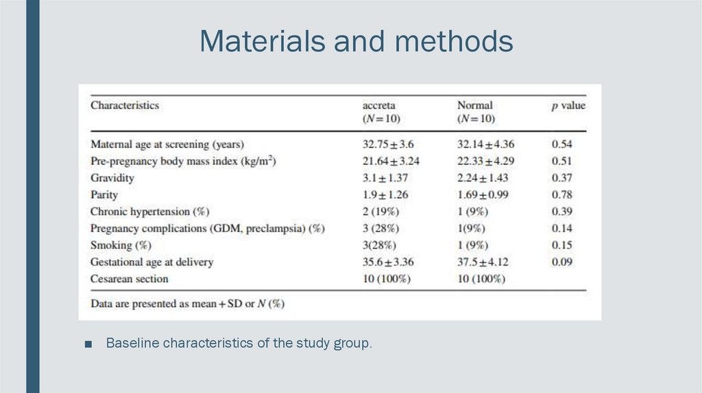
Materials and methods■ Baseline characteristics of the study group.
9.
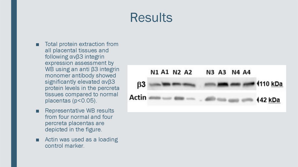
Results■ Total protein extraction from
all placental tissues and
following αvβ3 integrin
expression assessment by
WB using an anti β3 integrin
monomer antibody showed
significantly elevated αvβ3
protein levels in the percreta
tissues compared to normal
placentas (p<0.05).
■ Representative WB results
from four normal and four
percreta placentas are
depicted in the figure.
■ Actin was used as a loading
control marker.
10.
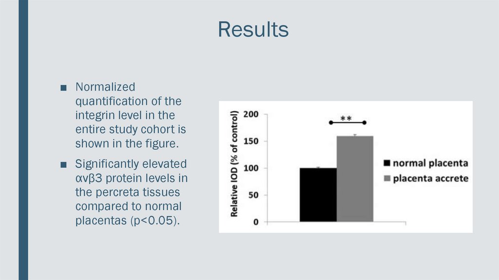
Results■ Normalized
quantification of the
integrin level in the
entire study cohort is
shown in the figure.
■ Significantly elevated
αvβ3 protein levels in
the percreta tissues
compared to normal
placentas (p<0.05).
11.
Results12.
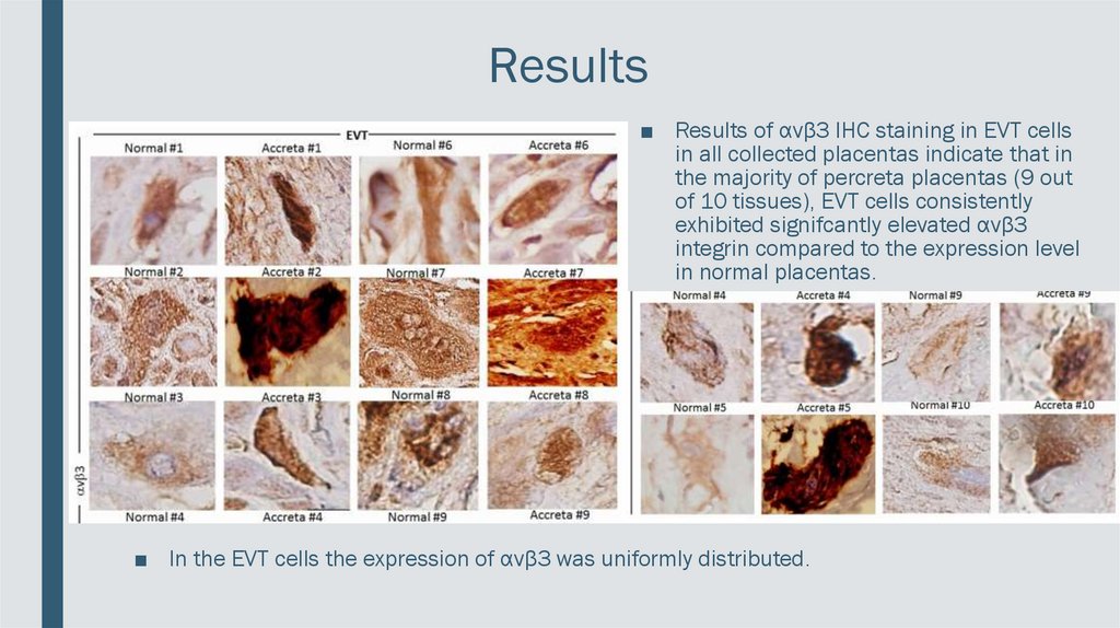
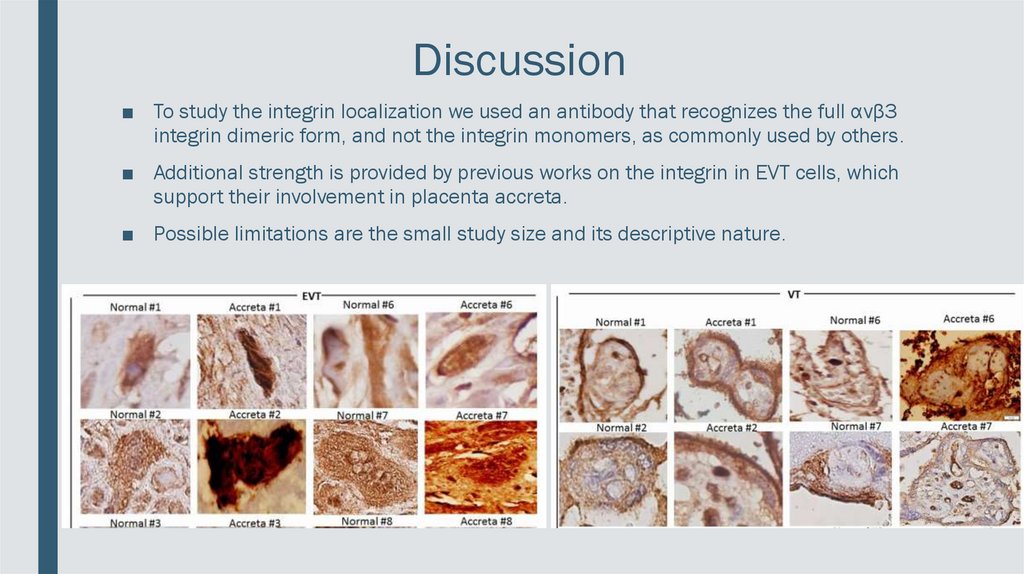
Results■ Results of αvβ3 IHC staining in EVT cells
in all collected placentas indicate that in
the majority of percreta placentas (9 out
of 10 tissues), EVT cells consistently
exhibited signifcantly elevated αvβ3
integrin compared to the expression level
in normal placentas.
■ In the EVT cells the expression of αvβ3 was uniformly distributed.
13.
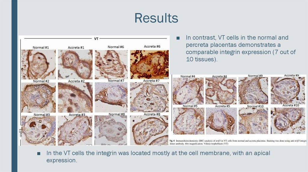
Results■ In contrast, VT cells in the normal and
percreta placentas demonstrates a
comparable integrin expression (7 out of
10 tissues).
■ In the VT cells the integrin was located mostly at the cell membrane, with an apical
expression.
14.
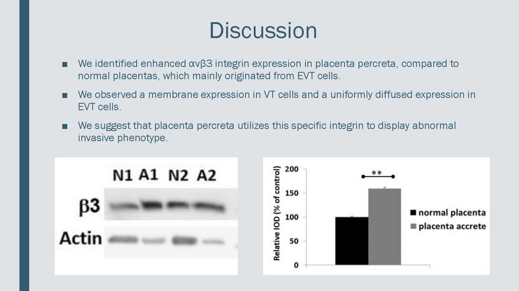
Discussion■ We identified enhanced αvβ3 integrin expression in placenta percreta, compared to
normal placentas, which mainly originated from EVT cells.
■ We observed a membrane expression in VT cells and a uniformly diffused expression in
EVT cells.
■ We suggest that placenta percreta utilizes this specific integrin to display abnormal
invasive phenotype.
15.
Discussion■ To study the integrin localization we used an antibody that recognizes the full αvβ3
integrin dimeric form, and not the integrin monomers, as commonly used by others.
■ Additional strength is provided by previous works on the integrin in EVT cells, which
support their involvement in placenta accreta.
■ Possible limitations are the small study size and its descriptive nature.
16.
Conclusion■ αvβ3 integrin is overexpressed in placenta percreta tissues, originating mainly from
EVT cells, and suggest for a potential function of this membrane receptor in the
pathogenesis of this condition.
■ Due to rarity of this condition, additional studies are needed to validate these
findings in a larger study cohort. In addition, more work is merited in order to fully
elucidate the biological role of αvβ3 integrin using in vitro and in vivo models.
17.
Questions for discussion1. What are the potential benefits of using knowledge of αvβ3 integrin
overexpression in treatment and diagnosis of placenta accreta?
2. Does the discovery of αvβ3 integrin overexpression in EVT cells in
placenta percreta significantly improves the understanding of the
pathogenesis of placenta accreta?
3. What are the new perspectives and potential targets in studying the
pathogenesis of placenta accreta?