


")


")
 Weening JJ et al. J Am Soc Nephrol 15: 241-50, 2004")


Похожие презентации:
")


Systemic lupus erythematosus
1. Systemic lupus erythematosus
2. General characteristics
Unknown etiology, multifactorial diseaseInvolve joints, kidneys, mucous
membranes, the central nervous system
Variety of antibodies
Symptoms vary greatly from person to
person
Lupus tends to be chronic
Alternation between remission and relapse
3. Epidemiology
Prevalence: 50-100 /100.000Incidence: 2-7 /100.000/year
age at onset: 20-30
Female vs. male ratio: 9-10:1
4. Etiology
Family history (1st degree relatives 1%)Genetic predisposition
MHC genes: HLA DR2,DR3
DR4-DIL, DR5-APS
non-MHC genes: complement
component, complement receptor, Fc
receptors, CRP, cytokines, apoptotic genes
(e.g., FAS)
5. Etiology
Provoking factorsSunlight, UV light
Infections
Hormonal status:estrogen, prolactin
Drugs
Isoniacid
Hidantoin
Hydralazin
Procainamid
D penicillinamin
Penicillins
Sulphonamids
TNF alpha blockers
6. PATHOGENESIS
Disturbed immune regulation:•Pathologic antigen presentation
•Increased MHC expression
•Enhanced co-stimulation
•Cytokine imbalance (Th1/Th2)
•Decrease of regulatory T cells
1. Polyclonal B cell activation
•Distrubed apoptosis
2. Pathologic autoantibody
production
3. Impaired clearance of
immune complexes
4. Accumulation of IC
5. Complement activation
7. Pathogenesis of SLE
Provoking factors: Genetics predisposition (MHC and non-MHC genesTriggering factors (UV, drugs, infections)
Hormonal status
Immune disregulation
Defect of clearance
DNA,
Apoptotic cells
APCs
Decreased Regulatory T cells activity
Increased help:
citokines,
co-stimulation
T cells
increased CD4+
actitivity
Autoreactive
B cells
Production
of Auto-AB
Increased IC
C’ activation
Organ damages
ADCC
aPL
8.
9. Antigen targets for autoantibodies in SLE
Nuclear antigens: ssDNA, dsDNA, histon,Sm, RNP
Cytoplasmic antigens: SS-A, SS-B,
ribosoma p protein, ANCA
Cells surface antigens: on endothel cells,
erythrocytes, neutrophils, lymphocytes,
platlets
Other antigens, plasma factors: Beta-2
glycoprotein I, phospholipids, immune
globulins
10. General symptoms
WeaknessFatigue
Tiredness
Fever
Weight loss
Hair loss
Lymphadenopathy
11. CLASSIFICATION OF SKIN SYMPTOMS IN SLE (Sontheimer RD.Lupus 6:84-95, 1997)
Lupus specificA. Acute cutan LE /ACLE/
Butterfly rush
Generalised ACLE
photosensitivity.
B. Subacute cutan LE /SCLE/
Annular
Psoriasiform
C. Chronic cutan LE /CCLE/
Classical discoid lesions
Hypertrophic DLE
Lupus panniculitis
Mucosal ulceration
Others (L.tumidus, Lichenoid)
non-specific for
Lupus
A. Cutan vascular
symptoms
Vasculitis
Vasculopathy
Raynaud’s syndrome
Livedo reticularis
B. Non-scarring diffuse
alopecia
C. Urticaria
D. Erythema exsudativum
multiforme
12. Lupus specific skin symptoms
Vespertilio=butterfly rashAcute cutan LE
13. Lupus specific skin symptoms
DLESCLE
14. Non-lupus specific skin symptoms
Raynaud phenomenonvasculitis
15. Musculosceletal involvment of lupus
Small joint symmetric non erosive polyarthritisAseptic femur neck necrosis
Osteoporosis
Myositis
16. Polyserositis
PleuritisPericarditis
Peritonitis
pleuritis
pericarditis
17. Respiratory involvment
PleuritisAlveolitis obliterans
Pulmonal fibrosis
Pulmonal hypertension
ARDS
Pulmonal embolism
18. Cardiovascular involvments
PericarditisMyocarditis
Cardiomyopathy
Endocarditis
non-infectious verrucosus endocarditis
(Libman-Sacks endocarditis)
subacute infectious endocarditis
Valvulopathy
Atherosclerosis of coronary
19.
Non-infectious endocarditisPericarditis
AMI
20. Nomenclature of neuro-psychiatric symptoms of SLE (ACR ad hoc Committee, Arthritis Rheum. 42:599-608, 1999.)
CNSAseptic meningitis
Cerebrovascular lesion
Demyelinating syndrome
Headache (migraine)
Chorea
Myelopathy
Convulsion
Psychosis
Acute confusing state
Cognitive dysfunction
PNS
Acute Guillain-Barré sy.
Disturbance in autonomic
nervous system
Mononeuritis
simplex/multiplex
Myasthenia gravis like
Cranial nerve lesion
Polyneuropathy
Brain infarct
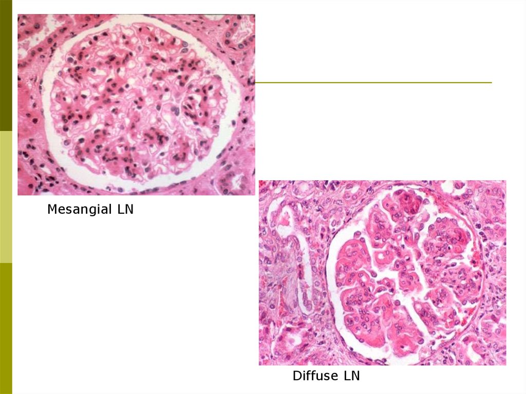
21. Histopathologic classification of lupus nephritis (ISN/RPS) Weening JJ et al. J Am Soc Nephrol 15: 241-50, 2004
Class I.Class II.
Class III.
A.
A/C.
C.
Class IV.
Minimal mesangial nephritis
Mesangial proliferative nephritis
Focalis lupus nephritis (<50% of glomeruli are involved)
Active lesions: focal proliferative GN
Active and chronic lesions: focal proliferativ and
sclerosing GN
Chronic inactive lesions with glomerular scarring: focal
sclerosing GN.
Diffuse lupus nephritis (>50% of glomeruli are involved)
diffuse segmental (IV-s) type, when only a part of the involved
glomeruli are affected
diffuse global GN (IV-G), when the entire glomeruli are affected
IV-S (A),IV-G (A),
IV-S (A/C), IV-G (C),
IV-S (C),
Class V.
Membranous lupus nephritis
May associate with findings characterised in class III/IV.
Class VI.
Sclerosing glomerulonephritis
90% of glomeruli are sclerotic
22.
Mesangial LNDiffuse LN
23. Other manifestations
Haematology● Leukopenia, lymphopenia
● AIHA/ Thrombocytopenia/Evans sy.
● Pancytopenia
● TTP, CAPS
● Lymphadenopathy/Splenomegaly
Othe
● Vasculitis
● Pancreatitis, lupus hepatitis
● Pepeticus ulcus/GI-bleeding
● Mesenterial thrombosis/vasculitis
● A./v. central retinae thrombosis
● Opticus neuritis
● Chorioretinitis
● Sicca sy.
24. Laboratory tests and findings in SLE
General inflammatory findings: ESR , normal CRPHaematology: pancytopenia,
Kidney tests: sodium, potassium, carbamide,
creatinine
enzymes: CK, AST/GOT, ALT/GPT, LDH
(haemolysis, myositis, hepatitis)
Haemostasis: Lupus anticoagulant
Immunserology: IgG, C3, C4, CH50, ANA, aDNA,
nukleosoma, histon, anti-Sm, anti-cardiolipin,
anti-beta2-glikoprotein I.
Urine (protein!) and urine sediment (WBC, RBC,
count)
24 hours urine collection: detection of protein
25. Radiology and other examinations in SLE
Chest X ray, CT (HRCT), breath test, scanAbdominal ultrasonograph
ECG, echocardiography
Neurology examination: EEG, ENG, EMG, CT,
MRI, test of cerebrospinal fluid
Biopsies: skin (vagy lupus band teszt)
kidney
muscles
n. suralis
26.
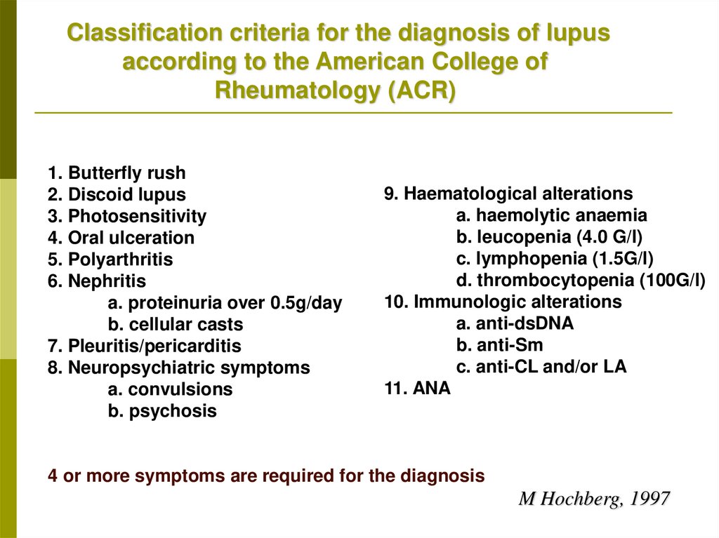
Classification criteria for the diagnosis of lupusaccording to the American College of
Rheumatology (ACR)
1. Butterfly rush
2. Discoid lupus
3. Photosensitivity
4. Oral ulceration
5. Polyarthritis
6. Nephritis
a. proteinuria over 0.5g/day
b. cellular casts
7. Pleuritis/pericarditis
8. Neuropsychiatric symptoms
a. convulsions
b. psychosis
9. Haematological alterations
a. haemolytic anaemia
b. leucopenia (4.0 G/l)
c. lymphopenia (1.5G/l)
d. thrombocytopenia (100G/l)
10. Immunologic alterations
a. anti-dsDNA
b. anti-Sm
c. anti-CL and/or LA
11. ANA
4 or more symptoms are required for the diagnosis
M Hochberg, 1997
27.
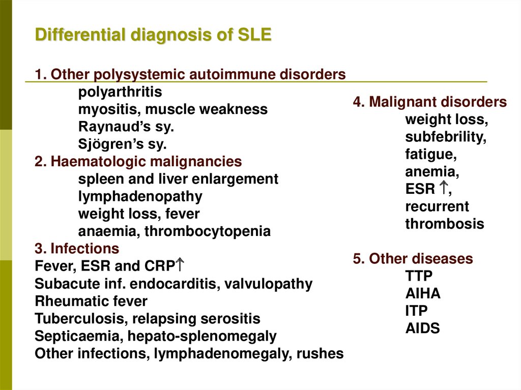
Differential diagnosis of SLE1. Other polysystemic autoimmune disorders
polyarthritis
4. Malignant disorders
myositis, muscle weakness
weight loss,
Raynaud’s sy.
subfebrility,
Sjögren’s sy.
fatigue,
2. Haematologic malignancies
anemia,
spleen and liver enlargement
ESR ,
lymphadenopathy
recurrent
weight loss, fever
thrombosis
anaemia, thrombocytopenia
3. Infections
5. Other diseases
Fever, ESR and CRP
TTP
Subacute inf. endocarditis, valvulopathy
AIHA
Rheumatic fever
ITP
Tuberculosis, relapsing serositis
AIDS
Septicaemia, hepato-splenomegaly
Other infections, lymphadenomegaly, rushes
28. Monitoring of activity in SLE disease activity index: DAI
ConvulsionPsychosis
Organic brain
syndrome
Visual field
defects
(retinopathy)
Cranial nerve lesion
Lupus headache
Stroke
Arthritis
Myositis
8
8
8
8
8
8
8
4
4
Casts in urine
Haematuria
Proteinuria
Pyuria
New rushes
Alopecia
Oral ulcer
Pleuritis
Pericarditis
Low complement
Elevated aDNA
Fever
Thrombopenia
Leucopenia
4
4
4
4
2
2
2
2
2
2
2
1
1
1
29. Subgroups in SLE
Subacute cutan lupus erythematosusNeonatal lupus erythematosus
Drug-induced lupus
SLE in elderly
SLE with APS
30.
SUBGROUPS IN SLE1. SUBACUTE CUTAN LUPUS (SCLE)
Clinical characteristics:
annular/psoriasiform skin eruptions
photosensitivity (60-70%)
less frequent kidney involvement (10%)
less common CNS symptoms (20%)
Laboratory signs:
aSSA/aSSB antibodies (60-70%)
Therapeutical considerations:
sun screens
topical steroids
systemic low dose steroid
antimalarial drugs
31.
SLE SUBGROUPS2. NEONATAL LUPUS (NLE)
Frequency:
Cause:
rare
maternal autoantibodies
passing through the placenta
Clinical characteristics: generalised skin eruptions
hepato-splenomegaly
transient thrombocytopenia
autoimmun haemolytic anaemia
congenital heart block
Laboratory signs:
aSSA/aSSB antibodies
ANA positivity
high a-dsDNA concentration
LE cell phenomenon
Special aspects of therapy:
Corticosteroids
HIVIG
pace maker
32.
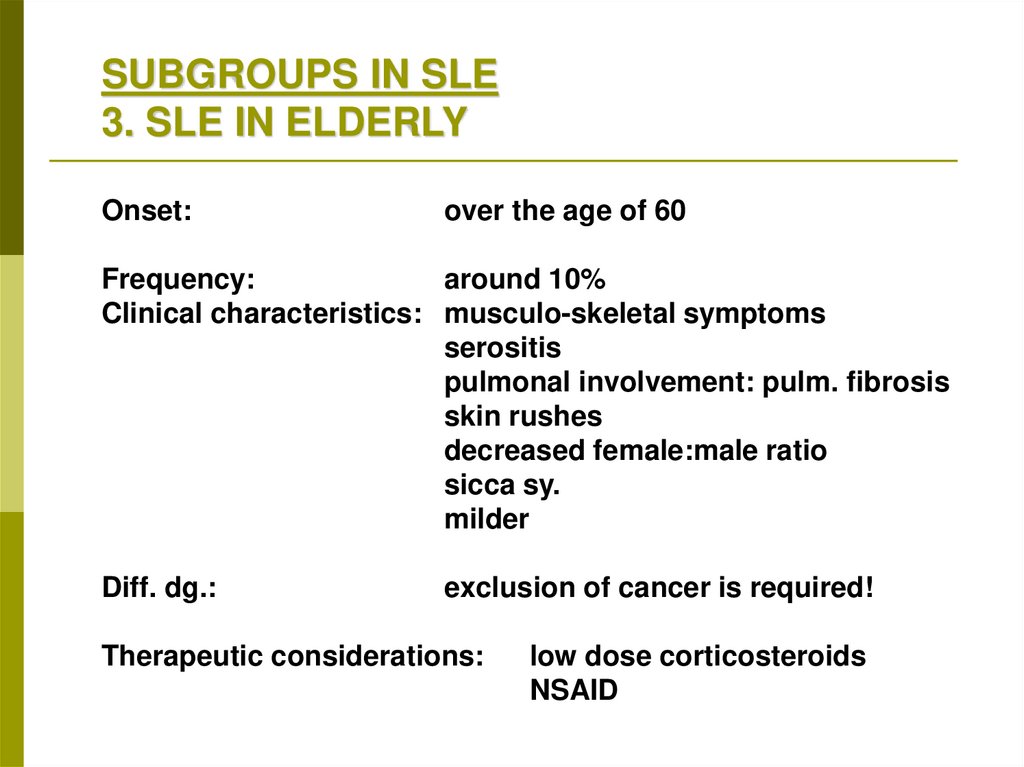
SUBGROUPS IN SLE3. SLE IN ELDERLY
Onset:
over the age of 60
Frequency:
around 10%
Clinical characteristics: musculo-skeletal symptoms
serositis
pulmonal involvement: pulm. fibrosis
skin rushes
decreased female:male ratio
sicca sy.
milder
Diff. dg.:
exclusion of cancer is required!
Therapeutic considerations:
low dose corticosteroids
NSAID
33.
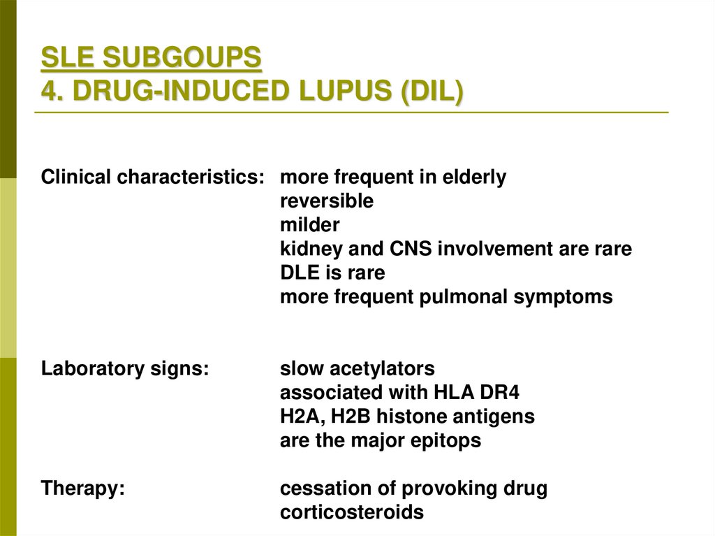
SLE SUBGOUPS4. DRUG-INDUCED LUPUS (DIL)
Clinical characteristics: more frequent in elderly
reversible
milder
kidney and CNS involvement are rare
DLE is rare
more frequent pulmonal symptoms
Laboratory signs:
slow acetylators
associated with HLA DR4
H2A, H2B histone antigens
are the major epitops
Therapy:
cessation of provoking drug
corticosteroids
34. Negative prognostic factors in SLE
Sex:maleAge under 20 or above 50
Diffuse proliferative lupus nephritis
CNS manifestations
Anti-phospholipid antibodies
Endocarditis
35. Causes of death
In the early phase of the disease processIn the later phase of the disease process
Kidney failer
Neurology involvement
SLE Activity
Cardiovascular event
Thromboembolism
Malignant disorders
In both:
infections
36. Therapy of lupus
General -proceduresAvoidance of UV lights
Sunscreens
Termination of the use of provoking drugs
Avoidance of contraceptive pills
Adequate antibiotics therapy
37. Therapy of SLE
Antimalarial drugs: hydroxichlorouin,chloroquin (Delagil)
In the cases of arthralgia, arthritis, skin
symptoms, serositis
Dosis: 200-400 mg/die
Side effect: ocular complications
38. Therapy of SLE
Steroids: methylpednisolon (Solu-Medrol, Medrol,Methypred)
In acute flares and relapses
in neonatal lupus: dexamethason 4 mg/die
Dosis: start with 0.5-1 mg/bwkg, then slowly
decreased dosis
Pulse steroid: 1 g/3 days
Side effects!
39. Immunosupressives
Methotrexat (Trexan)7.5-20 mg/week, treatment of polyarthritis, vasculitis
CAVE: bonemarrow and liver toxicity
Azathiorpin (Imuran)
1-2 mg/bwkg/day, multiorgan involvement
CAVE: bonemarrow and liver toxicity
Cyclophosphamid (Cytoxan)
500-1000 mg/m2/month for 6 months, then
same amount/3 months for 1.5 years (NIH
protocol)
In the cases of lupus nephritis, alveolitis,
vasculitis, CNS involvement
CAVE:bonemarrow and liver toxicity
40. Immunmodulation
Cyclosporin A (Sandimmun Neoral)In the cases of haematology involvement, membranous
lupus nephritis
dosis: 3 mg/bwkg/day
Mycophanolat mophetil/Mycofenol acid (Cellcept/Myfortic)
In the case of lupus nephritis max. 3 gr/day
Diamino diphenylsulphon (Dapson)
41. Potential targets in the therapy of SLE
42. Other
HIVIG 0.4 g/bwkg/day for 5 daysPlasmapheresis 100 ml/bwkg plasma
exchange synchronized with ISU
Stem cell transplantation