
 Медицина
Похожие презентации:


")

Radiological research methods and radiological semiotics of chronic nonspecific lung diseases
1.
Radiological research methods and radiologicalsemiotics of chronic nonspecific lung diseases
Prepared by the Department of "Visual Diagnostics" of KazNMU named
after S. D. Asfendiyarov
2.
Content• Radiological methods of chest organs
research
• Radiological anatomy of chest organs
Radiological semiotics of chronic nonspecific lung diseases :
• 1. Chronic bronchitis
• 2. Bronchiectatic disease
• 3. Emphysema
• 4. Pneumosclerosis
• 5. Chronic nonspecific pneumonia
3.
Radiological research methodschest organs
Fluorography
Radioscopy
Radiography
Tomography
Bronchography
Angiopulmonography
Ultrasound diagnostics
Computed tomography
Magnetic resonance imaging
Radionuclide diagnostics (Scintigraphy, PET / CT)
4.
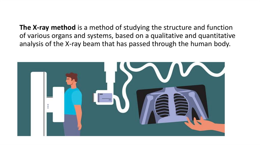
The X-ray method is a method of studying the structure and functionof various organs and systems, based on a qualitative and quantitative
analysis of the X-ray beam that has passed through the human body.
5.
Fluorography• the method of X-ray examination, which
consists in photographing an image from a
fluorescent X-ray screen (which is used more
often), the screen of an electron-optical
converter or systems designed for subsequent
digitization of images, on a small-format
photographic film-usually 110x110 mm,
100*100 mm.
6.
Radioscopy• Radioscopy (Greek. scopeo-to consider, observe) is an X-ray
examination in which a mobile X-ray image of the organ under study
is obtained on the screen
• The method makes it possible to examine the patient in various
positions, to assess the topographic and anatomical features of the
studied organs and the functional state of some organs and systems
(excursion of the diaphragm, heart contractions, the act of
swallowing, etc.).
7.
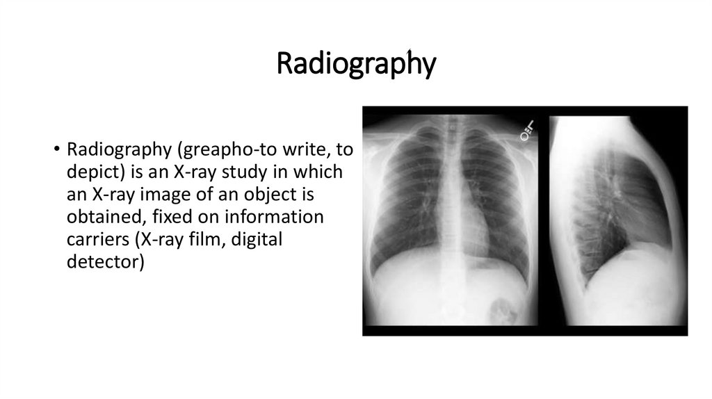
Radiography• Radiography (greapho-to write, to
depict) is an X-ray study in which
an X-ray image of an object is
obtained, fixed on information
carriers (X-ray film, digital
detector)
8.
Radiography• Overview radiography is an image of the entire organ under
study or the entire anatomical area.
• Targeted radiography is a selective fixation of the organ of
interest or its part, providing an optimal image of the
pathological focus.
9.
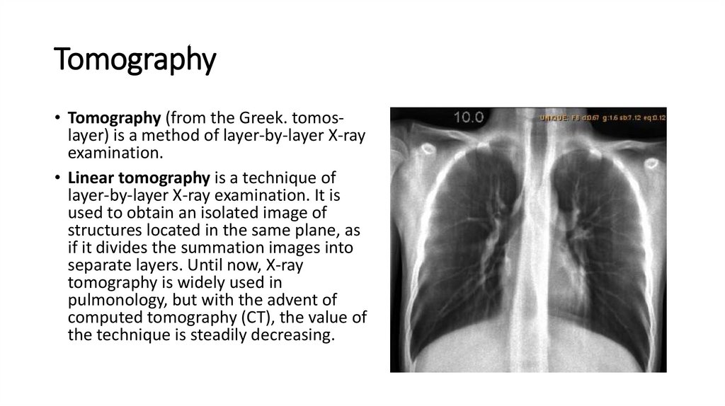
Tomography• Tomography (from the Greek. tomoslayer) is a method of layer-by-layer X-ray
examination.
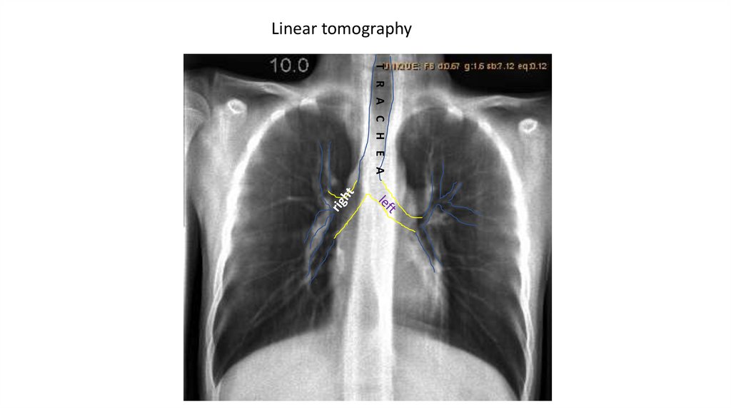
• Linear tomography is a technique of
layer-by-layer X-ray examination. It is
used to obtain an isolated image of
structures located in the same plane, as
if it divides the summation images into
separate layers. Until now, X-ray
tomography is widely used in
pulmonology, but with the advent of
computed tomography (CT), the value of
the technique is steadily decreasing.
10.
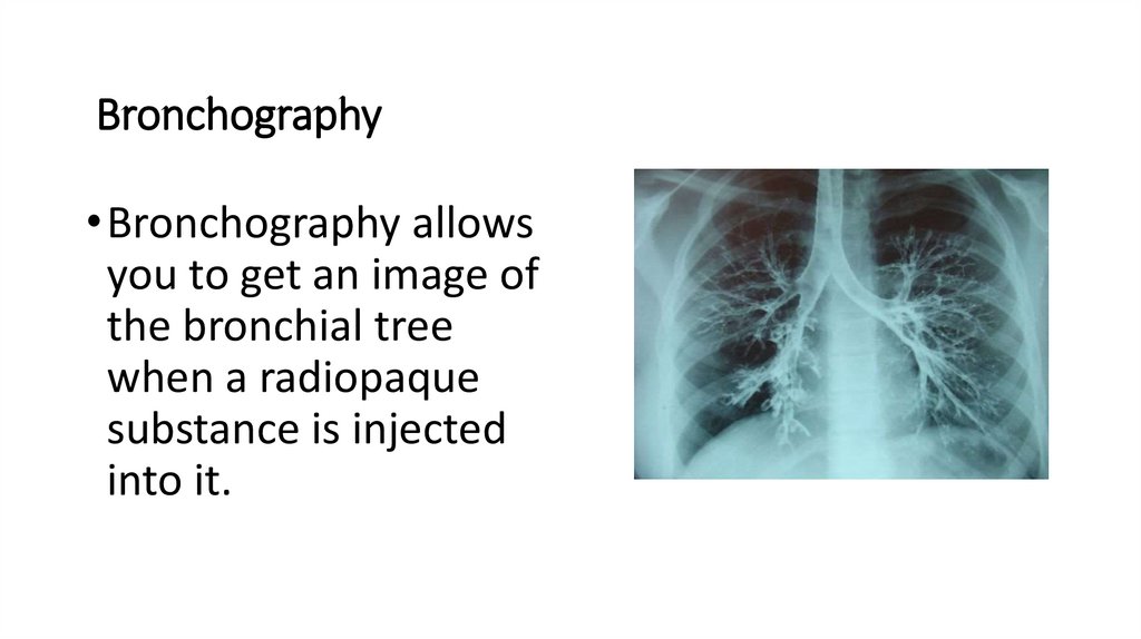
Bronchography• Bronchography allows
you to get an image of
the bronchial tree
when a radiopaque
substance is injected
into it.
11.
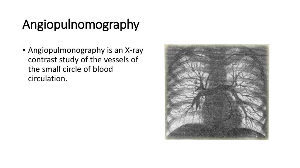
Angiopulnomography• Angiopulmonography is an X-ray
contrast study of the vessels of
the small circle of blood
circulation.
12.
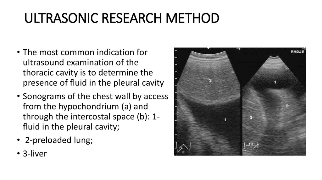
ULTRASONIC RESEARCH METHOD• The most common indication for
ultrasound examination of the
thoracic cavity is to determine the
presence of fluid in the pleural cavity
• Sonograms of the chest wall by access
from the hypochondrium (a) and
through the intercostal space (b): 1fluid in the pleural cavity;
• 2-preloaded lung;
• 3-liver
13.
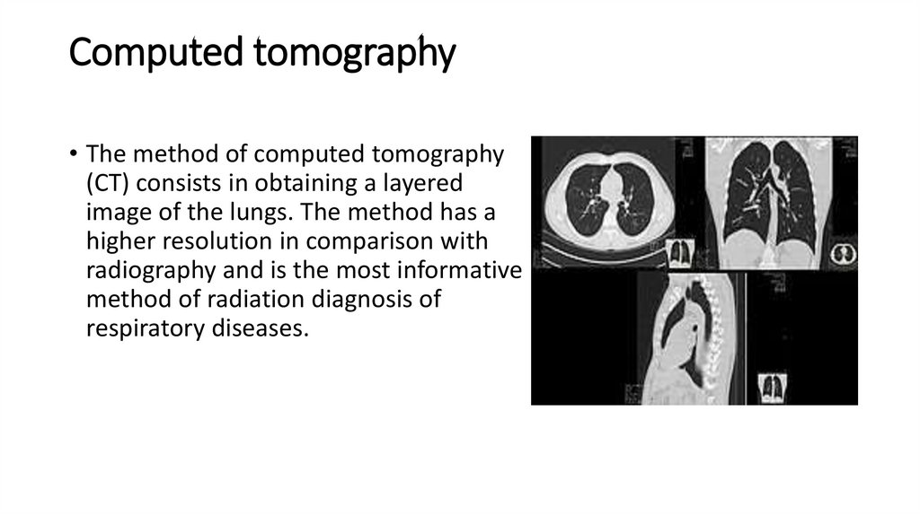
Computed tomography• The method of computed tomography
(CT) consists in obtaining a layered
image of the lungs. The method has a
higher resolution in comparison with
radiography and is the most informative
method of radiation diagnosis of
respiratory diseases.
14.
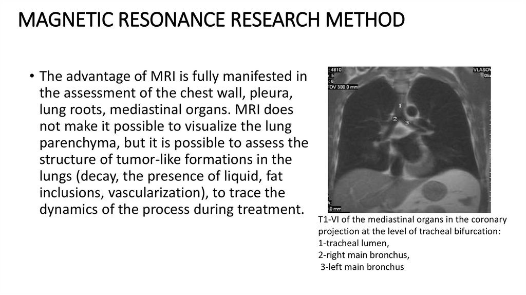
MAGNETIC RESONANCE RESEARCH METHOD• The advantage of MRI is fully manifested in
the assessment of the chest wall, pleura,
lung roots, mediastinal organs. MRI does
not make it possible to visualize the lung
parenchyma, but it is possible to assess the
structure of tumor-like formations in the
lungs (decay, the presence of liquid, fat
inclusions, vascularization), to trace the
dynamics of the process during treatment.
T1-VI of the mediastinal organs in the coronary
projection at the level of tracheal bifurcation:
1-tracheal lumen,
2-right main bronchus,
3-left main bronchus
15.
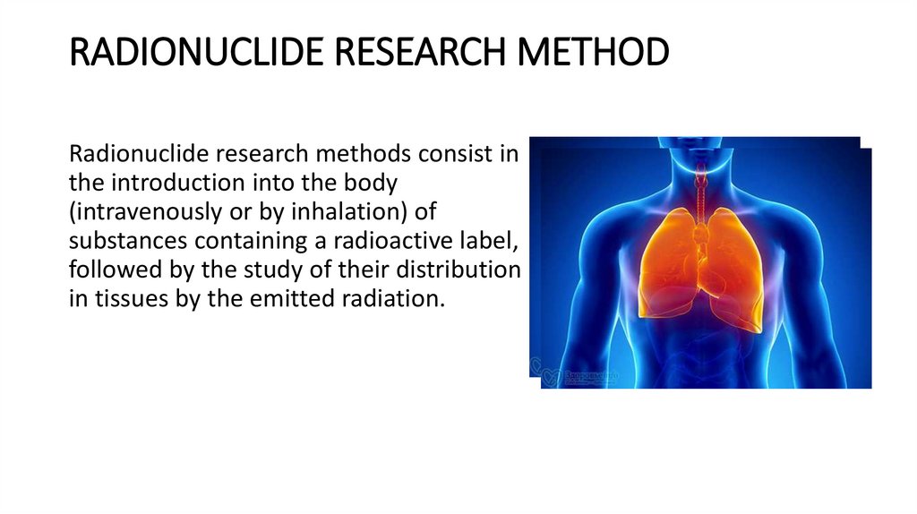
RADIONUCLIDE RESEARCH METHODRadionuclide research methods consist in
the introduction into the body
(intravenously or by inhalation) of
substances containing a radioactive label,
followed by the study of their distribution
in tissues by the emitted radiation.
16.
Radionuclide methods of lung research are carried out mainly intwo versions:
• - perfusion scintigraphy to
assess the state of blood
flow in the small circle of
blood circulation;
• - inhalation scintigraphy to
assess the fundation of
external radiation.
17.
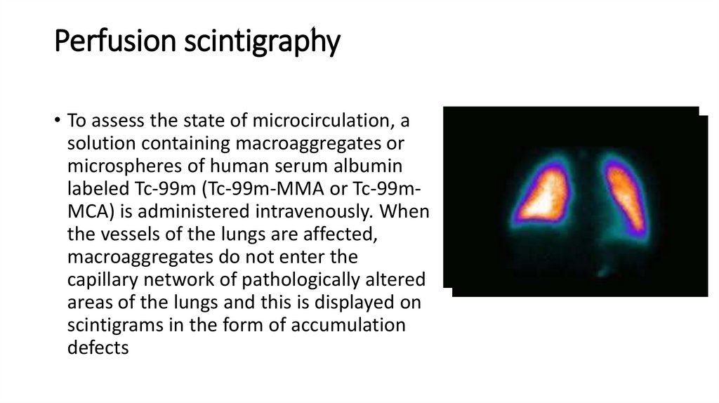
Perfusion scintigraphy• To assess the state of microcirculation, a
solution containing macroaggregates or
microspheres of human serum albumin
labeled Tc-99m (Tc-99m-MMA or Tc-99mMCA) is administered intravenously. When
the vessels of the lungs are affected,
macroaggregates do not enter the
capillary network of pathologically altered
areas of the lungs and this is displayed on
scintigrams in the form of accumulation
defects
18.
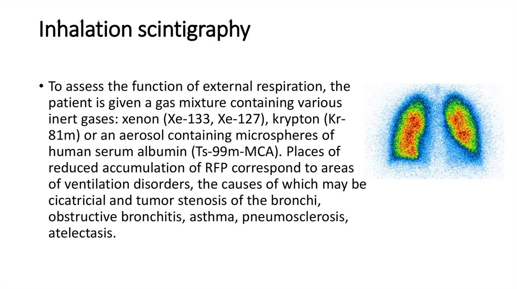
Inhalation scintigraphy• To assess the function of external respiration, the
patient is given a gas mixture containing various
inert gases: xenon (Xe-133, Xe-127), krypton (Kr81m) or an aerosol containing microspheres of
human serum albumin (Ts-99m-MCA). Places of
reduced accumulation of RFP correspond to areas
of ventilation disorders, the causes of which may be
cicatricial and tumor stenosis of the bronchi,
obstructive bronchitis, asthma, pneumosclerosis,
atelectasis.
19.
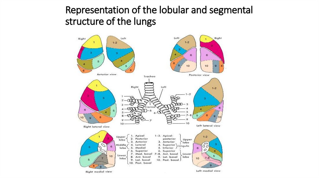
Representation of the lobular and segmentalstructure of the lungs
20.
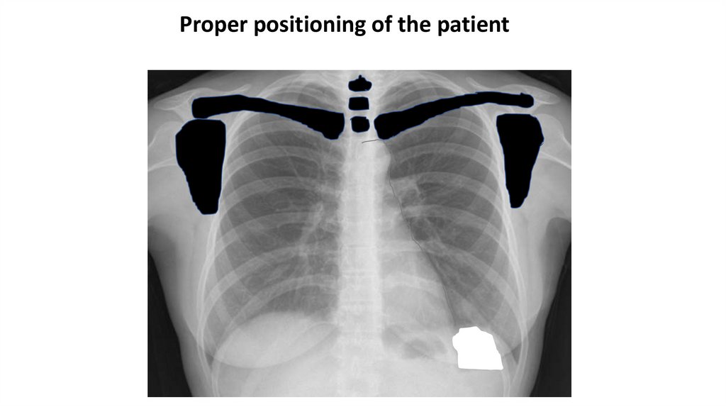
Proper positioning of the patient21.
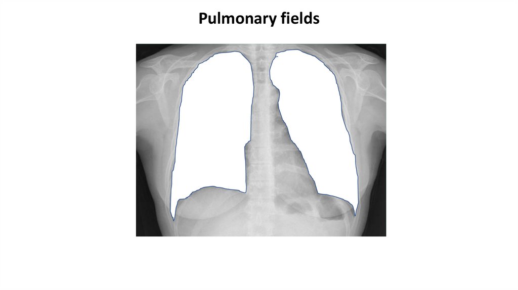
Pulmonary fields22.
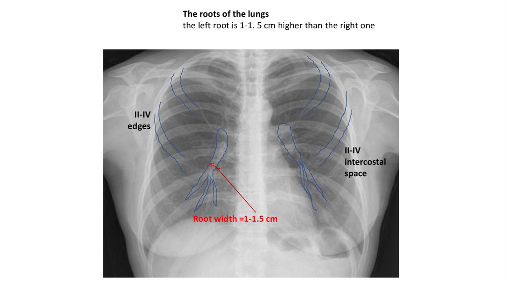
The roots of the lungsthe left root is 1-1. 5 cm higher than the right one
II-IV
edges
II-IV
intercostal
space
Root width =1-1.5 cm
23.
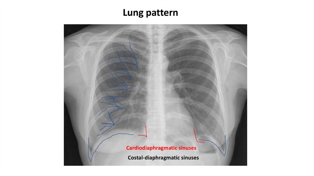
Lung patternCardiodiaphragmatic sinuses
Costal-diaphragmatic sinuses
24.
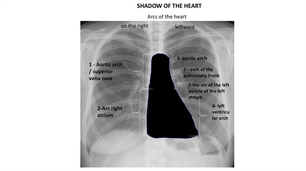
SHADOW OF THE HEARTArcs of the heart
on the right
leftward
1-aortic arch
1 - Aortic arch
/ superior
vena cava
2 - arch of the
pulmonary trunk
3-the arc of the left
auricle of the left
atrium
2-Arc right
atrium
4- left
ventricu
lar arch
25.
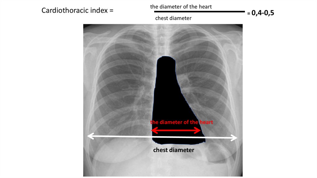
Cardiothoracic index =the diameter of the heart
chest diameter
the diameter of the heart
chest diameter
= 0,4-0,5
26.
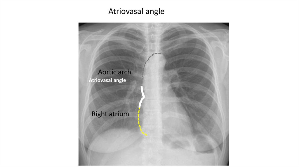
Atriovasal angleAortic arch
Atriovasal angle
Right atrium
27.
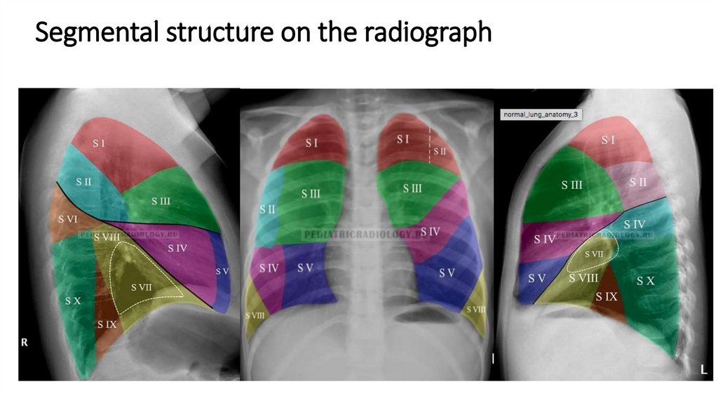
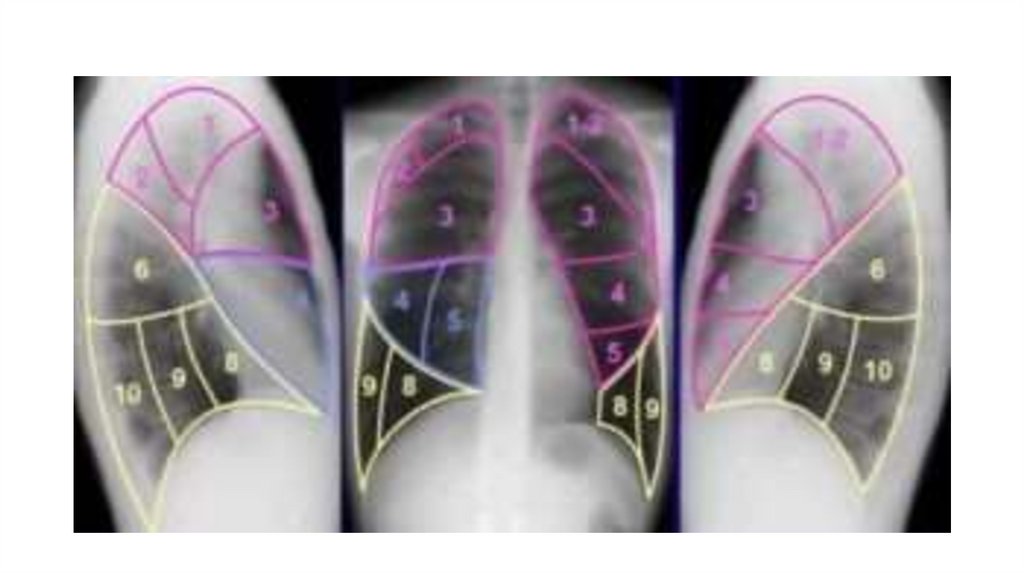
Segmental structure on the radiograph28.
29.
Linear tomographyT R A C H E A
30.
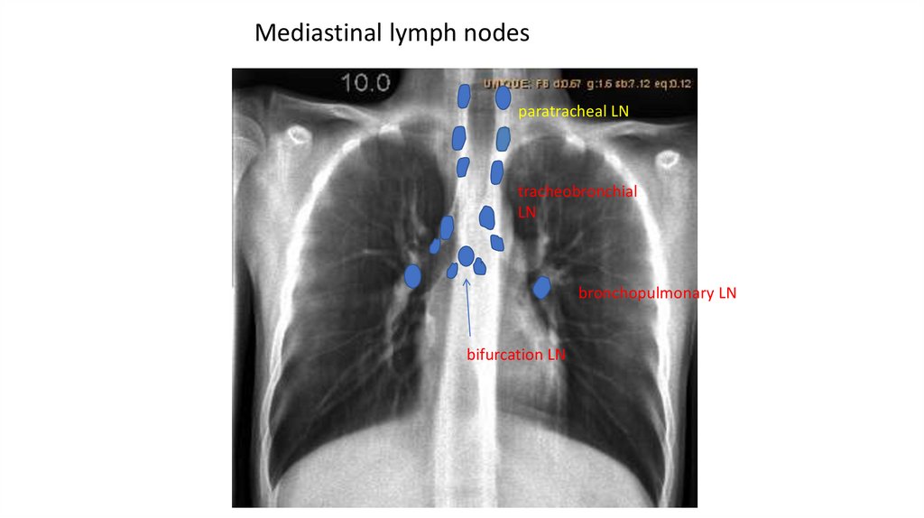
Mediastinal lymph nodesparatracheal LN
tracheobronchial
LN
bronchopulmonary LN
bifurcation LN
31.
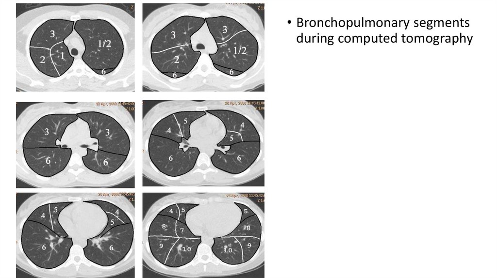
• Bronchopulmonary segmentsduring computed tomography
32.
CT at the level of the upper lobes in thepulmonary window: 1-contour of the upper
mediastinum; 2-trachea; 3, 4-pulmonary pattern
– various sections of small branches of blood
vessels
CT at the level of tracheal bifurcation in the pulmonary
window: 1-lumen of the right main bronchus;
2-right upper lobe bronchus;
3-anterior segmental bronchus of the upper lobe of
the right lung;
4-subsegmental bronchus;
5 – bronchial lumen of the V-th order;
6 – left main bronchus;
7-cross-sections of segmental bronchi;
8-sections of vessels in different planes
33.
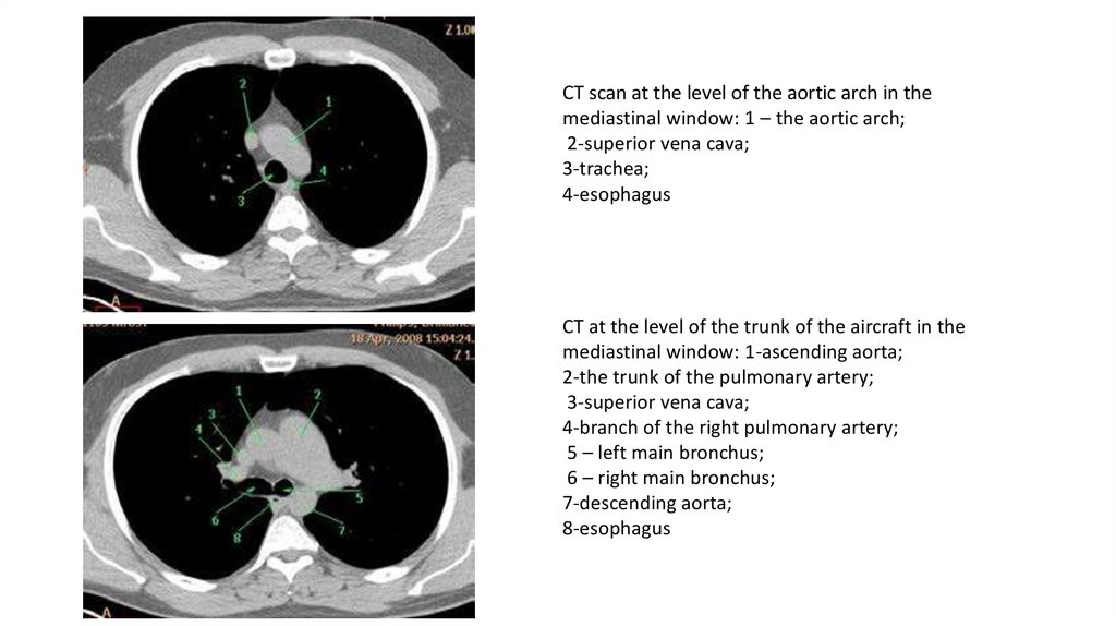
CT scan at the level of the aortic arch in themediastinal window: 1 – the aortic arch;
2-superior vena cava;
3-trachea;
4-esophagus
CT at the level of the trunk of the aircraft in the
mediastinal window: 1-ascending aorta;
2-the trunk of the pulmonary artery;
3-superior vena cava;
4-branch of the right pulmonary artery;
5 – left main bronchus;
6 – right main bronchus;
7-descending aorta;
8-esophagus
34.
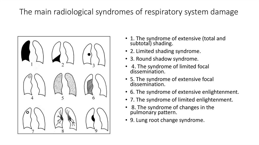
The main radiological syndromes of respiratory system damage• 1. The syndrome of extensive (total and
subtotal) shading.
• 2. Limited shading syndrome.
• 3. Round shadow syndrome.
• 4. The syndrome of limited focal
dissemination.
• 5. The syndrome of extensive focal
dissemination.
• 6. The syndrome of extensive enlightenment.
• 7. The syndrome of limited enlightenment.
• 8. The syndrome of changes in the
pulmonary pattern.
• 9. Lung root change syndrome.
35.
Chronic nonspecific lung diseases• Chronic nonspecific lung diseases(CNL), a group of chronic diseases of
the bronchopulmonary system, different in causes and mechanisms
of development, but having a number of common clinical, functional
and morphological manifestations: cough, shortness of breath,
violation of bronchial patency, fibrosis, combined with destructive
and inflammatory changes in the bronchi, blood vessels, lung
parenchyma.
36.
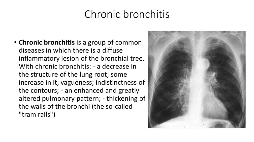
Chronic bronchitis• Chronic bronchitis is a group of common
diseases in which there is a diffuse
inflammatory lesion of the bronchial tree.
With chronic bronchitis: - a decrease in
the structure of the lung root; some
increase in it, vagueness; indistinctness of
the contours; - an enhanced and greatly
altered pulmonary pattern; - thickening of
the walls of the bronchi (the so-called
"tram rails")
37.
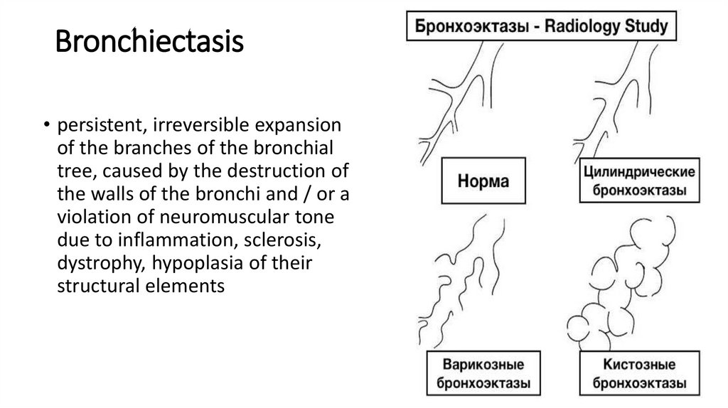
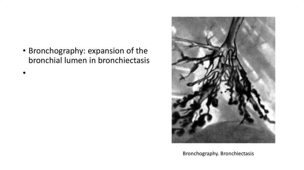
Bronchiectasis• persistent, irreversible expansion
of the branches of the bronchial
tree, caused by the destruction of
the walls of the bronchi and / or a
violation of neuromuscular tone
due to inflammation, sclerosis,
dystrophy, hypoplasia of their
structural elements
38.
• Bronchography: expansion of thebronchial lumen in bronchiectasis
Bronchography. Bronchiectasis
39.
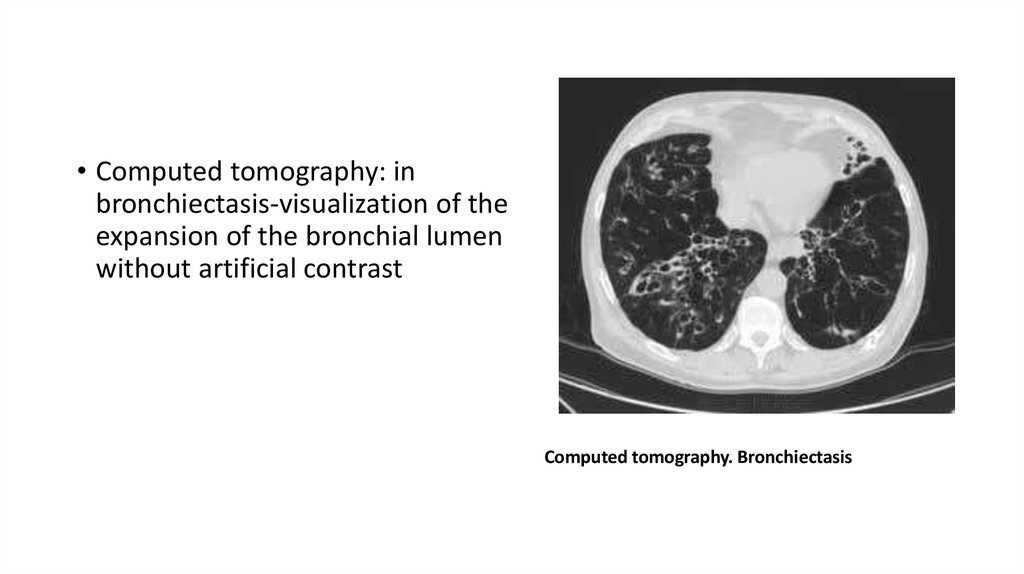
• Computed tomography: inbronchiectasis-visualization of the
expansion of the bronchial lumen
without artificial contrast
Computed tomography. Bronchiectasis
40.
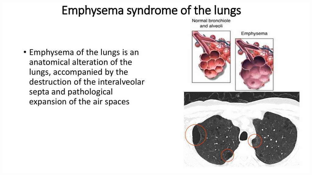
Emphysema syndrome of the lungs• Emphysema of the lungs is an
anatomical alteration of the
lungs, accompanied by the
destruction of the interalveolar
septa and pathological
expansion of the air spaces
41.
According to the degree of involvement in the pathological process ofacinus, the following types of emphysema of the lungs are
distinguished:
• panlobular (panacinar) - with the defeat of the whole acinus;
• centrilobular (centriacinar) – with a lesion of the respiratory alveoli in
the central part of the acinus;
• perilobular (periacinar) - with a lesion of the distal part of the acinus;
• okolorubtsovaya (irregular or uneven); bullous (bullous lung disease
in the presence of air cysts-bull).
• There are particularly distinguished congenital lobar emphysema of
the lungs and McLeod's syndrome-emphysema with an unclear
etiology, affecting one lung.
42.
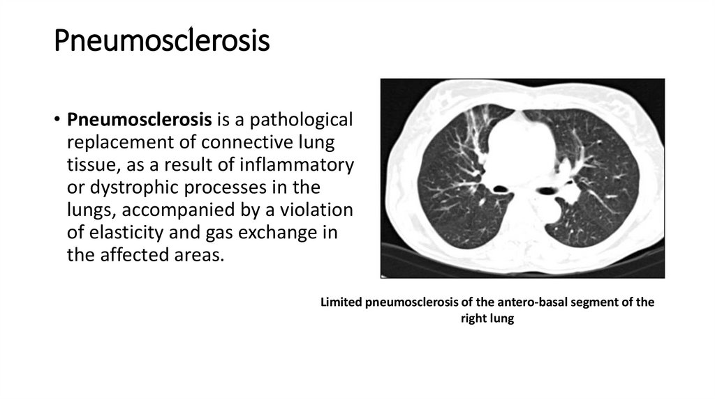
Pneumosclerosis• Pneumosclerosis is a pathological
replacement of connective lung
tissue, as a result of inflammatory
or dystrophic processes in the
lungs, accompanied by a violation
of elasticity and gas exchange in
the affected areas.
Limited pneumosclerosis of the antero-basal segment of the
right lung
43.
According to the degree of severity of the replacement of thepulmonary parenchyma with connective tissue, there are:
• pneumofibrosis - severe limited changes in the lung parenchyma,
alternating with air lung tissue
• pneumosclerosis (actually pneumosclerosis) - compaction and
replacement of the lung parenchyma with connective tissue;
• pneumocyrrosis is an extreme case of pneumosclerosis,
characterized by complete replacement of the alveoli, vessels and
bronchi with connective tissue, pleural compaction, displacement of
the mediastinal organs to the affected side.
44.
CHRONIC NONSPECIFIC PNEUMONIA• a limited inflammatory process of the lungs, characterized by the
development of purulent-necrotic foci, the growth of connective
tissue and foci of productive inflammation. The term "chronic
nonspecific pneumonia" refers to chronic inflammation of all
structures in the affected area, the proliferation of connective tissue
and abscessing (destruction).
45.
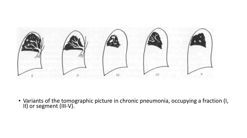
• Variants of the tomographic picture in chronic pneumonia, occupying a fraction (I,II) or segment (III-V).